Hyperthermia accelerates neuronal loss differently between the hippocampal CA1 and CA2/3 through different HIF 1α expression after transient ...
←
→
Page content transcription
If your browser does not render page correctly, please read the page content below
INTERNATIONAL JOURNAL OF MOlecular medicine 49: 55, 2022

          Hyperthermia accelerates neuronal loss differently between
             the hippocampal CA1 and CA2/3 through different
            HIF‑1α expression after transient ischemia in gerbils
                            TAE‑KYEONG LEE1*, DAE WON KIM2*, HYEJIN SIM3, JAE‑CHUL LEE3,
                          HYUNG IL KIM4,5, MYOUNG CHEOL SHIN4, JUN HWI CHO4, JOON HA PARK6,
                                CHOONG‑HYUN LEE7, MOO‑HO WON3 and JI HYEON AHN3,8

                    1
                Department of Food Science and Nutrition, Hallym University, Chuncheon, Gangwon 24252;
         2
       Department of Biochemistry and Molecular Biology and Research Institute of Oral Sciences, College of Dentistry,
  Gangnung‑Wonju National University, Gangneung, Gangwon 25457; 3Department of Neurobiology, School of Medicine,
             Kangwon National University, Chuncheon, Gangwon 24341; 4Department of Emergency Medicine,
 Kangwon National University Hospital, School of Medicine, Kangwon National University, Chuncheon, Gangwon 24289;
  5
    Department of Emergency Medicine, Dankook University Hospital, College of Medicine, Dankook University, Cheonan,
Chungnam 31116; 6Department of Anatomy, College of Korean Medicine, Dongguk University, Gyeongju, Gyeongbuk 38066;
      7
       Department of Pharmacy, College of Pharmacy, Dankook University, Cheonan, Chungnam 31116; 8Department of
     Physical Therapy, College of Health Science, Youngsan University, Yangsan, Gyeongnam 50510, Republic of Korea

                                      Received January 4, 2022; Accepted February 17, 2022

                                                  DOI: 10.3892/ijmm.2022.5111

Abstract. The hippocampus has a different vulnerability to         hyperthermia was induced for 30 min before and during
ischemia according to the subfields CA1 to CA3 (initials of        the TFI, and neuronal death and HIF‑1α expression were
cornu ammonis). It has been reported that body temperature         observed at 0, 3, 6 and 12 h, 1, 2 and 5 days after TFI. Under
changes during ischemia affect the degree of neuronal death        normothermia, TFI‑induced neuronal death of CA1 pyramidal
following transient ischemia. Hypoxia‑inducible factor 1α          neurons occurred on day 5 after TFI, but CA2/3 pyramidal
(HIF‑1α) plays a key role in regulating cellular adaptation to     neurons did not die. In contrast, under hyperthermia, the
low oxygen conditions. In the present study, we investigated the   death of CA1 and CA2/3 pyramidal neurons was observed
pattern of neuronal death (loss) in CA1 and CA2/3 following        on day 2 after TFI. Under normothermia, HIF‑1α expression
5 min transient forebrain ischemia (TFI) under hyperthermia        was significantly elevated in both CA1 and CA2/3 pyra‑
(39.5±0.2˚C) and the relationship between neuronal death           midal neurons at 12 h and 1 day after TFI, and the increased
and changes in HIF‑1α expression using western blot analysis       HIF‑1α immunoreactivity in CA1 was dramatically reduced
and immunohistochemistry in gerbils. Normothermia or               from 2 days after TFI, but not in CA2/3 pyramidal neurons.
                                                                   Under hyperthermia, the basal expression of HIF‑1α in the
                                                                   sham group was significantly higher in both CA1 and CA2/3
                                                                   pyramidal neurons at 0 h after TFI than in the normothermia
                                                                   group. HIF‑1 expression was continuously higher, peaked
Correspondence to: Professor Moo‑Ho Won, Department                at 12 h after TFI, and then significantly decreased from 1 day
of Neurobiology, School of Medicine, Kangwon National
                                                                   after TFI. Overall, the present results indicate that resistance
University, 1 Kangwondaehak‑gil, Chuncheon, Gangwon 24341,
Republic of Korea                                                  to ischemia in CA2/3 pyramidal neurons is closely associated
E‑mail: mhwon@kangwon.ac.kr                                        with the persistence of increased expression of HIF‑1α after
                                                                   ischemic insults and that hyperthermia‑induced exacerbation
Professor Ji Hyeon Ahn, Department of Physical Therapy, College
                                                                   of death of pyramidal neurons is closely related to decreased
of Health Science, Youngsan University, 288, Junam‑ro, Yangsan,
                                                                   HIF‑1α expression after ischemic insults.
Gyeongnam 50510, Republic of Korea
E‑mail: jh‑ahn@ysu.ac.kr
                                                                   Introduction
*
    Contributed equally
                                                                   It is well known that cerebral neurons have different suscep‑
Key words: body temperature, hippocampal subfields, low oxygen,    tibilities to deleterious conditions, such as ischemic injury
pyramidal neurons, resistance to ischemia, transient forebrain     and neurodegeneration, depending on brain area (1). The
ischemia                                                           hippocampus is one of the brain areas vulnerable to ischemic
                                                                   insults, and hippocampal neurons respond differently to isch‑
                                                                   emic damage according to its subfields (CA1‑3) and impact of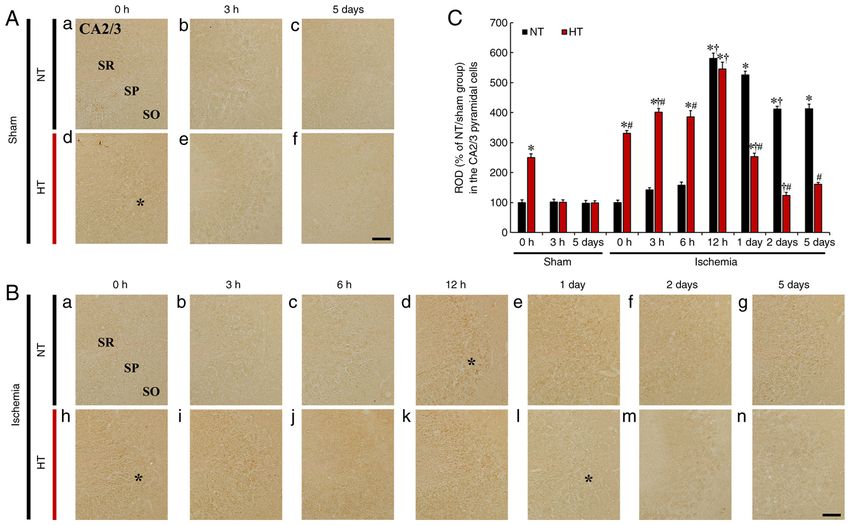
2              LEE et al: EFFECTS OF HYPERTHERMIA ON NEURONAL LOSS AND HIF‑1α AFTER TRANSIENT ISCHEMIA

ischemia. A short period (5 min) of transient forebrain ischemia         Experimental gerbils (total n=240) were divided into
(TFI) in gerbils induces death of pyramidal neurons located          four groups: i) sham‑operated gerbils under normothermia
in hippocampal CA1 at 4 or 5 days after TFI, but pyramidal           (NT/sham group; n=36), ii) TFI‑operated gerbils under normo‑
neurons of CA2/3 are resistant (2,3). Hippocampal neuronal           thermia (NT/ischemia group; n=84), iii) sham‑operated gerbils
vulnerability to cerebral ischemia can be altered by body            under hyperthermia (HT/sham group; n=36), iv) TFI‑operated
temperature or ischemic duration (4,5). Higher temperatures          gerbils under hyperthermia (HT/ischemia group; n=84).
cause more severe ischemic damage. It was reported that in               In each group, 12 gerbils (n=5 for western blotting; n=7 for
gerbils, hyperthermia before and during a brief episode (5 min)      histology) were sacrificed at 0 h (immediately after 5 min of
of TFI resulted in the accelerated death of hippocampal CA1          TFI operation), 3, 6 and 12 h, 1, 2 and 5 days after TFI (Fig. 1).
pyramidal neurons (6). Clinically, increased body temperature
during the first day after stroke is related to poor outcomes and    Induction of TFI under normothermia or hyperthermia. TFI
leads to devastating effects on mortality (7).                       induction was performed according to previously described
    Hypoxia‑inducible factor 1α (HIF‑1α), a well‑known               methods (16,17) with minor modification. The gerbils
isoform of hypoxia‑inducible factors, exerts hypoxia‑inducible       were anesthetized using inhalation anesthesia machine
transcriptional activity triggered by low oxygen condi‑              (Harvard Apparatus) with isoflurane (2.5%; Baxtor) in
tions, whereas HIF‑1β is an oxygen‑insensitive subunit (8).          oxygen (33%) and nitrous oxide (67%). Under anesthesia, the
HIF‑1α expression is localized in the cytosol of pyramidal           bilateral common carotid arteries, which are located in the
neurons located in all hippocampal subfields (CA1‑3) of rats         ventral neck, were ligated with aneurysm clips for 5 min.
and gerbils (9‑11). HIF‑1α plays a key role in regulating cellular   Before and during the TFI surgery, body temperature was
adaptation to low oxygen conditions by modulating gene               controlled using a heating pad, which was connected to a rectal
expression, targeting cell survival, angiogenesis, and glucose       thermistor (Harvard Apparatus) (Fig. 1). Rectal temperature
metabolism, which may contribute to alleviating ischemic             was controlled at 37.0±0.5˚C for normothermia and elevated
neuronal damage (12,13). Increased HIF‑1α expression signifi‑        up to 39.5±0.2˚C for hyperthermia for 30 min. After the TFI
cantly attenuates post‑ischemic damage of hippocampal CA1            surgery, the gerbils were kept in thermal incubators (tempera‑
pyramidal neurons by increasing the expression of nuclear            ture, 22±2˚C; humidity, 57±5%) to adjust body temperature on
factor‑κ B (NF‑κ B) and vascular endothelial growth factor           a normothermic level. The gerbils in the sham groups with
(VEGF) following ischemic preconditioning (9). In addition,          normothermia or hyperthermia received the same TFI surgery
systemic administration of a HIF‑1α activator (ML228) was            without ligation of the arteries.
found to alleviate neuronal apoptosis in hippocampal CA1
by attenuating the amplified expression of pro‑inflammatory          Western blot analysis. To analyze HIF‑1α protein level
cytokines and their receptors following cardiac arrest‑induced       in CA1 and CA2/3, the gerbils of each group were sacri‑
cerebral ischemia (14).                                              ficed under profound anesthesia by intraperitoneal (i.p.)
    Although previous studies have reported the effects of           injection of pentobarbital sodium (200 mg/kg) (18) (JW
HIF‑1α‑mediated neuroprotection and its related signaling            Pharmaceutical Co., Ltd.) at the designated time (0, 3, 6
pathways, few studies have been conducted on changes in              and 12 h, 1, 2 and 5 days after TFI). The death of animals
HIF‑1α expression in hippocampal subfields induced by TFI            was confirmed according to vital signs including heart
under hyperthermic conditions that cause more severe neuronal        beats, pupillary response, and respiratory pattern (lack of
damage. Therefore, the present study aimed to investigate the        cardiac activity for 5 min by cardiac palpation, unrespon‑
pattern of neuronal death and expression of HIF‑1α in hippo‑         siveness to light with dilated pupils using light into the eyes
campal subfields (CA1‑3) after 5 min TFI under hyperthermic          of the animal, and lack of spontaneously breathing pattern
conditions in gerbils.                                               with shallow and irregular breathing pattern). CA1 and
                                                                     CA2/3 tissues were respectively collected from the hippo‑
Materials and methods                                                campi using brain matrix. Proteins of CA1 and CA2/3 were
                                                                     extracted according to a previously published method (9)
Experimental animals and groups. In the present study, a total       using rabbit anti‑HIF‑1α (cat. no. ab228649, diluted 1:5,000,
of 240 male gerbils (age, 6 months; body weight, 70±5 g) were        Abcam) and anti‑β ‑actin (cat. no. ab8227, diluted 1:20,000,
used at the start of the experiment. The gerbils were bred and       Abcam) as primary antibodies.
kept in the Experimental Animal Center at Kangwon National               For quantification of the bands, densitometry analysis was
University located in Chuncheon (Korea). The animals                 performed using Scion Image software, Ver 4.0 (Scion Corp.).
were housed in a conventional room (temperature, 22±2˚C;             The ratio, as relative density, of HIF‑1α protein was cali‑
humidity, 57±5%; light/dark cycle, 12:12) with freely accessible     brated with the corresponding expression rate of β‑actin and
water and food. All experimental procedures were approved            normalized to that in NT/sham group at 0 h.
(approval no. KW‑200113‑1) by the Institutional Animal Care
and Use Committee on January 13, 2020. The Animal care               Tissue processing for histology. For histology, the gerbils of
and handling conformed to the NIH Guide for the Care and             each group were profoundly anesthetized with 200 mg/kg (i.p.)
Use of Laboratory Animals (The National Academies Press,             of pentobarbital sodium (18) (JW Pharmaceutical Co., Ltd.) at
8th edition, 2011) (15). All efforts were taken at each stage        the designated time (0, 3, 6 and 12 h, 1, 2 and 5 days after
of the experiments to minimize the numbers of animals used           TFI) and sacrificed after confirmation of death according to
and to limit any discomfort to which the animals might be            the method described above. As previously described (16,17),
exposed.                                                             in short, the gerbils were perfused transcardially with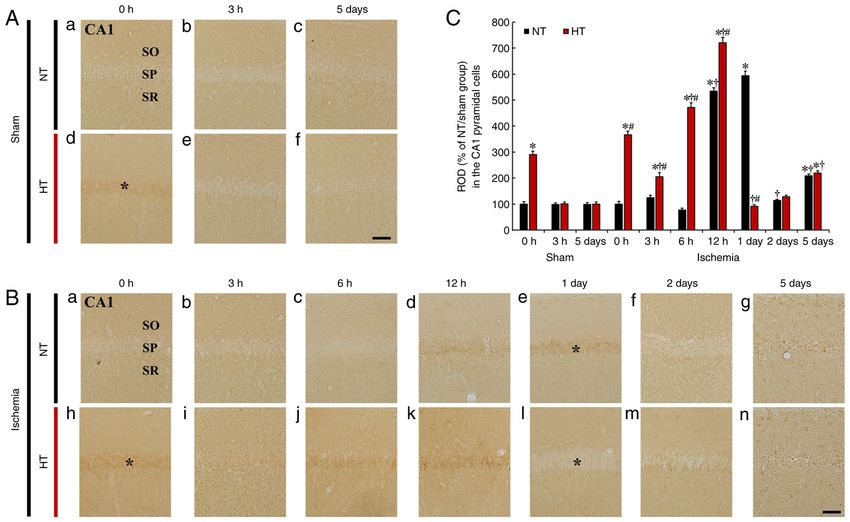
INTERNATIONAL JOURNAL OF MOlecular medicine 49: 55, 2022 3
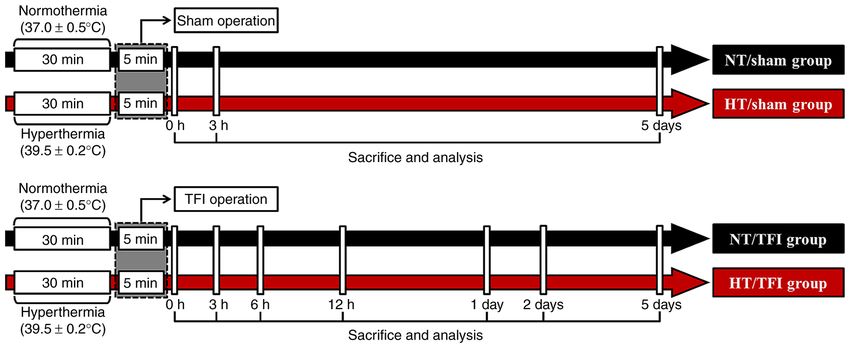
Figure 1. Timeline of the experiment. Body temperature was controlled before TFI for 30 min and during TFI. The gerbils of the sham groups were sacrificed
for western blot and histological analyses at 0 and 3 h and 5 days, and those of the TFI groups were sacrificed at 0, 3, 6 and 12 h, 1, 2 and 5 days after TFI.
TFI, transient forebrain ischemia.
4% paraformaldehyde and their brains were extracted from Immunohistochemistry. To examine changes in neurons and
the skulls. The brains were post‑fixed in the same fixative for HIF‑1α expression, immunohistochemistry was performed
5 h. The fixed brain tissues were infiltrated with 30% sucrose according to previously published methods (9,19,20). In short,
as cryoprotective agent when the brain tissues were cut. To the brain sections were immersed in 0.3% hydrogen peroxide
prepare brain sections containing the hippocampus, using a (H2O2) and soaked in 5% normal goat serum. Thereafter, the
cryostat, the frozen tissues were frontally cut into a 30‑µm brain sections were incubated with each primary antibody,
thickness. Thereafter, the sections were stored in 6‑well plates rabbit anti‑neuronal nuclei (cat. no. MAB377, NeuN; diluted
containing PBS for histological staining. 1:1,100, Chemicon International) and rabbit anti‑HIF‑1α
(cat. no. ab8366, diluted 1:500, Abcam) overnight at 4˚C.
Histochemistry using cresyl violet (CV). To examine the Subsequently, they were incubated in secondary antibodies,
morphology and distribution of hippocampal cells in all biotinylated goat anti‑rabbit IgG (cat. no. BA‑1000‑1.5, diluted
groups, CV histochemical staining was conducted as previ‑ 1:200, Vector Laboratories, Inc.) and streptavidin peroxidase
ously described (9). In brief, the sections were stained with complex (diluted 1:200, Vector Laboratories, Inc.). Finally, the
1.0% CV acetate (Sigma‑Aldrich; Merck KGaA) containing sections were visualized using 0.05% 3,3'‑diaminobenzidine
0.28% glacial acetic acid. The stained sections were tetrahydrochloride.
dehydrated using ethanol series and mounted with Canada Analyses of the numbers of NeuN immunoreac‑
balsam. tive neurons and HIF‑1α immunoreactivity in CA1 and
For analysis of hippocampal change following TFI, CA2/3 were conducted according to previously published
CV‑stained structures were observed using AxioM1 light methods (9,19‑21). In short, images of NeuN immunoreac‑
microscope at x4 and x20 magnifications (Carl Zeiss). In this tive structures and HIF‑1α immunoreactive structures were
examination, six sections per gerbil were selected at 120‑µm captured using an AxioM1 light microscope at x20 magni‑
intervals. fication. To quantitatively analyze the numbers of NeuN
immunoreactive neurons, the neurons were counted in
Histofluorescence using Fluoro‑Jade B (FJB). To examine 400 µm 2 at the middle of CA1 and CA2/3. To quantitatively
TFI‑induced neuronal death (loss) in CA1 and CA2/3, histo‑ analyze HIF‑1α immunoreactivity, the immunoreactive
fluorescence using FJB, a representative marker of neuronal structures were captured at the same area of interest and
degeneration/death, was conducted according to previously calibrated into an array of 512x512 pixels. Finally, the
published studies (16,17). In brief, the brain tissues were density of the HIF‑1α immunoreactive structures was rela‑
soaked in 0.06% potassium permanganate and incubated in tively evaluated using Adobe Photoshop version 8.0 and
0.0004% FJB (Histochem). Thereafter, the stained sections NIH Image J 1.59 software.
were prepared for observation.
For analysis of FJB‑positive cells, images of FJB‑positive Statistical analysis. Data obtained in this study are expressed
cells were captured using a fluorescence microscope as the mean ± SEM. We statistically analyzed the differences
(Carl Zeiss) with blue excitation f luorescence filter of the means between all groups by analysis of variance
(450‑490 nm). The captured images (FJB‑positive cells) were (ANOVA) with a post hoc Bonferroni's multiple comparison
analyzed using image analyzing software (NIH Image J 1.59). tests using SPSS program (version 18.0, IBM SPSS Statistics).
Cell count was performed in 400 µm2 at middle of the stratum For all statistical analyses, P‑values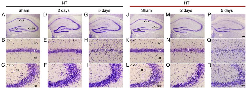
4 LEE et al: EFFECTS OF HYPERTHERMIA ON NEURONAL LOSS AND HIF‑1α AFTER TRANSIENT ISCHEMIA
Figure 2. (A‑R) Cresyl violet (CV) staining in CA1 and CA2/3 of the NT/sham (A‑C), NT/ischemia (D‑I), HT/sham (J‑L), and HT/ischemia (M‑R) groups on
day 2 and 5 after TFI. CV‑stained cells (asterisk in H) in the stratum pyramidale (SP) of CA1 of the NT/ischemia group were severely damaged on day 5 after
TFI. In the HT/ischemia group, decreased stainability in CV‑stained cells (asterisks in N, O) is shown in the SP of both CA1 and CA2/3 on day 2 after TFI.
On day 5 after TFI, CV‑stained cells (asterisks in Q, R) were apparently damaged in the SP of both CA1 and CA2/3. Scale bar, 200 µm (upper row) and 50 µm
(middle and lower rows). DG, dentate gyrus; SO, stratum oriens; SR, stratum radiatum; NT, normothermia; HT, hyperthermia.
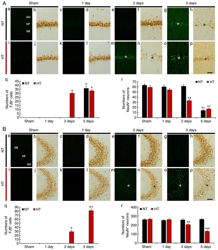
Results CA2/3. In the NT/sham group, FJB‑stained cells were not
observed in the SP of CA2/3 (Fig. 3B‑a). In the NT/ischemia
Differences in neuronal damage/death group, no FJB‑stained cells were observed at any point in time
Findings by CV histochemistry. CV‑stained cells were after TFI in CA2/3 (Fig. 3B‑c, e, g and q).
easily identified in all subfields of the gerbil hippocampus In the HT/sham group, FJB‑stained cells were not
of the NT/sham group. In particular, large pyramidal cells as detected in the SP of CA2/3 (Fig. 3B‑i). In the HT/ischemia
principal neurons consisted of the stratum pyramidale (SP) group, FJB‑stained cells were not found in the SP on day 1
(Fig. 2A‑C). In the NT/ischemia group, the distribution of after TFI (Fig. 3B‑k and q). However, many FJB‑stained
CV‑stained cells was not altered until 2 days after TFI in all cells were found in the SP on day 2 after TFI (29.4±2.8)
subfields (Fig. 2D‑F). However, on day 5 after TFI, CV‑stained (Fig. 3B‑m and q). Furthermore, on day 5 after TFI, the
pyramidal cells of the SP were apparently damaged in CA1, but number of FJB‑stained cells was significantly increased
those located in CA2/3 were similar to those in the NT/sham (81.7±4.2) (Fig. 3B‑o and q).
group (Fig. 2G‑I).
In the HT/sham group, the distribution of CV‑stained cells Findings by NeuN immunohistochemistry
in all subfields was not different from that in the NT/sham CA1. In the NT/sham group, NeuN immunoreactivity was
group (Fig. 2J‑L). In the HT/ischemia group, on day 2 after TFI, observed in the pyramidal cells of the SP in CA1 (Fig. 3A‑b). In the
most of the pyramidal cells in CA1 were weakly stained by NT/ischemia groups, the distribution of NeuN‑immunostained
CV (Fig. 2N), and CV‑stained cells in CA2/3 were not signifi‑ pyramidal cells in CA1 was not different from that of the
cantly different from those of the HT/sham group (Fig. 2O). NT/sham group on days 1 and 2 after TFI (Fig. 3A‑d and f).
On day 5 after TFI, most of the CV‑stained pyramidal cells However, on day 5 after TFI, NeuN‑immunostained pyra‑
in CA1 were severely damaged, and CA2/3 pyramidal cells midal cells were rarely observed in the SP. The percentage of
showed weak CV staining or were damaged (Fig. 2P‑R). remaining NeuN‑immunostained pyramidal cells was 22.2%
in the NT/sham group (Fig. 3A‑h and r).
Findings by FJB histofluorescence In the HT/sham group, the distribution of NeuN-
CA1. FJB fluorescence staining revealed no FJB‑stained degen‑ immunostained pyramidal cells was similar to that observed
erating (or dead) cells in the SP of CA1 in the NT/sham group in the NT/sham group (Fig. 3A‑j). In the HT/ischemia group,
(Fig. 3A‑a). In the NT/ischemia group, FJB‑stained cells were the NeuN immunoreactivity of NeuN‑immunostained
not found in the SP on days 1 and 2 after TFI (Fig. 3A‑c and e). pyramidal cells was weak on day 1 after TFI (Fig. 3A‑l).
However, a significant increase in the number of FJB‑stained On days 2 and 6 after TFI, NeuN‑immunostained pyra‑
cells (36.2±2.7) was observed in the SP on day 5 after TFI midal cells were significantly decreased (55.9 and 28.8% of
(Fig. 3A‑g and q). sham, respectively) (Fig. 3A‑n, p and r), showing that
In the HT/sham group, similar to the NT/sham group, NeuN‑immunostained pyramidal cells had significant
FJB‑stained cells were not found in the SP (Fig. 3A‑i). In the morphological alterations (pyknotic and tangle‑like appear‑
HT/ischemia group, a few FJB‑stained cells were detected ance) on day 5 after TFI (Fig. 3A‑p).
in the SP on day 1 after TFI (Fig. 3A‑k and q). In addition,
abundant FJB‑stained cells were found in the SP on days 2 CA2/3. In the NT/sham group, NeuN‑immunostained pyra‑
and 5 (30±1.5 and 33.9±2.1, respectively) after TFI (Fig. 3A‑m, midal cells were typically distributed in the SP of CA2/3
o and q). (Fig. 3B‑b). In the NT/ischemia group, the distribution and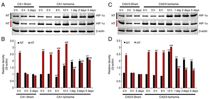
INTERNATIONAL JOURNAL OF MOlecular medicine 49: 55, 2022                                                             5

Figure 3. (A and B) Fluoro‑Jade B (FJB) histofluorescence staining and anti‑neuronal nuclei (NeuN) immunohistochemistry in CA1 (A) and CA2/3 (B) of
the NT/sham (a and b), NT/ischemia (c‑h), HT/sham (i and j) and HT/ischemia (k‑p) groups on day 1, 2 and 5 after TFI. In the NT/ischemia group, many
FJB‑stained cells (white asterisk in A‑g) were observed in the SP on day 5 after TFI in CA1, but not in CA2/3. However, in the HT/ischemia group, many
FJB‑stained cells (white asterisks in m and o) were detected in both CA1 and CA2/3 from 2 days after TFI. The numbers of NeuN‑immunostained pyramidal
cells of the NT/ischemia group were significantly reduced (black asterisk in A‑h) only in CA1 on day 5 after TFI. In the HT/ischemia group, the numbers of
NeuN‑immunostained pyramidal cells were decreased (black asterisks in n and p) in both CA1 and CA2/3 from 2 days after TFI. Scale bar, 50 µm. (q) Numbers
of FJB‑stained cells in CA1 (A) and CA2/3 (B). (r) Numbers of NeuN‑immunostained cells in CA1 (A) and CA2/3 (B). *P
6                LEE et al: EFFECTS OF HYPERTHERMIA ON NEURONAL LOSS AND HIF‑1α AFTER TRANSIENT ISCHEMIA

Figure 4. (A and C) Western blot analysis of HIF‑1α in CA1 (A) and CA2/3 (C) of the NT/sham, NT/ischemia, HT/sham, and HT/ischemia groups at 0, 3,
6 and 12 h, 1, 2 and 5 days after TFI. (B and D) Relative density of the immunoblot bands is represented. Protein levels of HIF‑1α normalized to β‑actin in CA1
(B) and CA2/3 (D) *P
INTERNATIONAL JOURNAL OF MOlecular medicine 49: 55, 2022                                                           7

Figure 5. (A and B) HIF‑1α immunohistochemistry in CA1 of the (A) NT/sham (a‑c), (B) NT/ischemia (a‑g), (A) HT/sham (d‑f) and (B) HT/ischemia (h‑n)
groups at 0, 3, 6 and 12 h, 1, 2 and 5 days after TFI. In the NT/sham group, HIF‑1α immunoreactivity in the SP was very weak. In the NT/ischemia group,
HIF‑1α immunoreactivity in the SP was significantly increased at 12 h and 1 day (asterisk), markedly reduced at 2 days and again increased at 5 days after
TFI. In the HT/sham group, HIF‑1α immunoreactivity was very high (asterisk) at 0 h and similar to that of the NT/sham group from 3 h after sham TFI. In
the HT/ischemia group, increased HIF‑1α immunoreactivity was gradually increased until 12 h after TFI and thereafter was lower (asterisk) than that in the
HT/sham group. Scale bar, 50 µm. (C) Relative optical density (ROD) (% of NT/sham group) of HIF‑1α immunoreactivity in CA1. *P
8                LEE et al: EFFECTS OF HYPERTHERMIA ON NEURONAL LOSS AND HIF‑1α AFTER TRANSIENT ISCHEMIA

Figure 6. (A and B) HIF‑1α immunohistochemistry in CA2/3 of the (A) NT/sham (a‑c), (B) NT/ischemia (a‑g), (A) HT/sham (d‑f) and (B) HT/ischemia (h‑n)
groups at 0, 3, 6 and 12 h, 1, 2 and 5 days after TFI. In the NT/sham group, HIF‑1α immunoreactivity in the SP was very weak. In the NT/ischemia group,
HIF‑1α immunoreactivity was significantly increased at 12 h (asterisk) and thereafter slightly reduced until 5 days after TFI. In the HT/sham group, HIF‑1α
immunoreactivity was very high at 0 h and thereafter reduced to the level of the NT/sham group. In the HT/ischemia group, increased HIF‑1α immunoreac‑
tivity was continuously increased until 1 day, and thereafter HIF‑1α immunoreactivity was significantly decreased until 5 days after TFI. Scale bar, 50 µm.
(C) Relative optical density (ROD) (% of NT/sham group) of HIF‑1α immunoreactivity in CA2/3. *P
INTERNATIONAL JOURNAL OF MOlecular medicine 49: 55, 2022                                                9

present study, HIF‑1α expression peaked at 12 h in both CA1         Ethics approval and consent to participate
and CA2/3 pyramidal cells of the HT/ischemia group and
then significantly decreased on day 1 after TFI (just before        Experimental procedures for the present study were approved
neuronal cell death), showing earlier neuronal death despite        by the Institutional Animal Care and Use Committee at
the initial high expression of HIF‑1α as compared with the          Kangwon National University (approval no. KW‑200113‑1).
NT/ischemia group. Although the exact reason for the early          The procedures of handling and caring animals conformed to
increase in HIF‑1α expression remains unclear, it may be due        the guidelines of the current international laws and policies
to a protective mechanism against greater damage, or another        (NIH Guide for the Care and Use of Laboratory Animals, The
mechanism may be involved in hyperthermia‑induced severe            National Academies Press, 8th ediiton, 2011).
neuronal damage. Therefore, based on our and previous studies,
it is likely that the reduction of increased HIF‑1α expression in   Patient consent for publication
the hippocampus precedes ischemic neuronal cell death under
normothermic and hyperthermic conditions.                           Not applicable.
     In summary, the present study showed that pyramidal cells
in CA1 and CA2/3 had differences in ischemia sensitivity            Competing interests
under normothermia, and the relatively high resistance of
CA2/3 pyramidal cells may be closely related to the main‑           The authors declare that they have no competing interests.
tenance of high expression of HIF‑1α after TFI. In addition,
hyperthermia accelerated ischemic neuronal death in both            Authors' information
CA1 and CA2/3, showing that hyperthermia‑induced neuronal
death is closely related to a reduction in increased HIF‑1α         Professor Moo‑Ho Won: ORCID: 0000‑0002‑7178‑6501;
expression in both ischemic CA1 and CA2/3. Therefore, we            Professor Ji Hyeon Ahn, ORCID: 0000‑0002‑5304‑0714.
should consider the environment or conditions of the brain in
which ischemic insults occur because ischemic damage in the         References
brain is not the same in all patients with ischemic stroke.
                                                                     1. Wang X and Michaelis EK: Selective neuronal vulnerability to
                                                                        oxidative stress in the brain. Front Aging Neurosci 2: 12, 2010.
Acknowledgements                                                     2. Kirino T: Delayed neuronal death in the gerbil hippocampus
                                                                        following ischemia. Brain Res 239: 57‑69, 1982.
                                                                     3. Yu DK, Yoo KY, Shin BN, Kim IH, Park JH, Lee CH, Choi JH,
The authors would like to acknowledge Mr. Seung Uk Lee                  Cho YJ, Kang IJ, Kim YM and Won MH: Neuronal damage
and Ms. Hyun Sook Kim for their technical assistance in this            in hippocampal subregions induced by various durations of
study.                                                                  transient cerebral ischemia in gerbils using Fluoro‑Jade B histo‑
                                                                        fluorescence. Brain Res 1437: 50‑57, 2012.
                                                                     4. Minamisawa H, Smith ML and Siesjo BK: The effect of mild
Funding                                                                 hyperthermia and hypothermia on brain damage following 5,
                                                                        10 and 15 min of forebrain ischemia. Ann Neurol 28: 26‑33, 1990.
                                                                     5. Dietrich WD, Busto R, Valdes I and Loor Y: Effects of normo‑
This research was supported by Basic Science Research                   thermic versus mild hyperthermic forebrain ischemia in rats.
Program through the National Research Foundation                        Stroke 21: 1318‑1325, 1990.
of Korea (NRF) funded by the Ministry of Education                   6. Kim MJ, Cho JH, Park JH, Park JH, Ahn JH, Tae HJ, Cho GS,
                                                                        Yan BC, Hwang IK, Lee CH, et al: Impact of hyperthermia
(NRF‑2021R1A2C1094224, NRF‑2020R1F1A1052380, and                        before and during ischemia‑reperfusion on neuronal damage and
NRF‑2020R1I1A1A01070897).                                               gliosis in the gerbil hippocampus induced by transient cerebral
                                                                        ischemia. J Neurol Sci 348: 101‑110, 2015.
                                                                     7. de Jonge JC, Wallet J and van der Worp HB: Fever worsens
Availability of data and materials                                      outcomes in animal models of ischaemic stroke: A systematic
                                                                        review and meta‑analysis. Eur Stroke J 4: 29‑38, 2019.
The data presented in this study are available on request from       8. Albadari N, Deng S and Li W: The transcriptional factors HIF‑1
                                                                        and HIF‑2 and their novel inhibitors in cancer therapy. Expert
the corresponding author.                                               Opin Drug Discov 14: 667‑682, 2019.
                                                                     9. Lee JC, Tae HJ, Kim IH, Cho JH, Lee TK, Park JH, Ahn JH,
Authors' contributions                                                  Choi SY, Bai HC, Shin BN, et al: Roles of HIF‑1α, VEGF, and
                                                                        NF‑κ B in ischemic preconditioning‑mediated neuroprotection
                                                                        of hippocampal CA1 pyramidal neurons against a subsequent
Conceptualization of the study concept was achieved by MHW              transient cerebral ischemia. Mol Neurobiol 54: 6984‑6998, 2017.
and JHA. Investigations in regards to western blot analysis and     10. Long Q, Fan C, Kai W, Luo Q, Xin W, Wang P, Wang A, Wang Z,
                                                                        Han R, Fei Z, et al: Hypoxia inducible factor‑1α expression is
immunohistochemistry were carried out by TKL, DWK and                   associated with hippocampal apoptosis during epileptogenesis.
HS. The study methodology, use of the software, validation              Brain Res 1590: 20‑30, 2014.
of the data collected and supervision of the study were the         11. Zhu T, Zhan L, Liang D, Hu J, Lu Z, Zhu X, Sun W, Liu L and
                                                                        Xu E: Hypoxia‑inducible factor 1α mediates neuroprotection
responsibility of JCL, HIK, MCS, JHC, JHP, CHL, JHP and                 of hypoxic postconditioning against global cerebral ischemia.
JHC. Data curation was carried out by TKL, DWK and JHA.                 J Neuropathol Exp Neurol 73: 975‑986, 2014.
Writing of the original draft was carried out by TKL, DWK,          12. Shi H: Hypoxia inducible factor 1 as a therapeutic target in isch‑
                                                                        emic stroke. Curr Med Chem 16: 4593‑4600, 2009.
MHW and JHA. Writing of the review and editing were MHW             13. Chatzi C, Schnell E and Westbrook GL: Localized hypoxia
and JHA. Project administration was the responsibility of TKL           within the subgranular zone determines the early survival of
DWK, MHW and JHA. Funding acquisition was carried out                   newborn hippocampal granule cells. Elife 4: e08722, 2015.
                                                                    14. Xing J and Lu J: HIF‑1α activation attenuates IL‑6 and TNF‑α
by JHA, MHW and TKL. All authors have read and agreed to                pathways in hippocampus of rats following transient global isch‑
the published version of the manuscript.                                emia. Cell Physiol Biochem 39: 511‑520, 2016.
10 LEE et al: EFFECTS OF HYPERTHERMIA ON NEURONAL LOSS AND HIF‑1α AFTER TRANSIENT ISCHEMIA
15. National Research Council (US) Committee for the Update of 23. Azzimondi G, Bassein L, Nonino F, Fiorani L, Vignatelli L, Re G
the Guide for the Care and Use of Laboratory Animals: Guide for and D'Alessandro R: Fever in acute stroke worsens prognosis: A
the Care and Use of Laboratory Animals. 8th edition. National prospective study. Stroke 26: 2040‑2043, 1995.
Academies Press, Washington, DC, 2011. 24. Reith J, Jørgensen HS, Pedersen PM, Nakayama H, Raaschou HO,
16. Kim B, Ahn JH, Kim DW, Lee TK, Kim YS, Shin MC, Cho JH, Jeppesen LL and Olsen TS: Body temperature in acute stroke:
Kim YM, Park JH, Kang IJ, et al: Transient forebrain ischemia Relation to stroke severity, infarct size, mortality, and outcome.
under hyperthermic condition accelerates memory impairment Lancet 347: 422‑425, 1996.
and neuronal death in the gerbil hippocampus by increasing 25. Busija DW, Leffler CW and Pourcyrous M: Hyperthermia
NMDAR1 expression. Mol Med Rep 23: 256 2021. increases cerebral metabolic rate and blood flow in neonatal pigs.
17. Ohk TG, Ahn JH, Park YE, Lee TK, Kim B, Lee JC, Cho JH, Am J Physiol 255 (2 Pt 2): H343‑H346, 1988.
Park JH, Won MH and Lee CH: Comparison of neuronal death 26. Corbett D and Thornhill J: Temperature modulation (hypo‑
and expression of TNF‑α and MCT4 in the gerbil hippocampal thermic and hyperthermic conditions) and its influence on
CA1 region induced by ischemia/reperfusion under hyperthermia histological and behavioral outcomes following cerebral isch‑
to those under normothermia. Mol Med Rep 22: 1044‑1052, 2020. emia. Brain Pathol 10: 145‑152, 2000.
18. Kanda I: Exotic animal formulary 4 edition. Can Vet J 56: 736, 2015. 27. Kopach O, Maistrenko A, Lushnikova I, Belan P, Skibo G and
19. Kim DW, Cho JH, Cho GS, Kim IH, Park JH, Ahn JH, Chen BH, Voitenko N: HIF‑1α‑mediated upregulation of SERCA2b: The
Shin BN, Tae HJ, Hong S, et al: Hyperthermic preconditioning endogenous mechanism for alleviating the ischemia‑induced
severely accelerates neuronal damage in the gerbil ischemic intracellular Ca(2+) store dysfunction in CA1 and CA3 hippo‑
hippocampal dentate gyrus via decreasing SODs expressions. campal neurons. Cell Calcium 59: 251‑261, 2016.
J Neurol Sci 358: 266‑275, 2015. 28. Lushnikova I, Orlovsky M, Dosenko V, Maistrenko A and Skibo G:
20. Yang GE, Tae HJ, Lee TK, Park YE, Cho JH, Kim DW, Park JH, Brief anoxia preconditioning and HIF prolyl‑hydroxylase inhibi‑
Ahn JH, Ryoo S, Kim YM, et al: Risperidone treatment after tion enhances neuronal resistance in organotypic hippocampal
transient ischemia induces hypothermia and provides neuropro‑ slices on model of ischemic damage. Brain Res 1386: 175‑183,
tection in the gerbil hippocampus by decreasing oxidative stress. 2011.
Int J Mol Sci 20: 4621, 2019. 29. Jeon GW, Sheldon RA and Ferriero DM: Hypoxia‑inducible factor:
21. Lee JC, Cho JH, Lee TK, Kim IH, Won MH, Cho GS, Shin BN, Role in cell survival in superoxide dismutase overexpressing mice
Hwang IK, Park JH, Ahn JH, et al: Effect of hyperthermia on after neonatal hypoxia‑ischemia. Korean J Pediatr 62: 444‑449,
calbindin‑D 28k immunoreactivity in the hippocampal forma‑ 2019.
tion following transient global cerebral ischemia in gerbils.
Neural Regen Res 12: 1458, 2017. This work is licensed under a Creative Commons
22. Bartsch T and Wulff P: The hippocampus in aging and disease: Attribution-NonCommercial-NoDerivatives 4.0
From plasticity to vulnerability. Neuroscience 309: 1‑16, 2015. International (CC BY-NC-ND 4.0) License.You can also read


