Annals of Case Reports - Gavin Publishers
←
→
Page content transcription
If your browser does not render page correctly, please read the page content below
Annals of Case Reports
                                                                                                            Park KM, et al. Ann Case Report 6: 667.

Case Report                                                                                                                Ann Case Report 6: 667.

    Resolution of Baker’s Cyst with Knee Osteoarthritis by Bidirectional
           Therapy: Posterior Aspiration and Anterior Injection
Kyeong Mee Park1*, Tae Hwan Cho1, Dong Phill Cho2
1
    Cho Orthopaedic & Oriental Clinic, Seoul, South Korea
2
    Fuzopuncture Institute, Seoul, South Korea
        *
         Corresponding author: Kyeong Mee Park, Cho Orthopaedic & Oriental Clinic, Seoul 06979, South Korea
        Citation: Park KM, Cho TH, Cho DP. (2021) Resolution of Baker’s Cyst with Knee Osteoarthritis by Bidirectional Therapy:
        Posterior Aspiration and Anterior Injection. Ann Case Report 6: 667. DOI: 10.29011/2574-7754.100667
        Received Date: 15 May, 2021; Accepted Date: 21 May, 2021; Published Date: 25 May, 2021

            Abstract
                  Baker’s cyst, the popliteal cyst, is a swelling with synovial fluid located behind the knee joint. Popliteal cysts often
            cause pain and may need therapeutic interventions. A 68-year-old female with pain on the knee fossa and heavy discomfort
            on the calf visited our clinic. On physical examination, her left knee was swollen with a bulging mass in the popliteal region
            and showed a slight limitation in flexion movement compared to normal right knee. Ultrasonography showed anechoic images
            both in suprapatellar and posterior compartments of the left knee. Using an “in-plane needle technique” of ultrasonography, the
            cyst was punctured by a sterile syringe needle and cystic fluid was completely extracted. After shifting the position from prone
            to supine, she subsequently got a bolus of triamcinolone in the knee cavity. After the treatment, her uncomfortable symptoms
            disappeared and pain was decreased from VAS 7 to VAS 1. Up to 8 weeks from the treatment, her left knee did not show
            anechoic images in the suprapatellar and knee fossa compartments by ultrasonographic examination. The bidirectional therapy
            with posterior aspiration and anterior injection brought no recurrence during the 8 week-follow up period.

Keywords: Popliteal cyst; knee osteoarthritis; Ultrasonography;           anterior triamcinolone injection in popliteal cyst with knee OA
Aspiration; Injection; Steroid                                            patient.

Introduction                                                              Case
       Baker’s cyst, the popliteal cyst, is a synovial fluid-                   A 68-year-old female was admitted to Cho Orthopaedic
filled sac located between the tendons of gastrocnemius and               & Oriental Clinic with swollen popliteus and hardness to bend
semimembranosus muscles in the posteromedial region of knee               the left knee for 6 days. Her symptoms started 2 weeks ago and
fossa. Popliteal cyst arises as the result of accumulation and            worsened with time. She expressed her left calf became tight and
extrusion of synovial fluid, which is mostly implicated with              swollen. On physical examination, the left knee was swollen with
inflammatory knee joint diseases [1]. It is proposed that chronic         a bulging mass in the popliteal region. Passive dorsiflexion of the
joint effusion in knee osteoarthritis (OA) triggers a continuous          ankles was normal and dorsalis pedis pulses were palpable. The
unidirectional flow of synovial fluid from the cavity to the popliteal    range of motion of the left knee was slightly limited compared to
cyst [2]. Popliteal cyst in knee OA patients can cause a sensation        normal right knee. All ultrasound examinations were performed
of tightness, discomfort, or pain behind the knee. Treatment of           using a linear 3-16 MHz probe (HS40, Samsung Madison,
popliteal cyst is carried out only if it is symptomatic. If necessary,    Korea). With the patient in supine position and extended knees,
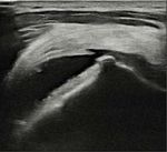
the cyst can be drained, injected with a steroid, or surgically           definite osteophytes and narrowing of joint space were identified
removed to relieve the symptoms. Open surgical excision surgery           in her left femorotibial joint by ultrasonography (Figure 1A). A
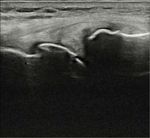
or arthroscopic surgery from posterior popliteal fossa sometimes          scoring system based on osteophytosis validated her as grade 2
discourages the patients due to a considerable recurrence rate [3].       of knee OA. Joint effusion was identified as compressible and
Scholars have insisted that popliteal cyst with knee OA patients          anechoic distension of the joint space, which was measured in the
is communicated with knee cavity and the cyst would disappear             suprapatellar recess (Figure 1B). With the patient lying in prone
automatically when joint effusion is ceased [4]. Here, we report a        position and extended knees, the Baker’s cyst was recognized as an
result of bidirectional therapy with posterior cystic aspiration and      anechoic cystic formation between the tendons of gastrocnemius

1                                                                                                                                Volume 6; Issue 03
Ann Case Rep, an open access journal
ISSN: 2574-7754
Citation: Park KM, Cho TH, Cho DP. (2021) Resolution of Baker’s Cyst with Knee Osteoarthritis by Bidirectional Therapy: Posterior Aspiration and
Anterior Injection. Ann Case Report 6: 667. DOI: 10.29011/2574-7754.100667
and semimembranosus muscles behind the left knee (Figure 2A). complained of a feeling of tightness behind the left knee and heavy
Under complete aseptic conditions, the cyst was punctured by an discomfort on the posterior side of the left calf. However, her
18-G, 1.5-inch syringe needle using an “in-plane needle approach” uncomfortable symptoms disappeared after posterior aspiration
of ultrasonography (Figure 2B). As the needle was introduced from from the cyst and anterior injection of triamcinolone into the knee
posteromedial direction, the intraluminal content was completely cavity. The VAS pain score was decreased from 7 points before the
extracted (Figure 2C). Once the cyst was thoroughly aspirated and treatment to 1 point after the treatment (Figure 3). Up to 8 weeks
decompressed, the puncture was covered by a sterile compression after the treatment, the left knee did not show anechoic images both
bandage. in suprapatellar and knee fossa compartments by ultrasonography.
Figure 1A: Ultrasonography of osteophytes in femorotibial joint Figure 2A: Ultrasonography of Baker’s cyst of the patient.
of the patient.
Figure 2B: Ultrasonography of in plane-needle approach to
Baker’s cyst.
Figure 1B: Ultrasonography of knee joint effusion in suprapatellar
recess.
The patient then shifted the position to supine and made her
knees bended at an angle of 45 by a triangle support. The knee
joint was determined by identifying and marking the dent between
the articular surfaces of lateral condyles of femur and tibia. Then, a
single intraarticular bolus of 40 mg triamcinolone (5 mL injection
volume in saline) was given to the patient without extraction of
knee effusion by a 23-gauge, 3.5-inch sterile syringe needle. She
was taught to perform knee compression by elastic bandages for
2 days following the treatment. The patient was followed up after
the treatment for 8 weeks. The VAS score and ultrasonographic
evaluation were compared between posttreatment and pretreatment.
Before the treatment, the patient had a limitation on flexion of Figure 2C: Ultrasonography of Baker’s cyst after the bidirectional
the left knee joint and suffered pain behind the knee. She also intervention.
2 Volume 6; Issue 03
Ann Case Rep, an open access journal
ISSN: 2574-7754Citation: Park KM, Cho TH, Cho DP. (2021) Resolution of Baker’s Cyst with Knee Osteoarthritis by Bidirectional Therapy: Posterior Aspiration and
Anterior Injection. Ann Case Report 6: 667. DOI: 10.29011/2574-7754.100667
with knee OA. According to our undisclosed data, a bidirectional
therapy consisting of posterior aspiration from the cyst and anterior
injection to the knee cavity was more efficient and longer lasting
than unidirectional one with posterior aspiration from the cyst and
posterior injection to the cyst. Further, the posterior aspiration
and anterior injection intervention is simpler and safer than open
surgery or arthroscopic resection.
This case study is presented to share our experience on the
bidirectional intervention to the popliteal cyst with knee OA. The
bidirectional intervention is worth being considered one of the first
choices which patients with Baker’s cyst consider a non-surgical
Figure 3: Effect of the bidirectional therapy on pain. and non-recurrent remedy.
Discussion References
In adults, popliteal cysts usually occur concomitantly with 1. Sansone V, De Ponti A. (1999) Arthroscopic treatment of popliteal cyst
knee OA which results in persistent and excess production of and associated intra-articular knee disorders in adults. Arthroscopy.
15: 368-372.
synovial fluid. Marti-Bonmati et al. [5] reported there was extremely
significant association between knee joint effusion and popliteal 2. Lindgren PG, Willen R. (1977) Gastrocnemio-semimembranosus
bursa and its relation to the knee joint. I Anatomy and histology. Acta
cyst using MRI examination. The bursa space is communicated Radiol Diagn (Stockh). 18: 497-512.
with knee cavity through one-way valvular mechanism covered by
posteromedial capsular fold. Accumulated evidence manifested a 3. Newsham KR. (2009) Recurrent popliteal cyst in an adult: a case
report and review. Orthop Nurs. 2009; 28: 11-14.
posteromedial injection of steroid after cystic aspiration induced a
rapid elimination of popliteal cyst associated with knee OA [6,7]. 4. Rupp S, Seil R, Jochum P, Jochum P, Kohn D. (2002) Popliteal cysts in
adults. Prevalence, associated intraarticular lesions, and results after
In a 56-year-old male with atraumatic left knee pain and swelling, arthroscopic treatment. Am J Sports Med. 30: 112-115.
Baker’s cyst aspiration with corticosteroid injection represented a
safe alternative treatment option for patients [8]. 5. Marti-Bonmati L, Molla E, Dosda R, Casillas C, Ferrer P. (2000)
MR imaging of baker cysts prevalence and relation to internal
However, there is a question of whether the in situ-injection derangements of the knee. MAGMA. 10: 205-210.
of triamcinolone at the site of cystic extraction is recommended 6. Mert K, Mehmet C, Hüseyin NE, Mustafa K, Meltem C, Mahmut Y, et
because of a substantial recurrency after the treatment. We al. (2012) Ultrasound guided percutaneous treatment and follow-up of
premised if a bolus of triamcinolone poured into the anterior knee Baker’s cyst in knee osteoarthritis. Eur J Radiol. 81: 3466-3471.
cavity, the synovial flow to the posterior popliteal cyst would be 7. Bandinelli F, Fedi R, Generini S, Porta F, Candelieri A, et al. (2012)
reduced, resulting in the extinction of the cyst. The result was just Longitudinal ultrasound and clinical follow-up of Baker’s cysts injection
with steroids in knee osteoarthritis. Clin Rheumatol. 31: 727-731.
as we expected. The anterior injection of triamcinolone after the
posterior cystic aspiration resulted in pain relief and disappearance 8. Fredericksen K, Kiel J. (2021) Bedside ultrasound‐guided aspiration
of the cyst during the 8-week follow-up period. We evaluate the and corticosteroid injection of a baker’s cyst in a patient with
osteoarthritis and recurrent knee pain. J Am Coll Emerg Physicians
bidirectional intervention is a valid method to treat popliteal cyst Open. 2: e12424.
3 Volume 6; Issue 03
Ann Case Rep, an open access journal
ISSN: 2574-7754You can also read


