Adhesion Molecule Profile and the Effect of Anti-VLA-4 mAb Treatment in Experimental Autoimmune Encephalomyelitis, a Mouse Model of Multiple Sclerosis
←
→
Page content transcription
If your browser does not render page correctly, please read the page content below

International Journal of
              Molecular Sciences

Article
Adhesion Molecule Profile and the Effect of Anti-VLA-4 mAb
Treatment in Experimental Autoimmune Encephalomyelitis, a
Mouse Model of Multiple Sclerosis
Grażyna Pyka-Fościak * , Grzegorz J. Lis                    and Jan A. Litwin

                                          Department of Histology, Jagiellonian University Medical College, 31-034 Kraków, Poland;
                                          grzegorz.lis@uj.edu.pl (G.J.L.); j.a.litwin@uj.edu.pl (J.A.L.)
                                          * Correspondence: gpfosciak@cm-uj.krakow.pl

                                          Abstract: In the course of multiple sclerosis (MS) and its animal model, experimental autoimmune
                                          encephalomyelitis (EAE), the infiltration of lymphocytes and other inflammatory cells across the
                                          blood–brain barrier is associated with interactions between adhesion molecules expressed by infiltrat-
                                          ing cells and vascular endothelium. Monoclonal antibodies (mAb) against the α4 subunit of α4-β1
                                          integrin (VLA-4) show beneficial effects in both MS and EAE. (1) Background: The aim of this study
                                          was to examine the expression of selected adhesion molecules: VLA-4, VCAM-1, LFA-1, ICAM-1
                                          and PECAM-1 in the successive phases of EAE and the effect of anti-VLA-4 mAb treatment on that
                                          expression. (2) Methods: EAE was induced in C57BL/6 mice by immunization with MOG35–55
                                          peptide. The animals were killed in three successive phases of the disease: onset (day 13), peak (day
                                          18) and chronic (day 28). Frozen sections of the lumbar spinal cord were examined by quantitative
                                          immunofluorescence microscopy. The expression of the studied molecules was quantified as the
                                          percentage of the cross-sectioned spinal cord lesion area occupied by immunopositive structures. (3)
Citation: Pyka-Fościak, G.; Lis, G.J.;
                                          Results: The expression of the studied molecules showed two temporal patterns: (1) an increase in the
Litwin, J.A. Adhesion Molecule
Profile and the Effect of Anti-VLA-4
                                          onset phase, a maximum in the peak phase and a decrease in the chronic phase, which corresponded
mAb Treatment in Experimental             to the temporal pattern of the clinical score, the number of lesions and the inflammation level (ICAM-
Autoimmune Encephalomyelitis, a           1, LFA-1 and PECAM-1), and (2) an increase in the peak phase and no significant change or further
Mouse Model of Multiple Sclerosis.        increase in the chronic phase (VCAM-1, VLA-4). Among the molecules studied, ICAM-1 and LFA-1
Int. J. Mol. Sci. 2022, 23, 4637.         exhibited the highest expression levels in the peak phase of EAE. Anti-VLA-4 mAb inhibited the
https://doi.org/10.3390/ijms23094637      expression of not only VLA-4 but also other adhesion molecules. (4) Conclusions: The interactions of
Academic Editors: Savina Apolloni,
                                          adhesion molecules governing the migration of leukocytes across the blood–brain barrier change in
Nadia D’Ambrosi and Cristoforo            the successive phases of EAE. The therapeutic mechanism of anti-VLA-4 mAb treatment seems to
Comi                                      include a complex influence on a variety of adhesion molecules expressed by infiltrating cells and
                                          vascular endothelium.
Received: 10 March 2022
Accepted: 20 April 2022
                                          Keywords: experimental autoimmune encephalomyelitis (EAE); anti-VLA-4 mAb; inflammation;
Published: 22 April 2022
                                          adhesion molecules; multiple sclerosis
Publisher’s Note: MDPI stays neutral
with regard to jurisdictional claims in
published maps and institutional affil-
iations.                                  1. Introduction
                                                The specific expression of adhesion molecules at the blood–brain barrier (BBB) level
                                          is a pathogenic symptom in neuroinflammatory diseases such as multiple sclerosis (MS)
Copyright: © 2022 by the authors.
                                          and its animal model, experimental autoimmune encephalomyelitis (EAE) [1–3]. When
Licensee MDPI, Basel, Switzerland.        autoreactive pathogenic Th cells break down the BBB, complex molecular interactions occur
This article is an open access article    between adhesion molecules in the cell membranes of the infiltrating cells and capillary
distributed under the terms and           endothelial cells [3,4]. The invasion of Th cells into the brain and spinal cord leads to the
conditions of the Creative Commons        formation of lesions exhibiting an inflammatory response, demyelination, gliosis (scarring),
Attribution (CC BY) license (https://     axonal injury and axonal loss.
creativecommons.org/licenses/by/                Studies on adhesion molecules and their expression patterns at the BBB during leuko-
4.0/).                                    cyte infiltration in the course of EAE have revealed that these molecules are a valuable

Int. J. Mol. Sci. 2022, 23, 4637. https://doi.org/10.3390/ijms23094637                                          https://www.mdpi.com/journal/ijms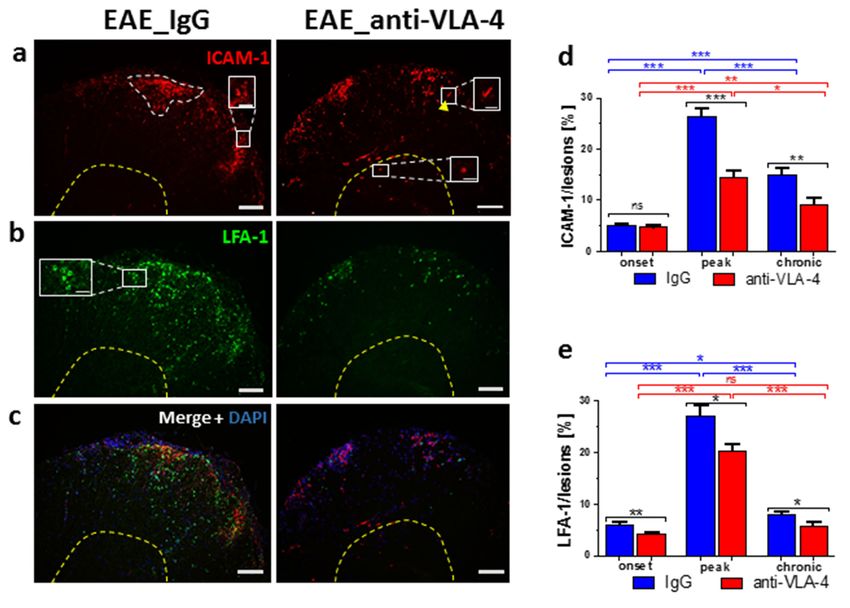
Studies on adhesion molecules and their expression patterns at the BBB during leu-
kocyte infiltration in the course of EAE have revealed that these molecules are a valuable
target for the evaluation of therapeutic interventions at the BBB. The interactions between
Int. J. Mol. Sci. 2022, 23, 4637 α4-β1 integrin (VLA-4), lymphocyte function-associated antigen (LFA-1), vascular 2 of 12cell ad-
hesion molecule (VCAM-1) and intercellular adhesion molecule (ICAM-1) influence the
adhesion and migration of infiltrating cells [5,6]. Another important adhesion molecule
involved
target for inthethe transendothelial
evaluation of therapeutic migration of leukocytes
interventions at the BBB. is The
platelet-endothelial
interactions between cell adhe-
sion molecule-1 (PECAM-1, CD31) [7]. It shows homophilic
α4-β1 integrin (VLA-4), lymphocyte function-associated antigen (LFA-1), vascular cell interaction through its extra-
cellular
adhesiondomain
molecule(sPECAM-1)
(VCAM-1) and and also binds
intercellular to integrin
adhesion molecule αv-β3, an adhesion
(ICAM-1) influence the molecule
adhesion
found on and migrationcells
endothelial of infiltrating
[8]. PECAM-1 cells [5,6]. Another
stabilizes BBB important adhesion
permeability and molecule
regulates in- vascu-
volved
lar in theand
integrity transendothelial
immune cellmigrationtrafficking of leukocytes
[7]. is platelet-endothelial cell adhesion
molecule-1
Blocking of adhesion molecules is an effectiveinteraction
(PECAM-1, CD31) [7]. It shows homophilic strategy tothroughprevent itsCNS
extracellular
inflammation
domain (sPECAM-1) and also binds to integrin αv-β3, an adhesion molecule found on
in MS and its animal model, EAE [4,9]. Natalizumab, used in the therapy of MS, is a mon-
endothelial cells [8]. PECAM-1 stabilizes BBB permeability and regulates vascular integrity
oclonal antibody (mAb) against the α4 subunit of α4-β1 (VLA-4) and α4-β7 integrins; its
and immune cell trafficking [7].
main Blocking
mechanism of action
of adhesion is to block
molecules is anthe binding
effective of lymphocyte
strategy to prevent CNS α4 integrins
inflammationto their en-
dothelial
in MS and receptors
its animal (VCAM-1),
model, EAE preventing inflammatory
[4,9]. Natalizumab, used cells
in thefrom
therapycrossing
of MS, theis BBB,
a en-
tering the CNS
monoclonal and attacking
antibody (mAb) against myelin the sheaths
α4 subunit [10,11].
of α4-β1 The(VLA-4)
results and
of studies on the effect of
α4-β7 integrins;
natalizumab
its main mechanism (anti-VLA-4)
of action therapy
is to block in EAE are partly
the binding confusing.α4Anti-VLA-4
of lymphocyte integrins to theirmAb treat-
endothelial
ment was found receptors (VCAM-1),
to suppress preventing inflammatory
inflammatory infiltration and cells from crossing
clinical the BBB,
scores during the pro-
entering the CNS and attacking myelin sheaths [10,11]. The
gression of EAE [10,12]. Preclinical administration of anti-VLA-4 mAb delayed the onset results of studies on the
effect of natalizumab (anti-VLA-4) therapy in EAE are partly confusing. Anti-VLA-4 mAb
and reduced the severity of the disease [12,13], but the treatment at the peak of acute dis-
treatment was found to suppress inflammatory infiltration and clinical scores during the
ease or during remission exacerbated disease relapses [13]. Administration of synthetic
progression of EAE [10,12]. Preclinical administration of anti-VLA-4 mAb delayed the
VLA-4
onset andantagonist
reduced during the chronic
the severity phase [12,13],
of the disease of EAEbut had theessentially
treatment no effect;
at the peakmoreover,
of
disease worsening
acute disease occurred
or during afterexacerbated
remission cessation ofdisease treatment. The[13].
relapses lackAdministration
of effect after of late anti-
VLA-4
synthetictreatment can be associated
VLA-4 antagonist during the with changes
chronic phasein oftheEAEexpression of adhesion
had essentially molecules
no effect;
atmoreover,
differentdisease
stagesworsening
of the disease development
occurred after cessation[14].of treatment. The lack of effect after
late The
anti-VLA-4
variabletreatment
activity ofcan be associatedinfiltrates
inflammatory with changes
in theinCNS,the expression
characteristic of adhesion
of MS and EAE,
molecules at different stages of the disease development [14].
probably reflects the differentiated expression of adhesion molecules associated with leuko-
The variable activity of inflammatory infiltrates in the CNS, characteristic of MS and
cyte migration. In this context, we used quantitative immunofluorescence on EAE spinal cord
EAE, probably reflects the differentiated expression of adhesion molecules associated with
sections to investigate the expression of VCAM-1/VLA-4 and ICAM-1/LFA-1 pairs of interact-
leukocyte migration. In this context, we used quantitative immunofluorescence on EAE
ing adhesion
spinal molecules,
cord sections and we the
to investigate compared
expression it with the expression and
of VCAM-1/VLA-4 of PECAM-1
ICAM-1/LFA-1 and studied
the effect
pairs of anti-VLA-4
of interacting adhesionmAb treatment
molecules, andonwethat expression
compared it withinthe
theexpression
successive phases of EAE.
of PECAM-
The spinal cord was chosen because it is the dominant location
1 and studied the effect of anti-VLA-4 mAb treatment on that expression in the successive of EAE lesions [15], and some-
times
phasesEAE affects
of EAE. Thethespinal
spinalcordcordwas butchosen
not thebecause
brain [16].it is the dominant location of EAE
lesions [15], and sometimes EAE affects the spinal cord but not the brain [16].
2. Results
2. Results
The outline of the study is presented in Figure 1.
The outline of the study is presented in Figure 1.
Figure 1. Outline of the study.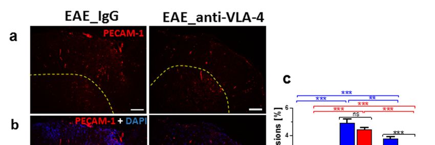
Int. J. Mol. Sci. 2022, 23, x FOR PEER REVIEW 3 of 12
Int. J. Mol. Sci. 2022, 23, 4637 3 of 12
Figure 1. Outline of the study.
2.1.
2.1. Effect
Effect of
of Anti-VLA-4
Anti-VLA-4 mAb
mAb and
and IgG
IgG Treatment on the
Treatment on the Development
Development of of Progressive
Progressive EAE
EAE
The clinicalcourse
The clinical courseofofEAE
EAEmice
mice was
was attenuated
attenuated by by anti-VLA-4
anti-VLA-4 mAb mAb
andand
IgG IgG treat-
treatment.
ment. Anti-VLA-4
Anti-VLA-4 mAb treatment
mAb treatment significantly
significantly delayeddelayed
the onsetthe
in onset in immunized
immunized mice andmice and
reduced
reduced the severity of clinical EAE compared to the IgG group (Figure
the severity of clinical EAE compared to the IgG group (Figure 2a). The experimental 2a). The experi-
mental
timelinetimeline is diagramed
is diagramed in Figurein2a.
Figure 2a.
Figure 2. EAE mice were immunized with MOG and CFA (day 0) and PTx (day 0 and 1) (a, brown
Figure 2. The
arrows). EAEclinical
mice were immunized
scores of EAE werewithassessed
MOG and CFA (day 0) and
in experiments PTx (day
in which 0 and 1)mice
immunized (a, brown
were
arrows). The clinical scores of EAE were assessed in experiments in which immunized mice were
treated with IgG (a, blue points) or anti-VLA-4 mAb (a, red points) every third day for 12 days
treated with IgG (a, blue points) or anti-VLA-4 mAb (a, red points) every third day for 12 days (a,
(a, red arrows). Days of tissue collection and disease phases are indicated (a, upper part). Histograms
red arrows). Days of tissue collection and disease phases are indicated (a, upper part). Histograms
show the
show the cumulative
cumulativeclinical
clinicalscore
score(means
(means ± SEM)
± SEM) for for
IgGIgG
and and anti-VLA-4
anti-VLA-4 micemice (b)mean
(b) and and mean
score
score
in in onset,
onset, peakchronic
peak and and chronic
phasesphases
of EAEof(c).
EAE (c). immunostaining
CD45 CD45 immunostaining of cord
of spinal spinalsections
cord sections
shows
shows inflammatory
inflammatory infiltration
infiltration in thephase
in the peak peakof phase
EAEof forEAE
IgGfor
andIgG and anti-VLA-4
anti-VLA-4 groups groups (in all
(in all micro-
graphs, yellowyellow
micrographs, dasheddashed
line marks the border
line marks betweenbetween
the border white and grayand
white matter)
gray (d). In order
matter) (d). to
In assess
order
inflammation degree, the
to assess inflammation area occupied
degree, by CD45 by
the area occupied immunopositive cells wascells
CD45 immunopositive quantified as percent-
was quantified as
age of cross-sectioned
percentage cord surface
of cross-sectioned area (e).area
cord surface Anti-VLA-4 treatment
(e). Anti-VLA-4 attenuated
treatment the disease
attenuated clinical
the disease
scores
clinical(a–c),
scoresthe area
(a–c), theofarea
inflammatory lesions
of inflammatory compared
lesions to IgG
compared treatment
to IgG (e) and
treatment the the
(e) and number
number of
lesions (f). Data are presented as means ± SEM; n = 5 per group. nf—not found. Statistical signifi-
of lesions (f). Data are presented as means ± SEM; n = 5 per group. nf—not found. Statistical
cance was verified using a two-sided Mann–Whitney test at 0.05 confidence level (*** p < 0.001; ** p
significance was verified using a two-sided Mann–Whitney test at 0.05 confidence level (*** p < 0.001;
< 0.01; * p < 0.05; ns—not significant). Magnification is indicated by scale bars (=100 µm).
** p < 0.01; * p < 0.05; ns—not significant). Magnification is indicated by scale bars (=100 µm).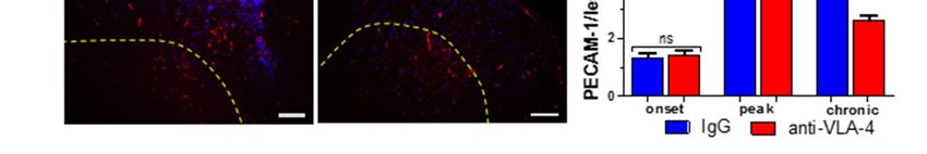
Int. J. Mol. Sci. 2022, 23, 4637                                                                                           4 of 12

                                        On the basis of the protocols included in the Hooke Kits™ EAE Emulsion (Hooke
                                   Laboratories, Lawrence, MA, USA), the three phases of EAE were characterized by the
                                   following mean clinical scores: onset: 1.02 ± 0.05 for IgG group vs. 0.4 ± 0.02 for anti-VLA-
                                   4 group; peak: 2.64 ± 0.13 for IgG group vs. 1.09 ± 0.05 for anti-VLA-4 group; chronic:
                                   1.5 ± 0.08 for IgG group vs. 0.94 ± 0.05 for anti-VLA-4 group (Figure 2a,c). In the chronic
                                   phase, the clinical scores decreased in the IgG group, but they remained at the same level
                                   in the anti-VLA-treated mice. The cumulative EAE score was significantly higher in the
                                   IgG group than in anti-VLA-4 mice (30.31 ± 1.49 and 13.53 ± 0.66, respectively; Figure 2b).

                                   2.2. CD45 Expression
                                        CD45, also known as leukocyte common antigen (LCA), expressed by the vast majority
                                   of hematolymphoid lineage cells, is widely used for the detection of leukocytes. Hence, a
                                   CD45 antibody was employed in this study to check if anti-VLA-4 mAb treatment affected
                                   inflammatory infiltrates, and the immunostained area was quantified by a morphometric
                                   analysis (Figure 2d,e). Immunoreactivity for CD45 showed large accumulations of leuko-
                                   cytes, mainly in the white matter of the spinal cords of the immunized mice. In all phases
                                   of the disease, leukocytes formed local aggregates with high cell densities, indicative of the
                                   presence of inflammatory foci. The degree of inflammation, expressed as the percentage of
                                   the immunopositive section surface area (Figure 2e) in both anti-VLA-4 and IgG groups,
                                   increased from the onset to the peak phase and decreased in the chronic phase; however,
                                   the values in the latter phase were higher than those in the onset phase, indicating that
                                   inflammation did not fully retreat. In all EAE phases, the values were significantly lower
                                   in anti-VLA-4-treated mice compared to IgG-treated mice (onset phase: 1.09 ± 0.06% vs.
                                   3.58 ± 0.09%; peak phase: 3.85 ± 0.15% vs. 12.8 ± 0.36%; chronic phase: 2.82 ± 0.15% vs.
                                   7.3 ± 0.15%).
                                        CD45 immunostaining allowed for assessing the number of inflammatory lesions per
                                   group and phase. In IgG-treated mice, during the successive phases, it increased from
                                   3.42 ± 0.18 to 10.92 ± 0.45 and then slightly decreased to 8.15 ± 0.22. Anti-VLA-4 treatment
                                   had no effect in the onset phase, reduced the number of lesions by nearly a half in the peak
                                   phase and caused further, although weak, reduction in the chronic phase (Figure 2f).

                                   2.3. ICAM-1/LFA-1 Expression
                                         Generally, anti-VLA-4 mAb treatment lowered both ICAM-1 and LFA-1 expression
                                   in inflammatory lesions. This effect was absent only in the case of ICAM-1 in the onset
                                   phase (Figure 3d). During the progression of EAE in IgG-treated mice, ICAM-1 expres-
                                   sion increased from 5.04 ± 0.39% to 26.27 ± 1.67% and then dropped to 14.92 ± 1.18%.
                                   The respective values for anti-VLA-4-treated mice were 4.68 ± 0.32%, 14.42 ± 1.4% and
                                   9.2 ± 1.11%. ICAM-1 was expressed by inflammatory cells, capillaries and occasional
                                   nerve-like cells in gray matter (Figure 3a,c).
                                         A similar pattern, i.e., a rise from onset to peak (from 6.04 ± 0.52% to 27.01 ± 2.11%)
                                   and a decrease in the chronic phase (to 7.97 ± 0.47%), was observed in spinal cords
                                   immunostained for LFA-1. Anti-VLA-4 mAb treatment slightly but significantly inhibited
                                   LFA-1 expression in all phases of EAE (Figure 3e). LFA-1 immunopositive inflammatory
                                   cells were located exclusively in the white matter (Figure 3b,c).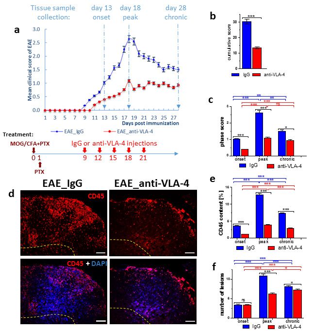
Int.
Int. J.
J. Mol. Sci. 2022,
Mol. Sci. 23, x4637
2022, 23, FOR PEER REVIEW 55of
of 12
12
Figure 3. Immunostaining
Figure 3. Immunostaining of of ICAM-1
ICAM-1 (a),(a), LFA-1
LFA-1 (b)(b) and
and their
their overlapping
overlapping fluorescence
fluorescence with
with DAPI
DAPI
nuclear staining
staining(c)
(c)inincross-sectioned
cross-sectioned spinal
spinal cords
cords of IgG-
of IgG- andand anti-VLA-4-treated
anti-VLA-4-treated micemice
in theinpeak
the
phasephase
peak of EAE, as well
of EAE, asas their
well asquantitative measurements.
their quantitative ICAM-1
measurements. and LFA-1
ICAM-1 expression
and LFA-1 was quan-
expression was
tified as percentage
quantified of lesion
as percentage area
of lesion occupied
area occupiedbyby immunopositive
immunopositivestructures
structures(d,e);
(d,e);white
white dotted
dotted line
marks an
marks an exemplary
exemplary lesion
lesion in
in which
which expression
expression of of adhesion
adhesion molecules
molecules waswas measured.
measured. Localization
Localization
of LFA-1
of LFA-1 is
is associated
associated with
with inflammatory
inflammatory cellscells (b,c),
(b,c), and
and localization
localization ofof ICAM-1
ICAM-1 is is associated
associated with
with
inflammatory cells (a, inset), a few nerve cells in gray matter (a, inset) and capillaries (a, inset, ar-
inflammatory cells (a, inset), a few nerve cells in gray matter (a, inset) and capillaries (a, inset,
rowhead). Data are presented as means ± SEM; n = 5 per group. Statistical significance was verified
arrowhead). Data are presented as means ± SEM; n = 5 per group. Statistical significance was verified
using a two-sided Mann–Whitney test at 0.05 confidence level (*** p < 0.001; ** p < 0.01; * p < 0.05;
using
ns—not a two-sided Mann–Whitney
significant). Magnification test at 0.05 confidence
is indicated by scale bars level (***=p100
((a–c) < 0.001; ** p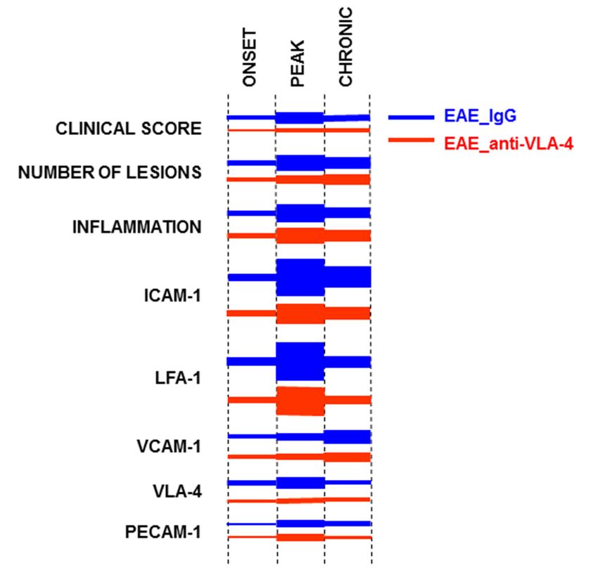
Int. J. Mol. Sci. 2022, 23, x FOR PEER REVIEW                                                                                                          6 of 12
Int. J. Mol. Sci. 2022, 23, 4637                                                                                                                        6 of 12

                                    Figure 4. Immunostaining of VCAM-1 (a), VLA-4 (b) and overlapping fluorescence with DAPI (c) in
                                     Figure 4. Immunostaining of VCAM-1 (a), VLA-4 (b) and overlapping fluorescence with DAPI (c)
                                    cross-sectioned
                                     in cross-sectioned spinal cordscords
                                                             spinal    of IgG-
                                                                             of and
                                                                                 IgG-anti-VLA-4-treated
                                                                                       and anti-VLA-4-treated mice inmice
                                                                                                                        the peak
                                                                                                                              in thephase
                                                                                                                                      peakofphase
                                                                                                                                              EAE, ofas well
                                                                                                                                                        EAE,asas
                                    their  quantitative    measurements.      VCAM-1        and  VLA-4     expression    was   quantified
                                     well as their quantitative measurements. VCAM-1 and VLA-4 expression was quantified as percent-        as  percentage    of
                                    lesion
                                     age ofarea   occupied
                                              lesion          by immunofluorescent
                                                      area occupied                       structuresstructures
                                                                         by immunofluorescent          (d,e). VCAM-1(d,e).isVCAM-1
                                                                                                                             mainly localized
                                                                                                                                       is mainly inlocalized
                                                                                                                                                    capillariesin
                                     capillaries
                                    (a, inset), and(a,localization
                                                       inset), and localization
                                                                    of VLA-4 isof      VLA-4 is with
                                                                                    associated     associated    with inflammatory
                                                                                                         inflammatory                    cells
                                                                                                                            cells in white     in white
                                                                                                                                             matter   (b, matter
                                                                                                                                                          inset)
                                     (b, inset)
                                    and   in someand   in some neuron-like
                                                     neuron-like   cells in graycells in gray
                                                                                    matter   (b,matter
                                                                                                  inset).(b, inset).
                                                                                                           Data   areData    are presented
                                                                                                                      presented     as means as± means
                                                                                                                                                   SEM;±nSEM;
                                                                                                                                                            =5
                                     n =group;
                                    per   5 per group;
                                                  nf—notnf—not
                                                            found. found.    Statistical
                                                                    Statistical            significance
                                                                                  significance             was verified
                                                                                                  was verified    using a using    a two-sided
                                                                                                                            two-sided             Mann–Whit-
                                                                                                                                        Mann–Whitney        test
                                    atney  test
                                        0.05    at 0.05 confidence
                                             confidence     level (***level (*** p
Int. J. Mol. Sci. 2022, 23, x FOR PEER REVIEW 7 of 12
Int. J. Mol. Sci. 2022, 23, 4637 7 of 12
Int. J. Mol. Sci. 2022, 23, x FOR PEER REVIEW 7 of 12
Figure
Figure 5. ImmunostainingofofPECAM-1
5. Immunostaining PECAM-1 (a)(a)
and and its overlapping
its overlapping fluorescence
fluorescence withwith
DAPIDAPI(b) in(b) in
cross-
Figure 5. Immunostaining
sectioned spinalspinal
cross-sectioned ofofIgG-
cords cords PECAM-1
ofand
IgG- (a) and its overlapping
anti-VLA-4-treated
and micefluorescence
anti-VLA-4-treated in mice
the peak with
phase
in the DAPI
peak (b) inas
of phase
EAE, cross-
ofwell
EAE, asas
its
sectioned
well as itsspinal
quantitative cords of IgG-
measurements.
quantitative and anti-VLA-4-treated
PECAM-1
measurements. localizationmice
PECAM-1 in the peak
is associated
localization phasescarce
with of EAE,
is associated asscarce
cells
with well
and as its and
capillaries.
cells
quantitative
Percentagemeasurements.
values in (c)valuesPECAM-1
concern localization is associated with scarce cells and capillaries.
capillaries. Percentage in lesion area occupied
(c) concern lesion area by occupied
immunopositive structures.
by immunopositive Data are pre-
structures.
Percentage
sented as values
means in
± (c) concern
SEM; n = lesion
5 per area
group.occupied by
Statistical immunopositive
significance wasstructures.
verified Data
using are
a pre-
two-sided
Data are
sented as presented
means ± SEM; n=5±
as means perSEM;
group.n =Statistical
5 per group. Statistical
significance wassignificance wasaverified
verified using two-sided using a
Mann–Whitney test at 0.05 confidence level (*** p < 0.001; ** p < 0.01; ns—not significant). Magnifi-
two-sided Mann–Whitney
Mann–Whitney test at 0.05level
test at 0.05 confidence confidence
(*** p < 0.001; ** p p<Int. J. Mol. Sci. 2022, 23, 4637 8 of 12
3. Discussion
The expression of adhesion molecules at the brain–blood barrier (BBB) is of pathogenic
relevance in inflammatory diseases of the CNS such as MS and its animal model, EAE.
The upregulation of various adhesion molecules in the brain and spinal cord tissues of
MS/EAE has been demonstrated in several studies [2,3,6]. Moreover, blocking of adhesion
molecules was shown to be an effective strategy to ameliorate the severity of EAE [9,10].
The inhibition of VLA-4 integrin-mediated binding of leukocytes crossing the BBB to their
endothelial receptors is believed to be the basis of the therapeutic potential of natalizumab,
a monoclonal antibody against alpha4 beta1integrin (VLA-4), in reducing the inflammatory
and clinical symptoms of MS and EAE. A similar effect on T-cell adhesion and infiltration
was observed after treatment with an antibody against ICAM-1 [17].
In the present study, we used an EAE model in which the progression of clinical scores
allows for characterizing three successive phases of the disease: onset, peak and chronic
(recovery). The inflammation level was assessed by measuring the expression of CD45, a
general marker of proinflammatory leukocytes, which are predominant in the inflammatory
foci. These cells also have a regulatory function, contributing to the development and
intensity of inflammation [18].
In IgG-treated mice (treatment control), inflammatory infiltration in the spinal cord
increased during the first two phases of EAE and decreased, but still remained at an elevated
level, in the chronic phase. Such a pattern was also observed in our previous studies [12,19].
Anti-VLA-4 mAb treatment significantly suppressed inflammatory infiltration compared
to the IgG group, delayed the clinical disease onset and reduced clinical scores in the
subsequent phases.
Quantitative analysis of the temporal expression patterns of adhesion molecules in MS
and EAE can provide valuable information because many new and innovative therapeutic
strategies in MS are aimed at preventing the penetration of inflammatory cells across
the BBB, and this process depends on interactions between adhesion molecules. VLA-
4/VCAM-1, LFA-1/ICAM-1 and PECAM-1/PECAM-1 interactions play an important role
in the adhesion and migration of leukocytes across the endothelial barrier [20,21]. The
lowered expression of adhesion molecules in brain endothelial cells can lead to reduced
inflammatory infiltration and the inhibition of new lesion formation in EAE [6].
In order to quantify the expression of the studied adhesion molecules, we could
not assess their overall expression in the entire spinal cord sections, as we did in our
previous studies [12,19], because the result would depend on the number and area of lesions
characterized by high inflammatory cell density, and these parameters differ between
sections. Hence, we decided to measure the expression of adhesion molecules in lesions
only, which show comparable inflammatory cell density.
A predominant temporal expression pattern emerged in control, IgG-treated mice: an
increase in the onset phase, a maximum in the peak phase and a decrease in the chronic
phase (ICAM-1/LFA-1 and PECAM-1)—this pattern corresponded to that of the clinical
score, the number of lesions and the inflammation level (CD45). The pair VCAM-1/VLA-1
did not show a decrease in expression in the chronic phase—instead, an increase (VCAM-1)
or no change compared with the peak phase (VLA-1) was observed.
The expression of adhesion molecules in the course of EAE has been the subject of
several studies [3,22–27]. It is, however, difficult to compare the results of these studies
with ours because the authors used different EAE models in mice and rats, with different
methods to induce the disease, and EAE phases were not specified in a standardized manner.
Generally, the reported successive changes in adhesion molecule expression corresponded
to the predominant pattern described above. The maintenance of high expression levels of
some adhesion molecules in the chronic phase of EAE is a novel observation indicating the
differentiated involvement of adhesion molecules in the course of the disease.
In the present study, ICAM-1 and LFA-1 were found to be the dominant adhesion
molecules in the peak phase of EAE, as far as the quantitative expression of the protein was
concerned. Although VLA-4 upregulation in T cells is crucial for their migration into theInt. J. Mol. Sci. 2022, 23, 4637 9 of 12
CNS [28], lymphocytes have been reported to adhere to activated endothelium via VLA-4
and LFA-1, but their subsequent migration appears to be regulated mainly by ICAM-1 and
partly by PECAM-1 [17,19]. VLA-4 immunostaining of neuron-like cells observed in the
present study was surprising because, in the literature, it was reported only in some retinal
and peripheral neurons [29]. We suppose that this staining resulted from some non-specific
cross-reaction, especially since it was not abolished by anti-VLA-4 treatment.
Low ICAM-1 expression in the onset phase and its high levels in the peak and chronic
phases seem to contradict the opinion that this adhesion molecule is involved mainly in
the early phase of EAE [22,24,25]. However, it confirms the significance of ICAM-1 in the
pathogenesis of EAE. Mutant mice lacking the ICAM-1 gene showed attenuated EAE with
reduced T-cell infiltration in the spinal cord [27].
Upregulated expression of PECAM-1 was observed in MS lesions and could reflect
vascular repair mechanisms aimed at the restoration of BBB integrity and the inhibition
of T-cell migration across the BBB [7]. The importance of PECAM expression in EAE is
not clear. Earlier studies suggested that it did not influence the clinical severity of the
disease [26]. However, PECAM-1-deficient mice showed earlier inflammatory infiltration
of the CNS and higher permeability of CNS vessels as compared with wild-type EAE
mice [23]. A prominent increase in PECAM-1 expression observed in the peak phase of
EAE and a weak decrease in its level in the chronic phase seem to confirm an important
role of this molecule in the development of the disease.
The novel finding of this study is that anti-VLA-4 mAb treatment inhibits the expres-
sion of not only VLA-4 but also the other adhesion molecules studied. As expected, this
inhibition in the case of VLA-4 was the strongest and not phase-dependent. Interestingly, in
the case of ICAM-1, VCAM-1 and PECAM-1, the inhibitory effect of anti-VLA-4 treatment
was delayed and occurred in the peak and chronic phases of EAE (ICAM-1 and VCAM-1)
or even only in the chronic phase (PECAM-1). These adhesion molecules are at least partly
associated with endothelial cells; hence, it seems that the endothelium responds with some
delay to the blocking effect of anti-VLA-4 mAb.
Anti-VLA-4 mAb treatment attenuates proinflammatory mediators, increases the pro-
duction of anti-inflammatory IL-10 [30] and also—probably indirectly—downregulates
metalloproteinases as well as upregulates their inhibitors, counteracting the disintegra-
tion of the vascular basal laminae [12]. The results of the present study justify the sup-
position that the therapeutic mechanism of anti-VLA-4 mAb treatment also includes a
complex influence on a variety of adhesion molecules expressed by infiltrating cells and
vascular endothelium.
4. Materials and Methods
4.1. Animals
Pathogen-free C57BL/6 mice (female, 10–11 weeks old, weight 19–24 g) were pur-
chased from the Center for Experimental Medicine of Bialystok Medical University, Poland
(strain imported from Jackson Laboratory). Mice were housed, five per cage, in the animal
house of the Jagiellonian Centre for Experimental Therapeutics (JCET), Krakow, under
a 12 h light–dark cycle in temperature-controlled environment (22 ± 2 ◦ C, 55 ± 10% hu-
midity). Standard irradiated laboratory chow and water were available ad libitum. All
experiments were conducted in compliance with the Council Directive 2010/63EU of the
European Parliament and of the Council of 22 September 2010 on the protection of ani-
mals used for scientific purposes and approved by the First and the Second Local Ethics
Committees in Krakow, Poland (Permissions 118/2015 and 274/2018).
4.2. Induction of EAE
To induce EAE, naïve mice (n = 30) were subcutaneously immunized with 200 µL injec-
tion of Hooke Kits™ EAE Emulsion (Hooke Laboratories, Lawrence, MA, USA) containing
MOG35–55 peptide emulsified in Complete Freund’s Adjuvant (CFA) including 4 mg/mL
heat-killed Mycobacterium tuberculosis (H37Ra). On the day of MOG35–55 immunizationInt. J. Mol. Sci. 2022, 23, 4637 10 of 12
and 24 h later, the mice were also injected intraperitoneally (i.p.) with 340 µL of Borde-
tella pertussis pertussis toxin (PTx) dissolved in phosphate-buffered saline (PBS) (Hooke
Laboratories, Lawrence, MA, USA) (Figures 1 and 2a).
4.3. Evaluation of EAE
The mice were examined daily for clinical signs of EAE from day 0 to day 28, and
the clinical symptoms of EAE were scored as reported previously [12]. In short, disease
severity was evaluated using a scale ranging from 0 to 3: (0) no clinical disease; 0.5—limp
tip of tail; 1—limp tail; 1.5—limp tail and hind leg inhibition; 2—limp tail and weakness of
hind legs; 2.5—limp tail and dragging of hind legs; 3—limp tail and complete paralysis of
hind legs. The mice were examined and scored by one person blinded to the treatment.
4.4. Experimental Groups
The immunized EAE mice (n = 30) were divided into two experimental groups: anti-
VLA-4 group (n = 15) receiving i.p. injection of 5 mg/kg of anti-VLA-4 mAb (Natal-
izumab, Biogen Idec, Berkshire, UK) and IgG group (IgG control group, n = 15) receiving
5 mg/kg IgG (Sigma-Aldrich, St. Louis, MO, USA). The injections (every 3 days, on days
9, 12, 15, 18 and 21) were continued until the appearance of first remission symptoms
(Figures 1 and 2a) [17,20].
In MOG35–55 immunized mice, the initial symptoms of the disease were observed
between 9 and 14 post-immunization days (onset phase). The maximum scores (peak
phase) occurred between post-immunization days 15 and 20, and then the mice partially
recovered (chronic phase). In EAE mice treated with anti-VLA-4 mAb, the onset phase
began later (day 11), and the peak phase was shorter (days 15 to 18).
For histological analysis, EAE mice (anti-VLA-4 and IgG groups) were sacrificed at
three different time points representing three disease phases (n = 5 per group and phase):
onset phase (day 13), peak phase (day 18) and chronic phase (day 28) (Figures 1 and 2a).
4.5. Tissue Sampling and Processing
To collect spinal cords, mice were anesthetized with ketamine/xylazine cocktail
(100 mg/kg and 10 mg/kg, respectively, i.p.). Next, the animals were transcardially per-
fused with ice-cold PBS for 10 min, followed by 4% paraformaldehyde for the next 10 min.
Spinal cords were carefully removed from the vertebral canal and postfixed in the same
fixative for 4 h. After overnight incubation in 5% sucrose at 4 ◦ C, tissue was embedded in
OCT (Shandon Cryomatrix, Thermo Fisher Scientific, Rockford, IL, USA) and snap-frozen
at −80 ◦ C. The study area of the spinal cord included the lumbar part, a region commonly
and rapidly affected in EAE. Serial cryosections, 10 µm thick, were cut at 100 µm intervals,
collected on poly-L-lysine-coated slides and air-dried. The sections were fixed with acetone
for 20 min and air-dried again.
4.6. Immunohistochemistry
The following primary antibodies were used: rat anti-ITGA4 (VLA-4, Thermo Fisher
Scientific, Rockford, IL, USA, 1:100; cat. # MA5-70075), rat anti-CD11a (LFA-1α, Thermo
Fisher Scientific, Rockford, IL, USA, 1:200; cat. # 14-0111-85), rabbit anti-VCAM-1 (Thermo
Fisher Scientific, Rockford, IL, USA; 1:50; cat. # PA5-86042), rabbit anti-ICAM-1 (Thermo
Fisher Scientific, Rockford, IL, USA; 1:100; cat. # PA5-79430) and rat anti-PECAM-1 (CD31,
Sigma-Aldrich, St. Louis, MO, USA, 2 µg/mL, cat. # CBL 1337). Anti-CD45 primary
antibody for total leukocytes (Thermo Fisher Scientific, 1:100, MA1-81247) was applied to
analyze the degree of inflammatory infiltration. The secondary antibodies included goat
anti-rat Alexa488-conjugated antibodies (Jackson IR, West Grove, PA, USA, 1:100; cat. #
112-545-167), Cy3-conjugated goat anti-rabbit antibodies (Jackson IR, West Grove, PA, USA;
1:300; cat. # 111-165-144), goat anti-rabbit Alexa488-conjugated antibodies (Jackson IR, West
Grove, PA, USA, 1:100; cat. # 111-545-144) and Cy3-conjugated goat anti-rat antibodiesInt. J. Mol. Sci. 2022, 23, 4637 11 of 12
(Jackson IR, West Grove, PA, USA; 1:300; cat. # 112-165-167). DAPI staining was used to
detect nuclei (Thermo Fisher Scientific, Rockford, IL, USA; 1.5 ug/mL; cat. # 62248).
The spinal cord sections were preincubated for 40 min in a blocking solution: PBS
containing 5% normal goat serum (Sigma-Aldrich, St. Louis, MO, USA), 0.01% sodium
azide, 0.05% thimerosal, 0.1% bovine serum albumin, 0.5% Triton X-100 and 2% dry milk.
They were next incubated overnight at room temperature with primary antibodies and,
after a rinse in PBS, incubated for 90 min with the secondary antibodies. Then, sections
were washed three times in PBS and mounted in glycerol/PBS solution.
4.7. Microscopy, Morphometry and Image Collection
The spinal cord sections were examined using Olympus BX50 brightfield/epifluorescence
microscope (Olympus, Tokyo, Japan). All low magnification images were recorded with
the use of Olympus DP71 digital CCD camera, stored as TIFF files and processed for
quantitative analysis using ImageJ software (NIH, Bethesda, MD, USA).
In each image, inflammatory lesions characterized by high density of cells were
marked (example in Figure 2a), and the percentage of immunopositive areas was assessed
in the lesions. Only in the case of CD45, a general marker of inflammation, the percentage
of immunopositive areas was estimated for the whole spinal cord cross-sectional area
(Figure 2e).
A total of at least 20 sections were analyzed per experimental group (n = 5) and phase.
4.8. Data Analysis
GraphPad Prism 5.0 software (GraphPad, La Jolla, CA, USA) was used throughout
this study for statistical analyses. All values are expressed as mean ± standard error of
the mean (SEM). Statistical significance of the obtained results was verified using two-
sided Mann–Whitney test at a confidence level of 0.05 (*** p < 0.001; ** p < 0.01; * p < 0.05;
ns—not significant).
Author Contributions: Conceptualization: G.P.-F.; analysis of data and their interpretation: G.P.-F.
and J.A.L.; performance of experiments and preparation of results: G.P.-F.; manuscript preparation:
G.P.-F. and J.A.L.; supervision and funding acquisition: G.P.-F.; graphic and photographic documen-
tation: G.P.-F.; writing—review & editing: G.J.L. All authors have read and agreed to the published
version of the manuscript.
Funding: The study was supported by statutory grant N41/DBS/000564 from the Jagiellonian
University Medical College to GFP.
Institutional Review Board Statement: All experiments were conducted in compliance with the
Council Directive 2010/63EU of the European Parliament and of the Council of 22 September 2010
on the protection of animals used for scientific purposes and approved by the First and the Second
Local Ethics Committees in Krakow, Poland (Permissions 118/2015 and 274/2018).
Informed Consent Statement: Not applicable.
Data Availability Statement: Not applicable.
Conflicts of Interest: The authors declare no conflict of interest.
References
1. Constantinescu, C.S.; Farooqi, N.; O’Brien, K.; Gran, B. Experimental autoimmune encephalomyelitis (EAE) as a model for
multiple sclerosis (MS). Br. J. Pharmacol. 2011, 164, 1079–1106. [CrossRef] [PubMed]
2. Cannella, B.; Raine, C.S. The adhesion molecule and cytokine profile of multiple sclerosis lesions. Ann. Neurol. 1995, 37, 424–435.
[CrossRef] [PubMed]
3. Engelhardt, B.; Conley, F.K.; Butcher, E.C. Cell adhesion molecules on vessels during inflammation in the mouse central nervous
system. J. Neuroimmunol. 1994, 51, 199–208. [CrossRef]
4. Archelos, J.J.; Hartung, H.P. The role of adhesion molecules in multiple sclerosis: Biology, pathogenesis and therapeutic
implications. Mol. Med. Today 1997, 3, 310–321. [CrossRef]
5. Rice, G.P.; Hartung, H.P.; Calabresi, P.A. Anti-alpha4 integrin therapy for multiple sclerosis: Mechanisms and rationale. Neurology
2005, 64, 1336–1342. [CrossRef]Int. J. Mol. Sci. 2022, 23, 4637 12 of 12
6. Yusuf-Makagiansar, H.; Anderson, M.E.; Yakovleva, T.V.; Murray, J.S.; Siahaan, T.J. Inhibition of LFA-1/ICAM-1 and VLA-
4/VCAM-1 as a therapeutic approach to inflammation and autoimmune diseases. Med. Res. Rev. 2002, 22, 146–167. [CrossRef]
7. Wimmer, I.; Tietz, S.; Nishihara, H.; Deutsch, U.; Sallusto, F.; Gosselet, F.; Lyck, R.; Muller, W.A.; Lassmann, H.; Engelhardt, B.
PECAM-1 Stabilizes Blood-Brain Barrier Integrity and Favors Paracellular T-Cell Diapedesis Across the Blood-Brain Barrier
during Neuroinflammation. Front. Immunol. 2019, 10, 711. [CrossRef]
8. Paddock, C.; Zhou, D.; Lertkiatmongkol, P.; Newman, P.J.; Zhu, J. Structural basis for PECAM-1 homophilic binding. Blood 2016,
127, 1052–1061. [CrossRef]
9. Steinman, L. Blocking adhesion molecules as therapy for multiple sclerosis: Natalizumab. Nat. Rev. Drug Discov. 2005, 4, 510–518.
[CrossRef]
10. Yednock, T.A.; Cannon, C.; Fritz, L.C.; Sanchez-Madrid, F.; Steinman, L.; Karin, N. Prevention of experimental autoimmune
encephalomyelitis by antibodies against a4b1 integrin. Nature 1992, 356, 63–66. [CrossRef]
11. Clerico, M.; Artusi, C.A.; Di Liberto, A.; Rolla, S.; Bardina, V.; Barbero, P.; De Mercanti, S.F.; Durelli, L. Long-term safety evaluation
of natalizumab for the treatment of multiple sclerosis. Expert. Opin. Drug Saf. 2017, 16, 963–972. [CrossRef] [PubMed]
12. Pyka-Fosciak, G.; Lis, G.J.; Litwin, J.A. Effect of natalizumab treatment on metalloproteinases and their inhibitors in a mouse
model of multiple sclerosis. J. Physiol. Pharmacol. 2020, 71, 265–273.
13. Theien, B.E.; Vanderlugt, C.L.; Eagar, T.N.; Nickerson-Nutter, C.; Nazareno, R.; Kuchroo, V.; Miller, S. Discordant effects of
anti-VLA-4 treatment before and after onset of relapsing experimental autoimmune encephalomyelitis. J. Clin. Investig. 2001, 107,
995–1006. [CrossRef] [PubMed]
14. Cannella, B.; Gaupp, S.; Tilton, R.G.; Raine, C.S. Differential efficacy of a synthetic antagonist of VLA-4 during the course of
chronic relapsing experimental autoimmune encephalomyelitis. J. Neurosci. Res. 2003, 71, 407–416. [CrossRef] [PubMed]
15. Lassmann, H.; Bradl, M. Multiple sclerosis: Experimental models and reality. Acta Neuropathol. 2017, 133, 223–244. [CrossRef]
16. Linker, R.A.; Lee, D.H. Models of autoimmune demyelination in the central nervous system: On the way to translational medicine.
Exp. Transl. Stroke Med. 2009, 1, 5. [CrossRef]
17. Archelos, J.J.; Jung, S.; Mäurer, M.; Schmied, M.; Lassmann, H.; Tamatani, T.; Miyasaka, M.; Toyka, K.V.; Hartung, H.P. Inhibition
of experimental autoimmune encephalomyelitis by an antibody to the intercellular adhesion molecule ICAM-1. Ann. Neurol.
1993, 34, 145–154. [CrossRef]
18. Ran, Z.; Yue-Bei, L.; Qiu-Ming, Z.; Huan, Y. Regulatory B cells and its role in central nervous system inflammatory demyelinating
diseases. Front. Immunol. 2020, 11, 1884. [CrossRef]
19. Pyka-Fosciak, G.; Stasiolek, M.; Litwin, J.A. Immunohistochemical analysis of spinal cord components in mouse model of
experimental autoimmune encephalomyelitis. Folia Histochem. Cytobiol. 2018, 56, 151–158. [CrossRef]
20. Greenwood, J.; Wang, Y.; Calder, V.L. Lymphocyte adhesion and transendothelial migration in the central nervous system—The
role of LFA-1, ICAM-1, VLA-4 and VCAM-1. Immunology 1995, 86, 408–415.
21. Wong, D.; Prameya, R.; Dorovini-Zis, K. In Vitro adhesion and migration of T lymphocytes across monolayers of human brain
microvessel endothelial cells: Regulation by ICAM-1, VCAM-1, E-selectin and PECAM-1. J. Neuropathol. Exp. Neurol. 1999, 58,
138–152. [CrossRef] [PubMed]
22. Dopp, J.M.; Brenemann, S.M.; Olschowka, J.A. Expression of ICAM-1, VCAM-1, L-selectin, and leukosialin in the mouse central
nervous system during the induction and remission stages of experimental allergic encephalomyelitis. J. Neuroimmunol. 1994, 54,
129–144. [CrossRef]
23. Graesser, D.; Solowiej, A.; Bruckner, M.; Osterweil, E.; Juedes, A.; Davis, S.; Ruddle, N.H.; Engelhardt, B.; Madri, J.A. Altered
vascular permeability and early onset of experimental autoimmune encephalomyelitis in PECAM-1–deficient mice. J. Clin.
Investig. 2002, 109, 383–392. [CrossRef] [PubMed]
24. Cannella, B.; Cross, A.H.; Raine, C.S. Adhesion-related molecules in the central nervous system. Upregulation correlates with
inflammatory cell influx during relapsing experimental autoimmune encephalomyelitis. Lab. Investig. 1991, 65, 23–31. [PubMed]
25. Ledeboer, A.; Wierinckx, A.; Bol, J.G.; Floris, S.; Renardel de Lavalette, C.; De Vries, H.E.; van den Berg, T.K.; Dijkstra, C.D.;
Tilders, F.J.; van Dam, A.M. Regional and temporal expression patterns of interleukin-10, interleukin-10 receptor and adhesion
molecules in the rat spinal cord during chronic relapsing EAE. J. Neuroimmunol. 2003, 136, 94–103. [CrossRef]
26. Williams, K.C.; Zhao, R.W.; Ueno, K.; Hickey, W.F. PECAM-1 (CD31) expression in the central nervous system and its role in
experimental allergic encephalomyelitis in the rat. J. Neurosci. Res. 1996, 45, 747–757. [CrossRef]
27. Bullard, D.C.; Hu, X.; Schoeb, T.R.; Collins, R.G.; Beaudet, A.L.; Barnum, S.R. Intercellular adhesion molecule-1 expression is
required on multiple cell types for the development of experimental autoimmune encephalomyelitis. J. Immunol. 2007, 178,
851–857. [CrossRef]
28. Baron, J.L.; Madri, J.A.; Ruddle, N.H.; Hashim, G.; Janeway, C.A., Jr. Surface expression of alpha 4 integrin by CD4 T cells is
required for their entry into brain parenchyma. J. Exp. Med. 1993, 177, 57–68. [CrossRef]
29. Hikita, S.T.; Cann, G.M.; Wingerd, K.L.; Mullick, L.H.; Wayne, W.C.; Webb, S.W.; Clegg, D.O. Integrin alpha4beta1 (VLA-4)
expression and activity in retinal and peripheral neurons. Mol. Cell Neurosci. 2003, 23, 427–439. [CrossRef]
30. Khademi, M.; Bornsen, L.; Rafatnia, F.; Andersson, M.; Brundin, L.; Piehl, F.; Sellebjerg, F.; Olsson, T. The effects of natalizumab
on inflammatory mediators in multiple sclerosis: Prospects for treatment-sensitive biomarkers. Eur. J. Neurol. 2009, 16, 528–536.
[CrossRef]You can also read