The Effect of Apium Graveolens L., Levisticum Officinale and Calendula Officinalis L. on Cell Viability, Membrane Integrity, Steroidogenesis, and ...
←
→
Page content transcription
If your browser does not render page correctly, please read the page content below
Physiol. Res. 70: 615-625, 2021 https://doi.org/10.33549/physiolres.934675 The Effect of Apium Graveolens L., Levisticum Officinale and Calendula Officinalis L. on Cell Viability, Membrane Integrity, Steroidogenesis, and Intercellular Communication in Mice Leydig Cells in Vitro Tomas JAMBOR1, Julius ARVAY2, Eva TVRDA3, Anton KOVACIK3, Hana GREIFOVA3, Norbert LUKAC3 1 BioFood Centre, Faculty of Biotechnology and Food Sciences, Slovak University of Agriculture in Nitra, Nitra, Slovak Republic, 2Department of Chemistry, Faculty of Biotechnology and Food Sciences, Slovak University of Agriculture in Nitra, Nitra, Slovak Republic, 3Department of Animal Physiology, Faculty of Biotechnology and Food Sciences, Slovak University of Agriculture in Nitra, Nitra, Slovak Republic Received March 9, 2021 Accepted May 11, 2021 Epub Ahead of Print June 2, 2021 Summary hormones synthesis at 150 and 300 µg/ml. Finally, the Several plants have the potential to protect essential reproductive disturbance of GJIC was significantly affected at 300 µg/ml of processes such as spermatogenesis or steroidogenesis, however, Levisticum officinale (82.5±7.7 %) and Calendula officinalis L. effective concentrations and main mechanisms of action are still (79.8±7.0 %). The balanced concentration ratio may support the unknown. This in vitro study was aimed to assess the effects of Leydig cell function, steroidogenesis as well as all essential Apium graveolens L., Levisticum officinale, and Calendula parameters that may significantly improve reproductive functions. officinalis L. extracts on the structural integrity, functional activity and gap junctional intercellular communication (GJIC) in mice Key words Leydig cells. TM3 cells were grown in the presence of Leydig cells • Viability • Membrane integrity • Steroidogenesis • experimental extracts (37.5, 75, 150 and 300 µg/ml) for 24 h. GJIC For the present study, high-performance liquid chromatography analysis was used to quantify flavonoids or phenolic acids. Corresponding author Subsequently, Leydig cell viability was assessed by alamarBlue Tomas Jambor, BioFood centre, Faculty of Biotechnology and assay, while the cell membrane integrity was detected by Food Sciences, Slovak University of Agriculture in Nitra, Tr. A. 5-carboxyfluorescein diacetate-acetoxymethyl ester. The level of Hlinku 2, 949 76 Nitra, Slovak Republic. E-mail: steroid hormones production was determined by enzyme-linked tomasjambor1@gmail.com immunosorbent assay. Additionally, GJIC was assessed by scalpel loading/dye transfer assay. According to our results, Apium Introduction graveolens L. significantly increased the viability and cell membrane integrity at 75 µg/ml (109.0±4.3 %) followed by Reproduction is an essential part of our common a decline at 300 µg/ml (89.4±2.3 %). In case of Levisticum life, and the factors affecting it have always been a focus officinale and Calendula officinalis L. was observed significant of extensive and continuous research. Nowadays, we decrease at 150 µg/ml (88.8±11.66 %, 87.4±6.0 %) and recognize plenty of exogenous factors, which may 300 µg/ml (86.2±9.3 %, 84.1±4.6 %). Furthermore, Apium interact with human and wildlife reproductive health, graveolens L. significantly increased the progesterone and including heavy metals, endocrine disruptors, and other testosterone production (75 and 150 µg/ml) however, Levisticum xenobiotics (Sedeh et al. 2012, Jambor et al. 2019). The officinale and Calendula officinalis L. significantly reduced steroid majority of their negative effects, such as decreased testis PHYSIOLOGICAL RESEARCH • ISSN 1802-9973 (online) - an open access article under the CC BY-NC-ND 4.0 license 2021 Institute of Physiology of the Czech Academy of Sciences, Prague, Czech Republic Fax +420 241 062 164, e-mail: physres@fgu.cas.cz, www.biomed.cas.cz/physiolres
616   Jambor et al.                                                                                               Vol. 70

weights, prostate cancer, poor semen quality, and            knowledge about the consequences of their higher
insufficient production of steroid hormones, are             concentrations on the reproductive functions is poor and
frequently linked to damage of essential cellular            extremely limited. Simultaneously, specific molecular
organelles or disruptions to the processes responsible for   mechanisms of action by which medicinal plants could
normal reproductive functions (Smith 2007). In general,      modulate the reproductive processes and parameters are
most of the mentioned problems could be solved by            not sufficiently understood.
standard medical methods, especially surgical procedures,              There is significant evidence that gap junctional
hormone therapy, or assisted reproductive technology         intercellular communication (GJIC) is essential for
methods. Inversely, an alternative therapy mediated by       normal reproductive development. GJIC is made up of
medicinal herbs may be another effective way to protect      transmembrane proteins called connexins (Cx) and, they
the reproductive system. Several studies have confirmed      considered as major molecular regulators of male
the higher compatibility of these plants with the human      fertility. Namely, the most abundant expressed gap
body and weak side effects in comparison to chemical         junction protein connexin 43 (Cx43) it necessary for
drugs (Kooti et al. 2016). The most beneficial effect of     spermatogenesis,        steroidogenesis     and     healthy
medicinal herbs is related to the content of biologically    reproductive      functions.    Thus,    testicular   GJIC
active substances that are able to improve                   dysregulation caused by different stressors could affect
spermatogenesis, steroidogenesis, increase sperm count       the etiopathology of subfertility correlated with various
and motility, and in some cases, reverse the overall         reproductive       abnormalities      (Gilleron,     2015).
subfertility. However, properly balanced doses determine     Undoubtedly, there is a critical need to elucidate cellular
the potential effects of individual herbs. In many cases,    interactions and clearly define effective doses of
the significant positive and protective effect was           medicinal herbs for the reproductive system's proper
confirmed in the lower doses of medicinal plants, while      functioning (Abbas 2017).
the higher doses and long-term exposition could be                     The present in vitro study aims to investigate the
hazardous for normal reproductive functions in males         impact of ethanolic extract from Apium graveolens L.,
(Liu et al. 2004, Nantia et al. 2009).                       Levisticum officinale, and Calendula officinalis L. on
          Apium graveolens L. (Apiaceae) is one of the       mice TM3 Leydig cells during 24 h cultivation. The
most confronted herbs with a high level of bioactive         experiments had in view to determine whether the use of
components such as limonene, sedanolide, alpha-pinene,       the selected medicinal herbs of known composition
or coumarin. Apium has a broad spectrum of effects such      exhibits any positive or negative effects on the
as anti-cancer, anti-microbial anti-inflammatory, and        mitochondrial activity or membrane integrity, sexual
analgesic (Subhadradevi et al. 2011). Levisticum             hormones release, as well as intercellular communication
officinale from the same family as A. graveolens L.          in mice Leydig cells.
contains a variety of bioactive molecules, and many
previous studies confirmed anti-cancer, anti-bacterial, or   Material and Methods
spasmolytic effects. Extracts from Levisticum are also
commonly used to treat rheumatism and urethritis (Ekiert     Preparation of the herbal extracts
2000). Calendula officinalis L. (Asteraceae) is mainly                 The leaves from Apium graveolens L.,
known for its antitumor activity and cytotoxic effects on    Levisticum officinale, and flowers from Calendula
tumor cell lines. Besides, flowers from Calendula are        officinalis L. were collected at the local university´s field
traditionally used for their anti-inflammatory and           in Nitra (Slovak Republic). Plant material was dried in
antioxidant properties. They are also rich in                the shade, mechanically comminuted, weighed, and
pharmacologically      active     components,   including    subsequently extracted with 96 % ethanol (CentralChem,
coumarins, quercetin, beta-amyrin or narcissin (Preethi      Bratislava, Slovak republic) for 2 weeks. After that, the
et al. 2010). Lower experimental concentrations of all       ethanol was evaporated (Stuart RE300DB rotary
plants mentioned above have been reported to have            evaporator, Bibby Scientific Limited, United Kingdom
a significant impact on libido, spermatozoa quality,         and vacuum pump KNF N838.1.2KT.45.18) under
sexual hormone production or testis weight, and pituitary-   reduced pressure (0.5 bar/g) and elevated temperature
gonadal axis (Halo et al. 2019, Saha et al. 2019, Tvrdá      40 °C in order to remove any residual ethanol. The crude
et al. 2019, Jambor et al. 2020). Nevertheless, current      extract was dissolved in a standard organic solvent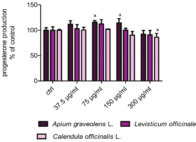
2021                                                          Higher Doses of Extracts Are Cytotoxic for Mice Leydig Cells   617

dimethylsulfoxide (DMSO, Sigma-Aldrich, St. Louis,           FBS (fetal bovine serum, BiochromAG, Berlin,
USA) and adjusted to 100 mg/ml as a starting solution        Germany) together with 2.5 mmol-1 L-glutamine (Sigma-
(Tvrdá et al. 2016).                                         Aldrich, St. Louis, USA) and 1 % penicillin/streptomycin
                                                             solution (Sigma-Aldrich, St. Louis, USA). Leydig cells
HPLC-DAD analysis of phenolic compounds                      were cultured at 37 °C with 5 % CO2 and 95 % saturated
         In the case of quantitative analysis of the         atmospheric humidity. Cells were regularly screened for
phenolic compounds, the aliquots of plant materials were     contamination. The Leydig cells density was determined
subjected to the high-performance liquid chromatography      using automated cell counter TC 20TM (Bio-Rad
(HPLC-DAD). One g of lyophilized leaves and flowers          Laboratories, California, USA) and adjust with culture
were dissolved in methanol (10 ml, 80 %, Sigma-Aldrich,      medium to a final concentration of 4 x 10³ cells per well.
St. Louis, USA). Afterward, the mixture was shaken on        The cells were grown in a 96-well plate followed by pre-
a horizontal shaker (25 °C, during 8 h, at 250 rpm) and      cultivation of the cells for 24 h until a monolayer was
filtered through 84 g/m2 filter paper (Munktell,             formed. Afterward, the medium was replaced to include
Germany). The samples were subsequently extracted in         varying concentrations of experimental extracts Apium
20 ml of 80 % (v/v) methanol by shaking horizontally         graveolens L., Levisticum officinale, and Calendula
(Unimax 2010, Heidolph Instrument, GmbH, Germany).           officinalis L. at 37.5, 75, 150 and 300 µg/ml. All treated
The high-performance liquid chromatograph (Agilent           groups were compared to the non-treated (control) Leydig
1260 Infinity HPLC Technologies, Waldbronn,                  cells cultured in cell-culture media. The applied
Germany) with quaternary solvent manager coupled with        concentration range was selected according to the results
degasser, sampler manager, Diode Array Detector, and         of our pilot range-finding experiments. The TM3 Leydig
column manager were used to analyse phenolic content in      cells remained in culture for 24 h. The time of exposition
the harvested leaves of Apium graveolens L., Levisticum      has been chosen regarding to previous pilot study with
officinale and from flowers of Calendula officinalis L.      bovine spermatozoa (Benko et al. 2019, Tvrdá et al.
HPLC measurements were performed on a Purosphere             2019). After the set time, cell viability, cell membrane
reverse phase C18 column (Darmstadt, Germany). The           integrity, steroid hormone production, and intercellular
mobile phase consisted of acetonitrile and 0.1 %             communication were evaluated.
phosphoric acid in double-deionized water (ddH2O). The
gradient elution was as follows: 0-1 min isocratic elution   Cell viability assay (alamarBlue)
(90 % C and 10 % D), 1-6 min linear gradient elution                  To determine the effect of experimental
(85 % C and 15 % D), 6-12 min (80 % C and 20 % D),           concentrations (37.5 – 300 µg/ml) of the herbal extracts
12-20 min (30 % C and 70 % D) and 20-25 min (30 % C          on the TM3 Leydig cell viability after 24 h exposure,
and 70 % D). The column thermostat was heated up to          alamarBlueTM assay was exploited. AlamarBlueTM cell
30 °C, while the samples were kept at 6 °C in the sampler    viability reagent (AB, ThermoFisher Scientific,
manager. The collected data were processed using the         Invitrogen, Vantaa, Finland) is a sensitive oxidation-
Agilent OpenLab ChemStation software for LC 3D               reduction indicator that fluoresces and changes the blue
Systems (Lukšič et al. 2016).                                colour of resazurin to a pink reduced form - resorufin
                                                             upon reduction by living cells mediated by mitochondrial
TM3 Leydig cell culture                                      enzymes (Hamid et al. 2004). Following respective
          The TM3 mouse Leydig cell line derived from        exposure, the culture medium was removed, the treated
the testis strain BALB/c nu/+ was obtained from the          cells were washed with PBS (phosphate-buffer saline,
American Type Culture Collection (ATCC, CRL-1714,            7.2 pH) and cultured with serum-free DMEM/F12
Manassas, USA). As a non-tumorigenic line, TM3               containing 5 % (v/v) alamarBlue solution at 37 °C under
Leydig cells are commonly used for a short-term in vitro     a humidified atmosphere of 95 % air and 5 % CO2. After
cultivation to reflect variance in steroid hormone           30 min incubation, the fluorescence was measured at
secretion. The cell culture medium consisted of              530 nm against 590 nm (excitation/emission)
DMEM/F12          (Dulbecco´s    Modified       Eagle´s      wavelengths by a microplate reader (GloMax®-Multi+,
Medium/Nutrient Mixture (Ham´s) F12, Sigma-Aldrich,          Promega Corporation, Madison, USA). The results are
St. Louis, USA) supplemented with 5 % HS (horse              expressed as a percentage of the control (non-treated)
serum, Gibco-Life Technologies, New Zealand), 2.5 %          group.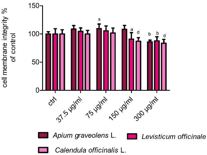
618   Jambor et al.                                                                                             Vol. 70

Cell membrane integrity assay (CFDA-AM)                     incubation, the cells were washed three times with
         To examine the impact of experimental              CaMg-PBS and fixed with a 4 % formaldehyde solution.
concentrations (37.5 – 300 µg/ml) of the herbal extracts    The images were captured by fluorescent microscope
on TM3 cells membrane integrity after 24 h incubation,      DMI 6000B (Leica Microsystems, Wetzlar, Germany)
5-carboxyfluorescein diacetate, acetoxymethyl ester         with DCF 345 FX camera. The area of cells stained with
(CFDA-AM, ThermoFisher Scientific, Invitrogen,              lucifer yellow was evaluated using ImageJ software
Vantaa, Finland) was used according to the previous         (Schneider et al. 2012). The results are expressed as
study (Schreer et al. 2005). In essence, culture media      a percentage of the control (non-treated) group.
supplemented with herbal extracts was replaced with         Statistics
fresh cultured media together with 4 µM CFDA-AM.                      The obtained data were statistically analysed
Subsequently, the TM3 cells were incubated for 30 min       using GraphPad Prism 5.0 (GraphPad Software
in the dark at 37 °C with 5 % CO2, and 95 % saturated       Incorporated, San Diego, California, USA). One-way
atmospheric humidity. The concentrations of the             analysis of variance (ANOVA) followed by Dunnett´s
fluorescent metabolites of CFDA-AM were measured at         multiple comparison test was used for statistical
wavelength 485 – 530 nm (excitation/emission) in            evaluations. Results were expressed as the mean ±
a microplate    reader     (GloMax®-Multi+,       Promega   standard deviation (S.D). All experiments were repeated
Corporation, Madison, USA). The results are expressed       at least three times. Statistical differences were expressed
as a percentage of the control (non-treated) group.         at a significance of P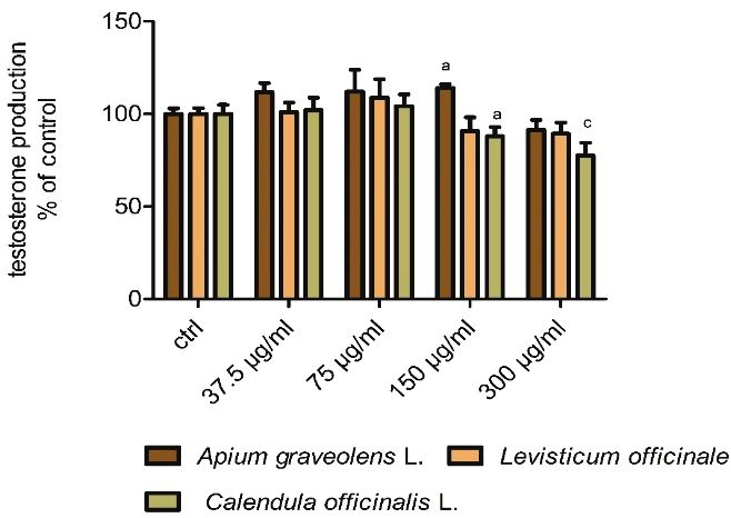
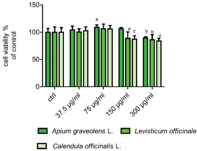
2021                                                                     Higher Doses of Extracts Are Cytotoxic for Mice Leydig Cells   619

effect on the cell viability of exposed cells compared to              effect up to 75 µg/ml on the presented parameter.
the control (100.0±6.7 %). The results showed that                     However, higher concentrations of Levisticum initiated
75 µg/ml (109.0±4.3 %) caused a significant (P
620 Jambor et al. Vol. 70
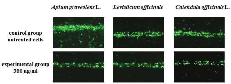
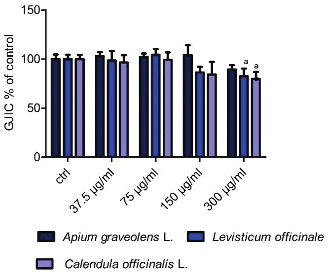
(P2021 Higher Doses of Extracts Are Cytotoxic for Mice Leydig Cells 621 Fig. 3.A Progesterone production in TM3 Leydig cells exposed to different concentrations of experimental extracts from Apium graveolens L., Levisticum officinale and Calendula officinalis L. in vitro after 24 h cultivation. ctrl – control group. Each bar Fig. 4.A Intercellular communication in TM3 Leydig cells exposed represents the mean (±S.D) progesterone production % of to different concentrations of experimental extracts from Apium control (untreated) and treated groups. Data were obtained from graveolens L., Levisticum officinale and Calendula officinalis L. in four (n=4) independent experiments. The level of significance vitro after 24 h cultivation. ctrl – control group. Each bar was set at (P
622   Jambor et al.                                                                                              Vol. 70

A high proportion of bioactive molecules was confirmed         cells. The beneficial effects of Calendula officinalis L.
by Yao et al. (2010). Their study identified major             were confirmed by many experimental studies focused on
phenolic acids in different cultivars of Apium graveolens      cancer diseases in most cases. However, only a few
such as p-coumaric acid (105 mg/kg) ferulic acid               studies provide information about the cytotoxic
(99.3 mg/kg), followed by flavonoids apigenin                  concentrations in non-carcinoma cells. Alnuqaydan et al.
(92.1 mg/kg), luteolin (90.5 mg/kg) or kaempferol              (2015) measured the cytotoxicity of the extract from C.
(94.6 mg/kg). Similar to our results, Złotek et al. (2019)     officinalis L. at di fferent concentrations for 4, 24, and
identified ferulic acid, ellagic acids, p-coumaric acid,       48 h on HaCaT cells in vitro. Calendula showed limited
caffeic acid, kaempferol, rutin, apterin, and quercetin-3-     toxicity with a significant effect in the highest
O-deoxhexoside-O-hexodside as the most abundant in             concentration. Only 4.4 and 4.2 mg/ml expressed as 2 %
Levisticum officinale L. Frum (2017) has monitored the         (v/v) and 5 % (v/v) showed a significant toxicity. The
level of polyphenols in Calendula officinalis L. where the     viability of HUVEC cells was monitored after 48 h in
highest concentrations of rutin, syringic acid, and gallic     vitro cultivation with C. officinalis L. (0.5 – 500 µg/ml)
acid were recorded. The lower amounts of cinnamic acid,        by MTT assay. The results suggest a gradual decline up
resveratrol, and ferulic acid were also detected. All          to 10 µg/ml, followed by a radical cytotoxic effect at 250
presented studies above confirmed similar levels of            and 500 µg/ml (Preethi et al. 2010). According to the
bioactive substances in our experimental medicinal herbs.      current knowledge, extract from selected medicinal herbs
We are convinced that their detailed identification and        used in our study could protect sensitive cellular
monitoring is definitely required for a better                 organelles and cell homeostasis in a concentration-
understanding of the physiological mechanism as well as        dependent manner. It is caused by the mutual ratio of
to help understand the potential changes in the male           bioactive molecules whose high levels have been
reproductive system.                                           confirmed by the previous part of our analysis. Obtained
          Mutual comparison of individual cellular models      results suggest that some experimental concentrations
confirmed different reactions to presented medicinal           may negatively affect basal cellular parameters what
herbs extracts. The vast majority of in vitro studies are      could result from higher toxic potential of selected
focused on tumorogenic cell lines where the increasing         extracts. Furthermore, we can assume that the cellular
concentrations of herbal extract inhibit cancer                membrane destruction or cell death could destroy
proliferation. In contrast, the result of our in vitro study   steroidogenesis enzymes activity resulting in decreased
confirmed that lower experimental concentrations might         hormone production. To resolve this issue, further
positively affect essential parameters of non-tumorigenic      investigations are required. At the same time, we are
cells, especially the cell viability and cell membrane         convinced that adequately applied dose settings could
integrity, but with increasing doses start at 150 to           improve males' reproductive functions. The cell structure
300 µg/ml are able to significantly damage these               and mitochondrial activity are closely related to the
parameters. Comparable consequences have previously            steroidogenic process ongoing in Leydig cells responsible
been reported by Subhadradevi et al. (2011). Mouse lung        for steroid hormone production.
fibroblast L929 cells were exposed to Apium graveolens                   Our in vitro study's data suggest that the
at concentrations ranging from 2 to 20 µg/ml during 48 h       secretion of progesterone and testosterone could be
and the number of viable cells was determined by the           positively affected by the lower doses (75 and 150 µg/ml)
MTT assay. The herbal extract statistically inhibited this     of Apium graveolens L. However, at the highest
parameter in a concentration-dependent manner. Sertel          concentration of Apium graveolens L., Levisticum
et al. (2011) evaluated the impact of Levisticum officinale    officinale, and Calendula officinalis L. has recorded
extract on the head and neck squamous carcinoma cells          a significant decrease in steroidogenic capacity resulting
(HNSCC) using XTT cytotoxicity assay. The biological           in a decline of progesterone and testosterone levels. The
model was cultured together with experimental                  efficacy of hydro-alcoholic extracts of A. graveolens L.
concentrations (0.0001 to 10 mg/ml) of extract for 72 h        on the serum levels of testosterone in male rats was
in vitro. The concentration-response curve showed              investigated by Kooti et al. (2016). Male Wistar rats were
a steady rise in the viability up to 0.1 mg/ml with            orally administered to 200 and 300 mg/kg of
a subsequent rapid decrease in cell viability to 4.7 %         A. graveolens L. for 20 days. The results showed a slight
(1 and 10 mg/ml) when compared to the untreated control        decrease in testosterone production at 300 mg/kg, but
2021 Higher Doses of Extracts Are Cytotoxic for Mice Leydig Cells 623
without significant changes. Similarly, Madkour (2014) in HCT116 cells following kaempferol treatment.
administered orally male albino rats at 200 mg/kg per day We are convinced, that dysregulation of GJIC
of A. graveolens L. oil for 8 weeks. The presented in our study could be an essential part of the
radioimmunoassay revealed an increased concentration of toxic mechanism related to the action of experimental
testosterone when compared to the control group. extracts. According to presenting data, the TM3 mice
Interestingly, Helal (2014) confirmed a slight decrease in Leydig cells are susceptible to the highest doses of
testosterone secretion in male Wistar rats after 6 weeks of applied medicinal herbs extracts with a toxic impact on
exposure to 50 µg/kg per body weight of A. graveolens L. essential cellular organelles and functions. However, as
Ghaedi et al. (2018) published an experimental study we mentioned before, the exact determination of proper
focused on the effect of Levisticum officinale extract on concentrations may definitely affect the activity of mice
the testis histology and testosterone production in Leydig cells and ensure sufficient production of male
diabetic rats. Treatment of rats with 500 mg/kg steroid hormones. Nowadays, the majority of
significantly increased the testis weight and serum experimental studies provide a broad spectrum of
testosterone levels. The authors assumed that effective information, which is not consistent. Therefore,
concentrations might reduce testicular tissue destructions. systematic and detailed research is definitely required for
The effect of Calendula on the male reproductive an exact conclusion formulation.
functions of rats was evaluated by Kushwaha et al.
(2007). Healthy male albino rats were orally Conclusion
administrated 200 mg/kg body weight of an extract from
C. officinalis for 60 days. The results confirmed Presented data revealed significant
a significant decrease in sperm motility and density as concentration-dependent effects of Apium graveolens L.
well as a significant reduction in serum testosterone level. Levisticum officinale and Calendula officinalis L. on cell
Gap junctional intercellular communication viability, membrane integrity, steroidogenesis, and
control testis functions at multiple steps such as testis intercellular communication of TM3 Leydig cells after
development, steroid hormone production or short time cultivation. It has been shown that although
spermatogenesis. At the same time, GJIC is extremely medically used plants have a strong potential to inhibit
sensitive to exogenous stressors, and in many cases could the onset of many pathological conditions as well as
partly participate in subfertility. Similarly, to our results support reproductive abilities, higher applied doses can
Gao et al. (2014) evaluated the effect of Apium encourage toxic effects mediated through reduced
graveolens L. seed extract on expression of gap viability, membrane integrity as well as GJIC inhibition.
junctional protein in human stomach cancer cell line – Given these in vitro observations, we assume that
Hs746T in vitro. Semi-quantitative RT-PCR, and a balanced concentration ratio may support the Leydig
Western blot analysis revealed an increase in endogenous cell function, steroidogenesis, and all essential parameters
Cx43 mRNA and protein expression following by Apium that may significantly improve reproductive capacity in
treatment, especially at 100 µg/ml after 72 h. Nakamura males.
et al. (2005) evaluated the effect of kaempferol, as
an important molecule of Calendula and Levisticum on Conflict of Interest
GJIC of MSU-2 human foreskin fibroblasts (HCT116) There is no conflict of interest.
and human colon cancer cells (KNC). GJIC was
measured 7 days after addition of experimental doses Acknowledgements
(5 and 10 µM). Kaempferol was found to enhance the This work was financially supported by the Scientific
level of GJIC in KNC cells to 1.33 times (5 µM) and Agency of the Slovak Republic VEGA no. 1/0083/21 and
1.29 times (10 µM) higher than control- untreated cells. Slovak Research and Development Agency Grant no.
On the other hand, no enhancement of GJIC was detected APVV-16-0289, APVV-15-0543.
References
ABBAS MA: Is the use of plants in Jordanian folk medicine for the treatment of male sexual dysfunction scientifically
based? Review of in vitro and in vivo human and animal studies. Andrologia 49: 1-21, 2017.
https://doi.org/10.1111/and.12619624 Jambor et al. Vol. 70
ALNUQAYDAN AM, LENEHAN CE, HUGHES RR, SANDERSON BJ: Extracts from Calendula officinalis offer in
vitro protection agains H2O2 induced oxidative stress cell killing of human skin cells. Phytother Res 29:
120-124, 2015. https://doi.org/10.1002/ptr.5236
BENKO F, PALKOVIČOVÁ V, ĎURAČKA M, ÁRVAY J, LUKÁČ N, TVRDÁ E: Antioxidant effects of marigold
(Calendula officinalis) flower extract on the oxidative balance of bovine spermatozoa. Contemporary
Agriculture 68: 92-102, 2019. https://doi.org/10.2478/contagri-2019-0015
EKIERT H: Medicinal plant biotechnology: the Apiaceae family as the example of rapid development. Pharmazie 55:
561-567. 2000.
FRUM A: HPLC determination of polyphenols from Calendula officinalis L. flowers. AUCFT 21: 97-101, 2017.
https://doi.org/10.1515/aucft-2017-0020
GAO LL, ZHOU CHX, SONG, XF, FAN KW, LI FR: Inhibition effects of celery seed extract on human stomach
cancer cell lines Hs746T. In: Frontier and Future Development of Information Technology in Medicine and
Education. S Li, Q Jin, X Jiang, JJ Park. (eds), Springer, Switzerland AG, 2014, pp 2553-2560.
https://doi.org/10.1007/978-94-007-7618-0_319
GHAEDI N, POURABOLI I, DABIRI S: Effect of Levisticum officinale extract on oxidative stress markers,
testosterone level and histology of testis tissue in diabetic rats. Ir J Physiol Pharmacol 2: 192-200, 2018.
GILLERON J: Connexins as potential therapeutic targets for testis pathologies. Cell Mol Med 1: 1-3, 2015.
https://doi.org/10.21767/2573-5365.100001
HALO JR M, MASSANYI P, GREN A, LASAK A, SLANINA T, ONDRUSKA L, MUCHACKA R, GALBAVY D,
IVANIC P, SCHNEIR R, FORMICKI G: Time and dose-dependent effect of Viscum album quercus on rabbit
spermatozoa motility and viability in vitro. Physiol Res 68: 955-972, 2019.
https://doi.org/10.33549/physiolres.934223
HAMID R, ROTSHTEYN Y, RABADI L, PARIKH R, BULLOCK P: Comparison of alamar blue and MTT assays for
high through-put screening. Toxicol In Vitro 18: 703-710, 2004. https://doi.org/10.1016/j.tiv.2004.03.012
HASAN A, SHAHRAKI A, SHAHRAKI J: The hepatoprotective effects of aquatic extract of Levisticum officinale
against paraquat hepatocyte toxicity. Pak J Pharm Sci 30: 2363-2368. 2017.
HELAL MAM: Celary oil modualtes DEHP-induced treproductive toxicity in male rats. Reprod Biol 14: 182-189,
2014. https://doi.org/10.1016/j.repbio.2014.04.002
JAMBOR T, KOVACIKOVA E, GREIFOVA H, KOVACIK A, LIBOVA L, LUKAC N: Assesment of the effective
impact of bishpenols on mitochondrial activity and steroidogenesis in a dose-dependency in mice TM3 Leydig
cells. Physiol Res 68: 689-693, 2019. https://doi.org/10.33549/physiolres.934200
JAMBOR T, ARVAY J, IVANISOVA E, TVRDA E, KOVACIK A, GREIFOVA H, LUKAC N: Investigation of the
properties and effects of Salvia Officinalis L. on the viability, steroidogenesis and reactive oxygen species
production in TM3 Leydig cells in vitro. Physiol Res 69: 661-673, 2020.
https://doi.org/10.33549/physiolres.934457
KOOTI W, FAROKHIPOUR M, ASADZADEH Z, ASHTARY-LARKY D, ASADI-SAMANI M: The role of
medicinal plants in the treatment of diabetes: a systematic review. Electron Physician 8: 1832-1842, 2016.
https://doi.org/10.19082/1832
KUSHWAHA S, AGARWAL M, MUTREJA A, CHAUHAN A: Impact of 50 % ethanolic extract of Calendula
officinalis (flower) on the reproductive funcion of male albino rats (Rattus norvegicus). Egypt J Biol 9: 1-5,
2007. https://doi.org/10.4314/ejb.v9i1.56552
LIU J, LIANG P, YIN C, WANG T, LI H, LI Y, YE Z: Effects of several Chinese herbal aqueous extracts on human
sperm motility in vitro. Andrologia 36: 78-83, 2004. https://doi.org/10.1111/j.1439-0272.2004.00607.x
LUKŠIČ L, ÁRVAY J, VOLLMANNOVÁ A, TÓTH T, ŠKRABANJA V, TRČEK J, GERM M, KREFT I.
Hydrothermal treatment of Tartary buckwheat grain hinders the transformation of rutin to quercetin. J Cer Sci
72: 131-134, 2016. https://doi.org/10.1016/j.jcs.2016.10.009.
MADKOUR NK: Beneficial role of celery oil in lowering the di(2-ethylhexyl) phtalata-induced testicular damage.
Toxicol Ind Health 30: 861-872. 2014. https://doi.org/10.1177/07482337124648082021 Higher Doses of Extracts Are Cytotoxic for Mice Leydig Cells 625
NAKAMURA Y, CHANG CH, MORI T, SATO K, OHTSUKI K, UPHAM BL, TROSKO JE: Augmentation of
differentiation of gap juncion function by kaempferol in partially differentiated colon cancer cells.
Carcinogenesis 26: 665-671, 2005. https://doi.org/10.1093/carcin/bgi003
NANTIA EA, MOUNDIPA PF, MONSEES TK, CARREAU S: Medicinal plants as potential male anti-invertility
agents: a review. Bas Clin Adrol 19: 148-158. 2009. https://doi.org/10.1007/s12610-009-0030-2
NOUR V, TRANDAFIR I, COSMELESCU S: Bioactive compounds, antioxidat activity and nutritional quality of
different culinar aromatic herbs. Not Bot Horti Agro Cluj Nap 45: 179-184, 2017.
https://doi.org/10.15835/nbha45110678
PREETHI KC, SIVEEN KS, KUTTAN R, KUTTAN G: Inhibition of metastasis of B16F-10 melanoma ce BL/6 mice
and extract of calendula officinalis L flowers. Asian Pac J Cancer Prev 11: 1773-1779, 2010.
SAHA R, ROYCHODHURY S, KAR K, VARGHESE AC, NANDI P, SHARMA GD, FORMICKI G, SLAMA P,
KOLESAROVA A: Coenzyme Q10 ameliorates cadmium induced reproductive toxicity in male rats. Physiol
Res 68: 141-145, 2019. https://doi.org/10.33549/physiolres.934000
SEDEH MA, SIAHPOUSH EA, DARVISHZADEH Z: The investigation of fertility increase and effective factors on it
among the kord clan in Andimeshk. JISDS 4: 81-98, 2012.
SERTEL S, EICHHORM T, PLINKERT PK, EFFERTH T: Chemical composition and antiproliferative activity of
essential oil from the leaves of a medicinal herb, Levisticum officinale, against UMSCC1 head and neck
squamous carcinoma cells. Anticancer Res 32: 185-191, 2011.
SCHNEIDER CA, RASBAND WS, ELICEIRI KW: NIH image to ImageJ: 25 years of image analysis. Nat Methods 9:
671-675, 2012. https://doi.org/10.1038/nmeth.2089
SCHREER A, TINSON CH, SHERRY JP, SCHIRMER K: Application of alamar blue/5-carboxylfluorescein diacetate
acetoxymethly ester as a noninvasive cell viability assay in primary hepatocytes from rainbow trout. Anal
Biochem. 344: 76-85, 2005. https://doi.org/10.1016/j.ab.2005.06.009
SMITH S: Drubs that cause sexual dysfuncion. Psychiatry 6: 111-114. 2007.
https://doi.org/10.1016/j.mppsy.2006.12.004
SUBHADRADEVI V, KHAIRUNISSA K, ASOKKUMAR K, SIVASHANMUGAM MUA, JAGANNAT P: Induction
of apoptosis and cytotoxic activities of Apium graveolnes Linn. using in vitro models. Mid East J Sci Res 9:
90-94, 2011.
SUN J, WANG H, LIU B, SHI W, SHI J, ZHANG Z, XING J: Rutin attenuates H2O2 -iduced oxidation damage and
apoptosis in Leydig cells by activating PI3K/Akt signal pathways. Biomed Pharm 88: 500-506, 2017.
https://doi.org/10.1016/j.biopha.2017.01.066
TVRDÁ E, MICHALKO J, MATUŠÍKOVÁ I, LUKÁČ N: In vitro effects of the Chlamydomonas reinhardtii extract on
bovine spermatozoa. J Microbiol Biotech Food Sci 6: 972-975, 2016.
https://doi.org/10.15414/jmbfs.2016/17.6.3.972-975
TVRDÁ E, VARGA A, SLÁVIK M, ÁRVAY J: Levisticum officinale and its effects on bovine spermatozoa activity.
J Microbiol Biotech Food Sci. 8: 1212-1216, 2019. https://doi.org/10.15414/jmbfs.2019.8.5.1212-1216
UPHAM BL, SOVADINOVA I, BABICA P: Gap juncional intercellualr communication: a funcional biomarker to
assess adverse effects of toxicants and toxins, and health benefits of natural product. J Vis Exp 118: 54281,
2016. https://doi.org/10.3791/54281
YAO Y, SANG W, ZHOU M, REN G: Phenolic composition and antioxidant activities of 11 celery cultivars. J Food
Sci 75: 9-13. 2010. https://doi.org/10.1111/j.1750-3841.2009.01392.xYou can also read