A computational pipeline to visualize DNA-protein binding states using dSMF data - AWS
←
→
Page content transcription
If your browser does not render page correctly, please read the page content below

ll
                                                                                                                OPEN ACCESS

Protocol
A computational pipeline to visualize DNA-
protein binding states using dSMF data

                                                                                                   Satyanarayan Rao,
                                                                                                   Srinivas
                                                                                                   Ramachandran
                                                                                                   satyanarayan.rao@
                                                                                                   cuanschutz.edu (S.R.)
                                                                                                   srinivas.ramachandran@
                                                                                                   cuanschutz.edu (S.R.)

                                                                                                   Highlights
                                                                                                   Pipeline that uses
                                                                                                   dSMF data to
                                                                                                   quantify DNA-protein
                                                                                                   binding states

                                                                                                   This pipeline can
                                                                                                   reliably map
                                                                                                   cobinding events on
                                                                                                   chomatinized DNA

                                                                                                   Unlike MNase- and
                                                                                                   DNase-seq, dSMF
                                                                                                   maps the unbound
                                                                                                   state of genomic
                                                                                                   DNA

Here, we present a pipeline to map states of protein-binding DNA in vivo. Our pipeline infers as
well as quantifies cooperative binding. Using dual-enzyme single-molecule footprinting (dSMF)
data, we show how our workflow identifies binding states at an enhancer in Drosophila S2 cells.
Data from cells lacking endogenous DNA methylation are a prerequisite for this pipeline.

                                                                                                   Rao & Ramachandran, STAR
                                                                                                   Protocols 3, 101299
                                                                                                   June 17, 2022 ª 2022 The
                                                                                                   Author(s).
                                                                                                   https://doi.org/10.1016/
                                                                                                   j.xpro.2022.101299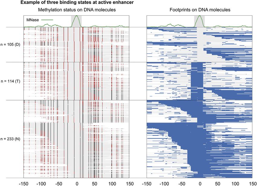
ll
                                                                                                                                       OPEN ACCESS

Protocol
A computational pipeline to visualize DNA-protein
binding states using dSMF data
Satyanarayan Rao1,2,3,* and Srinivas Ramachandran1,2,4,*
1Department    of Biochemistry and Molecular Genetics, University of Colorado School of Medicine, Aurora, CO 80045, USA
2RNA    Bioscience Initiative, University of Colorado School of Medicine, Aurora, CO 80045, USA
3Technical   contact
4Lead   contact
*Correspondence: satyanarayan.rao@cuanschutz.edu (S.R.), srinivas.ramachandran@cuanschutz.edu (S.R.)
https://doi.org/10.1016/j.xpro.2022.101299

SUMMARY
Here, we present a pipeline to map states of protein-binding DNA in vivo. Our
pipeline infers as well as quantifies cooperative binding. Using dual-enzyme sin-
gle-molecule footprinting (dSMF) data, we show how our workflow identifies
binding states at an enhancer in Drosophila S2 cells. Data from cells lacking
endogenous DNA methylation are a prerequisite for this pipeline.
For complete details on the use and execution of this protocol, please refer to
Rao et al. (2021) and Krebs et al. (2017).

BEFORE YOU BEGIN
Overview
This pipeline facilitates end-to-end analysis of dSMF reads for analyzing protein-DNA binding. Apart
from standard processing of raw sequencing reads, the pipeline implements a footprint calling algo-
rithm and maps footprints to the genomic region of interest. The pipeline is built using Snakemake
(Köster and Rahmann, 2012). Snakemake is a scalable bioinformatics workflow engine that enables
execution of reproducible pipelines easily in a high-performance cluster.

Downloading the pipeline

        Timing: 5 min

1. If git command is available on user’s machine, then please use this command ‘‘git clone https://
   github.com/satyanarayan-rao/star_protocol_enhancer_cooperativity.git’’, otherwise, please visit
   https://github.com/satyanarayan-rao/star_protocol_enhancer_cooperativity and click on the
   ‘‘code’’ button and select ‘‘Download Zip’’ to download the pipeline.

Installing required software and tools

        Timing: 1 h

We recommend using Anaconda software to build a virtual environment on user’s machine to enable
smooth operation of the pipeline without interference with other python packages already present in
the system.

2. Download and install Anaconda (individual edition): https://www.anaconda.com/products/
   individual).

                                                                                     STAR Protocols 3, 101299, June 17, 2022 ª 2022 The Author(s).     1
                                   This is an open access article under the CC BY-NC-ND license (http://creativecommons.org/licenses/by-nc-nd/4.0/).
ll
     OPEN ACCESS
                                                                                                            Protocol

3. Once Anaconda is installed, create a virtual environment named ‘‘dsmf_analysis’’ or ‘‘dsmf_viz’’
   using the command ‘‘conda create -n dsmf_analysis python=3.6’’.
4. After successful creation of the virtual environment, activate this environment by using the com-
   mand ‘‘conda activate dsmf_analysis’’.
5. Run ‘‘install_required_packages.sh’’ to install required packages to run the pipeline.

Data collection

      Timing: days; factors that affect timing include internet speed and available computing
    resources

This pipeline comprises the specific steps required for visualizing protein-binding on DNA mole-
cules using dSMF data. To perform the analysis, the following data are needed: A reference genome
in FASTA format; genomic regions of interest in BED format and reads from high-throughput
sequencing of dSMF libraries:

6. Since the data we analyzed was generated in Drosophila S2 cells, we are using the Apr. 2006 as-
   sembly of D. melanogaster genome (dm3, BDGP Release 5). The reference genome FASTA file
   can be downloaded from: http://hgdownload.cse.ucsc.edu/goldenpath/dm3/bigZips/dm3.fa.
   gz. For convenience, we have provided a script ‘ download_reference_genome.sh’ to download
   and place genome FASTA file in appropriate directory.
7. Any region of interest can be chosen, although regulatory regions of the genome are most mean-
   ingful. Here, we used cis-regulatory enhancers in Drosophila determined by STARR-seq. The data
   can be downloaded from: https://data.starklab.org/publications/yanez-cuna_genomeRes_
   2014/S2_peakSummits.txt.
8. We are using published paired-end bisulfite sequencing data (Krebs et al., 2017) as dSMF
   libraries here, but the user can easily perform similar analyses using their own data. Publicly avail-
   able dSMF libraries used here can be downloaded using the URLs provided in the README.md
   file (see "Download reference genome and dSMF data" section in the README.md file). We have
   included demo data (subset of the bisulfite sequencing data from (Krebs et al., 2017)) to illustrate
   a quick run through the whole pipeline, starting from sequencing data to publication-quality
   figures.

      CRITICAL: The bisulfite sequencing data should be paired-end.

Preparing metadata

      Timing: 10 min

 9. Prepare metadata files: create a tab separated values (.tsv) file consisting of three columns with
    headers: ‘‘sample’’, ‘‘paired_read_1_path’’, ‘‘paired_read_2_path’’. For each sequenced sam-
    ple, write a convenient name (please refrain from using special characters and spaces in the
    name to avoid the pipeline from failing), path for read1 and path for read2 in corresponding col-
    umns (e.g.,: ‘‘S2_tandem_R1_1’’, ‘‘data_from_geo/SRR3133326_1.fastq.gz’’, ‘‘data_from_geo/
    SRR3133326_2.fastq.gz’’. TSV file referring to the test dataset can be found here: ‘‘data_from_
    geo/samples.tsv’’.
10. For single binding sites, prepare a file containing regions of interest (ROI) using bed format (see
    format here: https://genome.ucsc.edu/FAQ/FAQformat.html#format1). Example can be seen
    in the GitHub repository ‘‘input_bed/example.bed’’.
11. For a pair of binding sites, prepare a file containing ROI using bedpe format (see .bedpe
    format description here: https://bedtools.readthedocs.io/en/latest/content/general-usage.html#
    bedpe-format). This format will allow us to provide the centers of the two binding sites. For an
    example, see ‘‘input_bed/example_cobinding.bedpe’’.

2     STAR Protocols 3, 101299, June 17, 2022
ll
Protocol OPEN ACCESS
12. Prepare configuration file as a key-value pair (yaml) file. Please see ‘‘configs/config.yaml’’ for an
example.
KEY RESOURCES TABLE
REAGENT or RESOURCE SOURCE IDENTIFIER
Deposited data
dSMF bisulfite sequencing data (Krebs et al., 2017) GSE77369
(Drosophila S2 cells)
STARR-seq summits from (Yáñez-Cuna et al., 2014) https://data.starklab.org/publications/yanez-
Drosophila S2 cells cuna_genomeRes_2014/S2_peakSummits.txt
Software and algorithms
Snakemake (Köster and Rahmann, 2012) https://snakemake.readthedocs.io/en/stable/
Gnuplot N/A http://www.gnuplot.info/
Trim Galore N/A https://www.bioinformatics.babraham.ac.uk/
projects/trim_galore/
Bowtie2 (Langmead and Salzberg, 2012) http://bowtie-bio.sourceforge.net/bowtie2/
index.shtml
Bismark (Krueger and Andrews, 2011) https://www.bioinformatics.babraham.ac.uk/
projects/bismark/
Bamtools (Barnett et al., 2011) https://github.com/pezmaster31/bamtools
Samtools (Danecek et al., 2021) http://www.htslib.org/
Deeptools (Ramı́rez et al., 2016) https://deeptools.readthedocs.io/en/develop/
Anaconda N/A https://www.anaconda.com/
Bioconda (Grüning et al., 2018) https://bioconda.github.io/
Other
Operating System Linux/Unix N/A
MATERIALS AND EQUIPMENT
Hardware.
Local – Memory 8 GB required, 16 GB recommended; Processors: 1 required, 4 recommended,
Computational Cluster – Memory: >16 GB recommended, Processors: >8 recommended.
Software and packages: A detailed list of software and packages can be found in the README.md
file.
STEP-BY-STEP METHOD DETAILS
In a single command, you can visualize protein-DNA binding state of a region of your interest from
raw sequencing data:
snakemake --snakefile cooperative_binding_analysis.smk
plots/single_binding/suppressed_merged_demo_S2_to_example_spanning_lf_15_r
f_15_extended_left_150_right_150_roi_peak_229.fp.pdf
plots/single_binding/suppressed_merged_demo_S2_to_example_spanning_lf_15_r
f_15_extended_left_150_right_150_roi_peak_229.methylation.pdf --configfile
configs/config.yaml
Below we will discuss the steps executed by the pipeline to generate the final figures.
STAR Protocols 3, 101299, June 17, 2022 3ll
OPEN ACCESS
Protocol
CAUTION: This command will run relatively quickly because demo dataset has been used. If using
full raw sequencing data, it will take around 10–15 h for the first run to prepare close to final data-
sets required for visualization. After that, for any regions of your interest it will take less than 5 min for
visualization.
Adapter trimming and alignment
Timing: 5–10 h
In this step, we perform standard preprocessing of NGS data and alignment to the reference genome
of interest. We use ‘‘Trim Galore’’ (see rule ‘‘trim_galore_pe’’ in snakemakes/trim_galore_pe.smk) for
adapter trimming and ‘‘Bismark’’ (see rule ‘‘Bismark_align_pe’’ in snakemakes/Bismark_align_pe.smk)
for bisulfite sequence alignment.
Suppressing cytosine methylation in contexts other than CpG or GpC dinucleotides
Timing: 1 h
In this step, we refine the methylation calls by Bismark based on the methyl-transferases used in the
dSMF experiment.
Note: We should only be considering methylation in CpG and GpC context based on the en-
zymes used in dSMF. To ensure this, we suppress methylation calls that are in other contexts
(HCH; H: A,C,T) and (DGD; D: A,T,G).
Extracting adjacent or overlapping bisulfite reads
Timing: 1 h
In this step, we extract properly aligned paired-end reads based on their SAM flags (: or ) from the merged alignment file (see step 1.3), and generate longer
reads by concatenating mate to read in case of pair and vice-versa in the case of (for further details about the meaning of SAM flags, see https://broadinstitute.github.io/
picard/explain-flags.html).
Note: In overlapping regions, information on the leftmost read is kept. We then extract all
longer reads that are overlapping or adjacent in nature. These reads form ‘‘DNA molecules’’
on which we will map binding states. Standard Illumina paired-end sequencing with read
lengths 150 bp leads to typically long enough (median length of 269 bp) ‘‘DNA molecules’’
(Rao et al., 2021) to map transcription factor (TF) and nucleosomal binding.
Defining footprints
Timing: 1 h
In this step, we define footprints on individual DNA molecules.
Note: It can be assumed that exogenous CpG and GpC methyltransferases reach saturation
by methylating all accessible CpG and GpC dinucleotides. A footprint is called when one or
more unmethylated cytosines are found between two methylated cytosines. We consider
footprints of at least 10 bp long. If two footprints are separated by just one bp, we merge
them to define a longer footprint. A DNA molecule with all unmethylated cytosines in CpG
or GpC contexts, which results in no footprint is given special consideration because it could
4 STAR Protocols 3, 101299, June 17, 2022ll
Protocol OPEN ACCESS
arise due to complete occlusion by nucleosomes, thus a footprint size of the whole DNA mole-
cule length is assigned in this case.
CRITICAL: With Drosophila S2 cells dSMF data, in open enhancers, one should expect
about 40%–50% of naked-DNA, about 15%–20% of TF-bound and about 30%–40% of
nucleosomal DNA (Rao et al., 2021). We imagine a similar distribution in other organisms
based on the conservation of chromatin structure across eukaryotes.
Assigning states on DNA molecules at single binding sites
Timing: 1 h
In this step, we assign binding states on individual DNA molecules mapped to ROI listed in a bed file.
Note: An example file is available at the GitHub repository ‘‘input_bed/example.bed’’. The
ROI coordinates are midpoints of the broader regions one is interested in. For TFs, we
consider 15 bp upstream and downstream of the midpoint to be sufficient. We then apply con-
ditions to assign a footprint as naked-DNA, TF, or nucleosomal: No footprint in ROI+-15 or
footprint of length less than 10 bp is assigned as naked-DNA, footprints of length between
10 and 50 bp in ROI+-15 are assigned as TFs, and others are assigned as nucleosomal. We
discard < 50 bp sized footprints on the edge of DNA molecules because we only know the
starting point of the footprint in these cases and hence the real length of these footprints is
unknown.
Assigning binding states at a pair of binding sites
Timing: 5 min
In this step, we assign co-binding states on individual DNA molecules that span a pair of TF binding
sites. Prepare a bedpe formatted file (see .bedpe format description here: https://bedtools.
readthedocs.io/en/latest/content/general-usage.html#bedpe-format) to provide the centers of
the two binding sites. For an example, see ‘‘input_bed/example_cobinding.bedpe’’. This step can
be executed by running the following command:
snakemake -np --snakefile cooperative_binding_analysis.smk
plots/cobinding_bedpe/suppressed_merged_S2_to_example_cobinding_lf_15_rf_
15_extended_left_300_right_300_roi_peak_110_4_and_peak_110_6.fp.pdf --
configfile configs/config.yaml
Note: Each TF binding site can have three states: naked or unbound, TF-bound, and nucleo-
some-bound. Thus, a pair of TF binding sites will have a total of nine states. States at individual
sites are assigned as described in step 6. Additionally, a footprint spanning both sites that
isll
OPEN ACCESS
Protocol
Figure 1. An example of three binding states observed at Peak 229 (position 0 is at chr2L:480305)
(Left) Each line in the heatmap is a DNA molecule (the grey fill). A red dot represents a methylated cytosine and a dark
gray dot represents an unmethylated cytosine. (Right) Blue lines are footprint calls on DNA molecules. These two
heatmaps are directly taken from the output of the pipeline and custom labeled. Number of DNA molecules
representing protein-DNA binding states is denoted on Y-axis. D: Naked DNA; T: TF-bound; N: Nucleosome-bound.
For optimal visualization, in each state, DNA molecules are sorted by their length and coordinates relative to position 0.
LIMITATIONS
This pipeline can only be used for experiments conducted in cells predominantly lacking endoge-
nous DNA methylation (for example Drosophila S2 cells). When regions of significant endogenous
methylation (>10% of a cytosine is methylated endogenously) are known, they should not be
included in the analysis (Raddatz et al., 2013; Takayama et al., 2014). The pipeline requires
paired-end datasets. This pipeline could be modified and extended for approaches that use nano-
pore and pacbio sequencing technologies that directly sequence methylated nucleotides and for
methods that involve adenine methylation.
TROUBLESHOOTING
Problem 1
Where else can I locate all binding state assignments for my region of interest after ‘‘assigning states
on DNA molecules at single binding sites’’, and ‘‘assigning binding states at a pair of binding sites’’
steps?
Potential solution
Files in ‘binding_states_fragments_extended/‘ and ‘cobinding_states_bedpe’ has binding state
information for single sites (‘input_bed/example.bed’) and pair of sites (‘input_bed/example_
cobinding.bedpe’) respectively. Please locate the corresponding file and search for your ROI.
The first column has binding state information (followed by #). 0: Naked-DNA; 1: TF 2: Nucleosome;
3: To be discarded.
Problem 2
I have fewer rows (DNA molecules) in methylation/footprint heatmap than the input matrix after the
steps: ‘‘assigning states on DNA molecules at single binding sites’’ and ‘‘assigning binding states at a
pair of binding sites’’.
6 STAR Protocols 3, 101299, June 17, 2022ll
Protocol OPEN ACCESS
Figure 2. An example of cobinding states observed at enhancer Peak 110 (position 0 is at chr2L: 19155173)
(Left) Each line in the heatmap is a DNA molecule (the grey fill). A red dot represents a methylated cytosine and a dark
gray dot represents an unmethylated cytosine. (Right) Blue lines are footprint calls on DNA molecules. These two
heatmaps are directly taken from the output of the pipeline and custom labeled. DNA molecules spanning the two
MNase peaks (shown in grey box at the top) are included in the plot. The MNase peaks are separated by 78 bp. Eight of
nine possible states are found at this locus. The number of DNA molecules mapped to each protein-DNA binding
state is denoted on Y-axis. D: Naked DNA; T: TF-bound; N: Nucleosome-bound. T-T, for example represents both
sites bound by TFs simultaneously. For optimal visualization, in each state, DNA molecules are sorted by their length
and coordinates relative to position 0.
Potential solution
We are only plotting valid protein-DNA and naked DNA states. Mapped DNA molecules with no
valid footprints (footprints less than 50 bp on the edge of DNA molecule) are discarded in heatmap
plotting. For instance, 25 DNA molecules are discarded in Figure 1.
Problem 3
The pipeline failed for my ROI named as ‘‘roi_peak_x" in my single site bed file when I ran the
following snakemake command (step: ‘‘assigning binding states at a pair of binding sites’’):
snakemake --snakefile cooperative_binding_analysis.smk
plots/single_binding/suppressed_merged_demo_S2_to_example_spanning_lf_15_
rf_15_extended_left_150_right_150_roi_roi_peak_229.fp.pdf --configfile
configs/config.yaml
How do I resolve this?
Potential solution
It is noteworthy that Snakemake builds workflow based on parsing the output filename. The example
peak_id written here as ‘‘roi_peak_x" overlaps with one of the string literals (‘‘roi’’) used by Snake-
make to retrieve wildcards. Thus, in this case rextend wildcard assigned a value ‘‘150_roi’’ in spite
of ‘‘150". Please refrain from using names and other parameters that overlaps with Snakemake
STAR Protocols 3, 101299, June 17, 2022 7ll
OPEN ACCESS
Protocol
wildcards. Please see the description of parameters here: https://github.com/satyanarayan-rao/
star_protocol_enhancer_cooperativity#interpreting-file-names.
Problem 4
I want to customize the pipeline, but I want to know which components of the pipeline will be
affected by my changes?
Potential solution
Snakemake facilitates visualization of pipeline up to your desired output file. To do this, you should
check if ‘dot’ command is available. If not, then please install graphviz (https://graphviz.org/). On
Linux machines, you can install this tool using:
sudo apt-get install graphviz
On macOS, you can install using:
brew install graphviz
Once graphviz is installed, you should be able to run the following:
snakemake -np --dag --snakefile cooperative_binding_analysis.smk
plots/single_binding/suppressed_merged_demo_S2_to_example_spanning_lf_15_
rf_15_extended_left_150_right_150_roi_peak_229.fp.pdf --configfile
configs/config.yaml | dot -Tpng > raw_sequencing_to_single_binding.png
This will visualize the pipeline after you make edits.
Problem 5
I ran jobs on HPC cluster, and one or more jobs aborted due to error, where should I look to trace-
back errors?
Potential solution
Please look at files in the directory ‘logs/cluster’. Look for the ID of aborted jobs in error files using
the command ‘grep -w *.err’ and then look for error(s) in these error files.
RESOURCE AVAILABILITY
Lead contact
Further information and requests for resources and reagents should be directed to and will be ful-
filled by the lead contact, Srinivas Ramachandran (srinivas.ramachandran@cuanschutz.edu).
Materials availability
This study did not generate new unique reagents.
Data and code availability
Data and code are available at the GitHub repository: https://github.com/satyanarayan-rao/
star_protocol_enhancer_cooperativity and at Zenodo: https://doi.org/10.5281/zenodo.5914775.
8 STAR Protocols 3, 101299, June 17, 2022ll
Protocol OPEN ACCESS
ACKNOWLEDGMENTS
This work was supported by the RNA Bioscience Initiative, University of Colorado School of Medi-
cine, and NIH grant R35GM133434 (S. Ramachandran).
AUTHOR CONTRIBUTIONS
S. Rao designed the project, generated all the code, performed the analysis, and wrote the manu-
script. S. Ramachandran designed and supervised the project and edited the manuscript.
DECLARATION OF INTERESTS
The authors declare no competing interests.
REFERENCES
Barnett, D.W., Garrison, E.K., Quinlan, A.R., Krebs, A.R., Imanci, D., Hoerner, L., Gaidatzis, D., Manke, T. (2016). deepTools2: a next generation
Strömberg, M.P., and Marth, G.T. (2011). Burger, L., and Schübeler, D. (2017). Genome-wide web server for deep-sequencing data analysis.
BamTools: a C++ API and toolkit for analyzing single-molecule footprinting reveals high RNA Nucleic Acids Res. 44, W160–W165.
and managing BAM files. Bioinformatics 27, polymerase II turnover at paused promoters. Mol.
1691–1692. Cell 67, 411–422.e4. Rao, S., Ahmad, K., and Ramachandran, S. (2021).
Cooperative binding between distant transcription
Danecek, P., Bonfield, J.K., Liddle, J., Marshall, J., Krueger, F., and Andrews, S.R. (2011). Bismark: a factors is a hallmark of active enhancers. Mol. Cell
Ohan, V., Pollard, M.O., Whitwham, A., Keane, T., flexible aligner and methylation caller for Bisulfite- 81, 1651–1665.e4.
McCarthy, S.A., Davies, R.M., et al. (2021). Twelve Seq applications. Bioinformatics 27, 1571–1572.
years of SAMtools and BCFtools. GigaScience 10, Takayama, S., Dhahbi, J., Roberts, A., Mao, G.,
giab008. Langmead, B., and Salzberg, S.L. (2012). Fast
Heo, S.-J., Pachter, L., Martin, D.I.K., and Boffelli, D.
gapped-read alignment with Bowtie 2. Nat.
(2014). Genome methylation in D. melanogaster is
Grüning, B., Dale, R., Sjödin, A., Chapman, Methods 9, 357–359.
B.A., Rowe, J., Tomkins-Tinch, C.H., Valieris, R., found at specific short motifs and is independent of
and Köster, J. (2018). Bioconda: Raddatz, G., Guzzardo, P.M., Olova, N., Fantappié, DNMT2 activity. Genome Res. 24, 821–830.
sustainable and comprehensive software M.R., Rampp, M., Schaefer, M., Reik, W., Hannon,
distribution for the life sciences. Nat. Methods G.J., and Lyko, F. (2013). Dnmt2-dependent Yáñez-Cuna, J.O., Arnold, C.D., Stampfel, G.,
15, 475–476. methylomes lack defined DNA methylation Bory n, Ł.M., Gerlach, D., Rath, M., and Stark, A.
patterns. PNAS 110, 8627–8631. (2014). Dissection of thousands of cell type-specific
Köster, J., and Rahmann, S. (2012). Snakemake—a enhancers identifies dinucleotide repeat motifs as
scalable bioinformatics workflow engine. Ramı́rez, F., Ryan, D.P., Grüning, B., Bhardwaj, V., general enhancer features. Genome Res. 24, 1147–
Bioinformatics 28, 2520–2522. Kilpert, F., Richter, A.S., Heyne, S., Dündar, F., and 1156.
STAR Protocols 3, 101299, June 17, 2022 9You can also read