Atomic structure of the predominant GII.4 human norovirus capsid reveals novel stability and plasticity
←
→
Page content transcription
If your browser does not render page correctly, please read the page content below
ARTICLE
                  https://doi.org/10.1038/s41467-022-28757-z                 OPEN

                  Atomic structure of the predominant GII.4 human
                  norovirus capsid reveals novel stability and
                  plasticity
                  Liya Hu 1,7, Wilhelm Salmen 2,7, Rong Chen1,7, Yi Zhou1, Frederick Neill2, James E. Crowe Jr.                                             3,4,

                  Robert L. Atmar2,5, Mary K. Estes 2,5,6 & B. V. Venkataram Prasad 1,2 ✉
1234567890():,;

                  Human noroviruses (HuNoVs) cause sporadic and epidemic viral gastroenteritis worldwide.
                  The GII.4 variants are responsible for most HuNoV infections, and GII.4 virus-like particles
                  (VLPs) are being used in vaccine development. The atomic structure of the GII.4 capsid in the
                  native T = 3 state has not been determined. Here we present the GII.4 VLP structure with
                  T = 3 symmetry determined using X-ray crystallography and cryo-EM at 3.0 Å and 3.8 Å
                  resolution, respectively, which reveals unanticipated novel features. A novel aspect in the
                  crystal structure determined without imposing icosahedral symmetry is the remarkable
                  adaptability of the capsid protein VP1 driven by the flexible hinge between the shell and the
                  protruding domains. In both crystal and cryo-EM structures, VP1 adopts a stable con-
                  formation with the protruding domain resting on the shell domain, in contrast to the ‘rising’
                  conformation observed in recent cryo-EM structures of other GII.4 VLPs. Our studies further
                  revealed that the resting state of VP1 dimer is stabilized by a divalent ion, and chelation using
                  EDTA increases capsid diameter, exposing new hydrophobic and antigenic sites and sug-
                  gesting a transition to the rising conformation. These novel insights into GII.4 capsid
                  structure, stability, and antigen presentation may be useful for ongoing vaccine development.

                  1 Verna and Marrs McLean Department of Biochemistry and Molecular Biology, Baylor College of Medicine, Houston, TX, USA. 2 Department of Molecular

                  Virology and Microbiology, Baylor College of Medicine, Houston, TX, USA. 3 The Vanderbilt Vaccine Center, Vanderbilt University Medical Center, Nashville,
                  TN, USA. 4 Department of Pediatrics, Vanderbilt University Medical Center, Nashville, TN 37232, USA. 5 Section of Infectious Diseases, Department of
                  Medicine, Baylor College of Medicine, Houston, TX, USA. 6 Section of Gastroenterology and Hepatology, Department of Medicine, Baylor College of
                  Medicine, Houston, TX 77030, USA. 7These authors contributed equally: Liya Hu, Wilhelm Salmen, Rong Chen. ✉email: vprasad@bcm.edu

                  NATURE COMMUNICATIONS | (2022)13:1241 | https://doi.org/10.1038/s41467-022-28757-z | www.nature.com/naturecommunications                                1
ARTICLE                                                                 NATURE COMMUNICATIONS | https://doi.org/10.1038/s41467-022-28757-z

N
         oroviruses (NoVs), belonging to the genus Norovirus in              assembles into T = 1 and T = 3 symmetries. Some GII VLPs,
         the Caliciviridae family, are nonenveloped, positive-sense          including GII.4 variants (Minerva, CHDC-1974, NSW-2012 and
         single-stranded RNA viruses capable of infecting many               the vaccine candidate GII.4c), show T = 4 symmetric organiza-
mammalian hosts, including humans, dogs, cats, pigs, mice,                   tion, with a diameter of ~500 Å, composed of 240 VP1 subunits.
sheep, and cattle1. In humans, these viruses (HuNoVs) are the                In addition, these studies showed that VP1 could exist in two
causative agents of epidemic and sporadic acute viral gastro-                distinct conformations similar to what was observed for VP1 of
enteritis worldwide, resulting in ~684 million illnesses and                 murine NoV21–23. In one conformation, the P domain rests on
~212,000 deaths annually2–4. The genome of NoVs is ~7.5 kb in                the S domain, referred to as a ‘resting’ conformation, whereas in
length and consists of three open reading frames (ORFs). ORF1                the other orientation, it is rotated and elevated from the S
codes for six non-structural proteins that are absent in the mature          domain, called a ‘raised’ conformation.
viral particles but play critical roles during virus replication.               The infectious GII.4 virion, similar to various animal calici-
ORF2 encodes the major capsid protein VP1 that encapsidates                  virus, has a smaller diameter of ~400 Å that is consistent with a
the (+) sense ssRNA genome and ORF3 codes for the minor                      T = 3 symmetry using negative stain EM20,24. Although the cryo-
structural protein VP2 that is suggested to be located internally in         EM structure of a GII.4 variant Minerva VLP with a
the capsid5.                                                                 T = 4 symmetry at 4.1 Å has been determined, the atomic
   Based on the amino acid sequences of VP1, NoVs are phylo-                 structure of a GII.4 HuNoV VLP capsid with T = 3 symmetry as
genetically classified into at least ten genogroups (GI-GX), which are        the native virus is not reported. Here we report the atomic
further subdivided into 49 genotypes (e.g., GII.4)6. NoVs of GI, GII,        structure of a GII.4 VLP determined using X-ray crystallography
GIV, GVIII, and GIX genogroups infect humans, and the genotype               and cryo-EM at 3.0 Å and 3.8 Å resolution, respectively. In both
GII.4 has been the predominant NoV genotype accounting for up to             structures, the GII.4 capsid exhibits the native T = 3 symmetry. A
80% of HuNoV infections. HuNoVs are highly contagious, and a                 unique aspect of our studies is that the crystallographic structure
few viral particles can lead to infection. Young children, immuno-           of the VLP is determined without explicitly imposing icosahedral
compromised patients, and the elderly are especially at high risk.           symmetry, which is rather unprecedented and allowed us to
Although there are candidate vaccines based on HuNoV virus-like              probe into conformational flexibility of the capsid protein that is
particles (VLPs) in clinical development7,8, as yet there are no             permissible within the context of an icosahedrally symmetric
approved vaccines or antivirals for prevention or treatment. It              organization. In addition to a detailed description of how GII.4
remains unknown whether the VLP-based vaccines can provide                   VP1 adapts to various quasiequivalent positions on a T = 3 ico-
long-term immunity against both homotypic and heterotypic                    sahedral lattice both in the crystalline state and in solution, our
infection with various epochally evolving GII.4 HuNoVs that are              studies revealed novel features, including identification of an ion-
genetically and antigenically distinct9. A deeper understanding of the       binding site that could impinge upon the stability and con-
capsid structures may provide a rational basis for developing more           formational flexibility of the GII.4 T = 3 capsid that may play a
effective vaccines.                                                          role in the cell entry processes and therefore provide informed
   Despite the recent success in the in vitro cultivation of                 strategies for vaccine development.
HuNoVs using stem cell-derived human enteroids10,11, it is still
challenging to obtain enough native viral particles for structural
studies. Therefore, VLPs, which are antigenically and structurally           Results
similar to native virions but lack the genome and are formed by              Crystal structure of GII.4 VLP shows a native T = 3 capsid
recombinant expression of the capsid protein VP1 with or                     organization. To elucidate the molecular details of the GII.4 VLP
without the minor structural protein VP2, have been extensively              (HOV, 2002 strain) structure, we expressed the capsid proteins
used as surrogates to characterize the structure of HuNoV viral              VP1 and VP2 using the baculovirus expression system. Previous
particles12–14. The first structure of a GI.1 VLP capsid was                  studies have shown that the co-expression of VP1 and VP2 results
determined using X-ray crystallography13. Following this, X-ray              in stable and structurally homogenous particles25. We initially
crystallographic structures of the native calicivirus of animal              determined the crystal structure of GII.4 HOV by imposing ico-
origins such as feline calicivirus and San Miguel sea lion virus,            sahedral symmetry using the protocols previously described13,15,16.
and cryo-EM structures of rabbit hemorrhagic disease virus and               Although the electron density representing the shell was clearly
receptor-bound feline calicivirus have been determined to near-              depicted, the electron density for the distal parts of the capsid was
atomic resolution15–18. All these structures showed that the                 poor, indicating that parts of the capsid structure deviated from
capsid exhibits T = 3 icosahedral symmetry, with a diameter of               strict icosahedral symmetry, perhaps affected by the packing of the
~400 Å, composed of 90 dimers of the capsid protein VP1 that                 GII.4 VLP in the crystal with the I222 space group. We then
consists of an internal N-terminal arm (NTA) and two distinct                resorted to determining the structure using only the non-
domains separated by a flexible hinge (Fig. 1a–c). In the capsid              crystallographic symmetry present within the crystallographic
architecture, the N-terminal domain ‘ties’ the VP1 subunits                  asymmetric unit (Supplementary Table 2). The crystallographic
through a network of interactions facing the interior of the capsid,         asymmetric unit in the I222 space group contains 45 copies of the
the S domain forms the icosahedral shell, and the P domain                   capsid protein VP1. The molecular replacement using GI.1 Nor-
protrudes from the S domain. The P domain is further divided                 walk VLP as the search model revealed the locations of all the 45
into P1 and P2 subdomains. The distally located P2 subdomain,                VP1 subunits in the asymmetric unit (Supplementary Fig. 1a).
which shows significant sequence variation among HuNoVs, is                   These subunits were refined using non-crystallographic symmetry
implicated in strain-dependent specific recognition of cellular               restraints without imposing the icosahedral 532 symmetry. The
glycans that function as both cell attachment and susceptibility             four asymmetric units of the unit cell related by the crystal-
factors19.                                                                   lographic 222 symmetry forms the complete capsid structure
   More recently, cryo-EM structures of various HuNoV VLPs                   consisting of 180 VP1 subunits with all the characteristics expected
representing GI.1, GI.7, GII.2, GII.4 HuNoV VLPs have been                   in a T = 3 organization (Supplementary Fig. 1b). For further
determined12,14,20. These structures reveal significant variations            analysis, these subunits were classified as 60 sets of traditionally
in the molecular organization of the capsid (Supplementary                   designated A, B, and C quasi-equivalent subunits representing the
Table 1). While GI.1 and GI.7 VLPs exhibit T = 3 icosahedral                 icosahedral asymmetric unit in a T = 3 lattice (Fig. 1d, e). These
symmetry consisting of 180 copies of VP1, the GII.2 VLP                      subunits cluster into 60 AB and 30 CC dimers in the capsid. The

2                NATURE COMMUNICATIONS | (2022)13:1241 | https://doi.org/10.1038/s41467-022-28757-z | www.nature.com/naturecommunications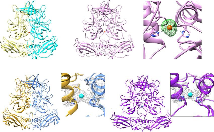
NATURE COMMUNICATIONS | https://doi.org/10.1038/s41467-022-28757-z                                                                       ARTICLE

              a                                                        b                                 c

                  NTA     S     Hinge P1       P2          P1                              P2

                                                                                                                                 C
                1 41            213 221 275          418        540                       P1              C

                                                                        S
                                                                                            Hinge
                                                                                                                                     N
                                                                         NTA                               N

              d                                             e

                                                                               5
                                        A
                                    B
                                   C                                     3     2      3

                                                                                                                  8
                                                                                                  8
                                                                                                  0
                                                                                                  0

                                                                                                              0
                                                                                                                  20
                                                                                               11
                                                                                               14
                                                                                               16

                                                                                                           18
                                                                                   Radius (Å)

Fig. 1 Crystal structure of GII.4 HOV VLP. a Schematic of VP1 primary structure colored by domain. b Ribbon representation of VP1 monomer. The NTA, S
domain, hinge, P1 subdomain, and P2 subdomain are colored corresponding to the schematic (a). c The VP1 dimeric formation. One subunit is colored as in
b, and the other subunit is shown in gray. The N- and C- termini of the structure are labeled. d GII.4 HOV VLP structure viewed along the icosahedral
twofold axis. The subunit A, B, and C are colored yellow, blue, and pink. e The surface and cut-away views of a GII.4 crystal surface colored by radial
distance showing an inner diameter of ~ 230 Å and an outer diameter of ~ 410 Å.

GII.4 VLP has an outer diameter of ∼410 Å and an inner diameter                 address whether these 15 copies maintained identical conforma-
of ∼230 Å (Fig. 1e). In the final model, following iterative cycles of           tions, as would be expected in the T = 3 capsid with strict ico-
refinement and model building, residues 26–531 of the A and C                    sahedral symmetry, or whether they showed structural
subunit and residues 10–532 of the B subunit were visualized.                   alterations, thereby providing insight into the allowed flexibility
Although the minor structural protein VP2 was co-expressed with                 within the T = 3 organization. Although the overall conformation
VP1 to obtain the VLPs, no electron density accounting for VP2                  of the S and P domains in these subunits is maintained, pairwise
residues was observed. This might be because of low copy numbers                superposition of A, B, and C subunits in the crystallographic
(1.5 - 8 copies) of VP2 per viral particle26,27, the intrinsic structural       asymmetric unit showed that while the subunits A and B
flexibility/disorder in VP2, or the lack of specific strong interac-              superimposed well with RMSD of 0.4 Å and 0.3 Å, respectively,
tions with the VP1 subunits.                                                    the C subunits showed significant variations with an RMSD of
   As expected, the structure of each VP1 subunit comprises the                 1.2 Å (Fig. 2a). In all the A and B subunits, the P domain making
NTA, the S domain, and the P domain (Fig.1b, c). The S domain                   close contact with the S domain is oriented in a similar manner;
(residue 41–213) has an eight-stranded β-barrel jellyroll structure             however, in the C subunits, as a result of the conformational
and forms the icosahedral shell. The P domain, linked to the S                  changes in the flexible hinge, there are noticeable alterations in
domain by a flexible hinge, comprises residues 222–540. The P                    the orientation between the S and P domains (Fig. 2b, c).
domain is further divided into P1 and P2 subdomains. The                        Examination of the crystal packing shows several of the C sub-
P1 subdomain, consisting of two segments of the polypeptide                     units in the capsid make close contact with the neighboring
(residues 222–275 and 419–540), folds into a structure similar to               capsid in the crystal and the flexible hinge allows their P domains
GI.1 Norwalk virus and other GII.4 P domain crystal structures.                 to suitably accommodate such a crystal packing (Supplementary
The S domains of the AB and CC dimers show bent and flat                         Fig. 1c). Interestingly, the icosahedral symmetry at the level of the
conformations to impart the required curvature to form the                      S domain is well maintained as well as the 5-fold symmetry that
icosahedral shell. Although there are small differences, as detailed            relates the AB dimers. These observations indicate that to
separately below, in all the subunits the P domain closely interacts            maintain the integrity of the T = 3 architecture, proper interac-
with the S domain (Fig. 2) (‘resting’ conformation) in stark                    tions between the S domains are critical and that the A-B dimers
contrast to what is observed in the T = 4 icosahedral structure of              surrounding the 5-fold axes have to be conformationally more
a GII.4 variant in which the P domain lacks any of the                          restrained; however, the C-C dimers can deviate from the strict
interactions with the S domain and is raised above by ~20 Å.                    icosahedral symmetry.

The intrinsic flexibility of VP1 subunits. The X-ray structure of                Cryo-EM structure shows GII.4 VLP adopts the same T = 3
the GII.4 VLP without explicitly imposing the icosahedral sym-                  capsid assembly in solution. To investigate if the conformational
metry allowed us to probe into the intrinsic flexibility in the                  flexibility observed in the crystal structure is indeed observed in
capsid protein within the crystallographic asymmetric unit con-                 solution and if the GII.4 VLP can conform to strict T = 3 ico-
taining 15 copies of the A, B, and C subunits. We wanted to                     sahedrally symmetric organization, we determined the cryo-EM

NATURE COMMUNICATIONS | (2022)13:1241 | https://doi.org/10.1038/s41467-022-28757-z | www.nature.com/naturecommunications                             3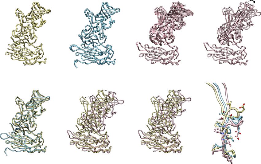
ARTICLE                                                                   NATURE COMMUNICATIONS | https://doi.org/10.1038/s41467-022-28757-z

        a                                                                                               N373
                                                                                                               12.3 Å                            8.2 °
                                                                                          7.9 Å
                                                                                       G392     G392'             N373'

                 Chain As                           Chain Bs                              Chain Cs                                 C11 vs. C12

        b                                                                                                12.0 Å
                                                                                                                        c
                                                               24.4 Å
                                                   17.1 Å                               9.3 Å
                                                                                                                                        Hinge

                                                                                                                            T219
                                                                                                                                                  E221

                 A1 vs. B1                         A1 vs. C11                           A1 vs. C12

Fig. 2 Structural comparison of VP1 subunits revealing its intrinsic flexibility. a Superimposition of 15 copies of subunit A, B, or C, respectively. The
structural change between Cs is indicated by red double-arrows. The distances between Cα atoms of residue G392 and the side chains of N373 are
measured and shown, respectively. b Superimposition of subunit A with B or C subunits using the S domain. The distance between a β-stand of P and S
domains is indicated by blue (subunit A) and red (subunit C) double-arrows. The distances between Cα atoms of residue G392 and the side chains of N373
are measured and shown, respectively, as in Fig. 2a. The black box indicates the hinge region of VP1. c Close-up view of the hinges of A, B, and C chains.
The stick model of residues 213–221 is shown and colored as in a.

structure using a single-particle reconstruction method by                     resolution of the cryo-EM map or the flexibility of the NTA in
imposing the icosahedral symmetry using cryo-SPARC (Supple-                    solution (Fig. 3c).
mentary Table 3). The cryo-EM images of the GII.4 VLP showed
that the majority of particles were of the same diameter of
~400 Å. The final resolution of the cryo-EM structure is ~3.8 Å as              Molecular interactions within and between VP1 subunits.
estimated by Fourier Shell Correlation (FSC) of 0.143 (Supple-                 Further analysis of the X-ray structure, in which the conforma-
mentary Fig. 2a). Our cryo-EM analysis shows that GII.4 VLPs in                tional details are resolved to a higher resolution, showed that the
solution also maintain the T = 3 capsid organization consisting of             A, B, and C subunits exhibit noticeable differences in their intra-
180 VP1 subunits with outer and inner diameters of ∼410 Å and                  domain interactions (Fig. 4). In the A subunit, the S and P
∼230 Å, respectively (Fig. 3a, b). The quality of the 3.8 Å cryo-EM            domains are closely associated via extensive molecular interac-
map allowed the unambiguous fitting of all the three quasi-                     tions, resulting in a more compact structure (Fig. 4a and c). The S
equivalent VP1 subunits in the T = 3 icosahedral capsid, which                 domain residues, such as P60-T65 and L76, interact with the
showed well-defined densities for their S and P domains (Sup-                   residues L474-R476 and S519-T526 of the P1 subdomain. The
plementary Figs. 2b, c). If, as in the crystal structure, the orien-           acidic residue E63 forms two salt bridges with the basic residue
tation of the P subunit with respect to the S domain (particularly             R476 (Fig. 4c). Whereas, in the B subunit, such S and P inter-
in the C subunit) showed variations, the density would not have                actions are somewhat less, resulting in a relatively more flexible
been as well defined because of icosahedral averaging. This                     structure. Compared to the A and B subunits, the C subunit
indicates that the C subunit adopts a singular conformation,                   assumes the most open conformation, with no molecular contacts
similar to one of the conformations in the crystal structure                   between the S and P domains (Fig. 4b). These differences con-
(Fig. 3c), to conform to the icosahedral symmetry. The con-                    tribute to the obligatory ‘bent’ conformation of the A/B dimers,
formations of the A and B subunits in the cryo-EM structure were               which surround the icosahedral 5-fold axes, and the ‘flat’ con-
the same as in the crystal structure. In all the three quasi-                  formation of the C/C dimers, at the icosahedral 2-fold axes,
equivalent subunits, the P domain rests on the S domain in the                 respectively (Fig. 4a, b). Such bent and flat conformations are a
resting conformation, as observed in the crystal structure                     common feature in all the T = 3 icosahedral structures and are
(Fig. 3a). This is also evident in the cut-away view of the cryo-EM            required for imparting the necessary curvature for the formation
map, which shows continuous density between the S domain and                   of the T = 3 capsid structure.
the P domain (Fig. 3b). In contrast to the crystal structure, the                 Despite the differences in the S-P1 interactions between A, B,
densities for the N-terminal 43 residues of NTA were not                       and C subunits, the interactions involving the S domain with the
observed in the cryo-EM map, perhaps because of the lower                      hinge and the neighboring residues are common in all these

4                  NATURE COMMUNICATIONS | (2022)13:1241 | https://doi.org/10.1038/s41467-022-28757-z | www.nature.com/naturecommunications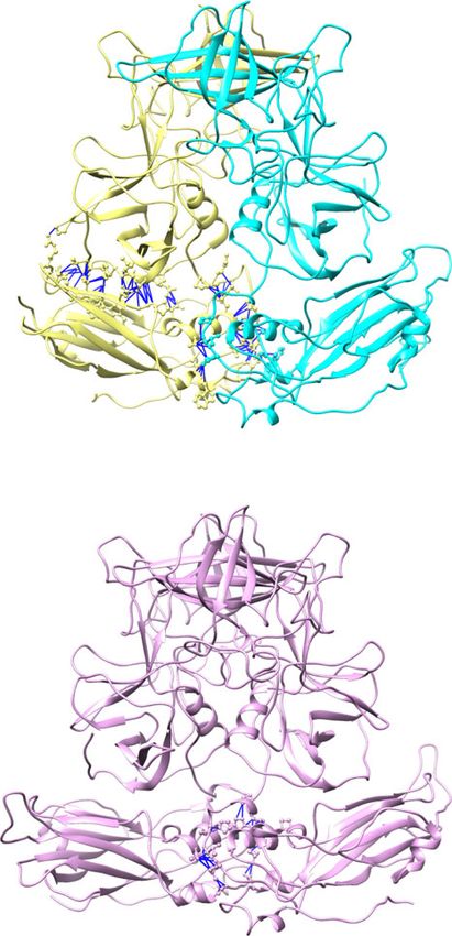
NATURE COMMUNICATIONS | https://doi.org/10.1038/s41467-022-28757-z                                                                      ARTICLE

               a                                              b

                                                                                5
                                          A
                                      B
                                     C                                    3     2       3

                                                                                                                8
                                                                                                 0
                                                                                                 8

                                                                                                 0

                                                                                                            0
                                                                                                                20
                                                                                              14
                                                                                              11

                                                                                              16

                                                                                                          18
                                                                                    Radius (Å)
               c

                                 N              N                                           N                        N             N
                                                                      N
                              Chain A                                         Chain B                                    Chain C
Fig. 3 Cryo-EM structure of GII.4 HOV VLP. a GII.4 HOV VLP cryo-EM structure aligned to the crystal structure shown in Fig. 1 and viewed along the
icosahedral twofold axis, shows the T = 3 symmetry of GII.4 HOV VLP in solution. The subunits A, B, and C are colored yellow, blue, and pink. b The
surface and cut-away views of a GII.4 cryo-EM map colored by radial distance, showing similar particle radius as observed in the crystal structure.
c Superimposition of subunit A (orange) with B (blue) or C (magenta) subunits of the cryo-EM structure with that of the VLP crystal structure (subunit A,
yellow; subunit B, light blue; subunit C, pink) using the S domain. The N-terminus of VP1 in the cryo-EM and crystal structure is labeled with colored and
black “N“s.

subunits (Fig. 4a, b, and d). The hydrophobic residues W51, I52,                 unbiased simulated annealing omit map. This density is present
and Y88 of the S domain closely associate with the hydrophobic                   between the P1 subdomains in all the VP1 dimers in the crys-
residues L215, V216, and P218 of the hinge (Fig. 4d). In addition                tallographic asymmetric units. Because the crystals of GII.4 VLP
to interacting with the S domain of the same subunit, the                        were obtained in a condition containing 100 mM cadmium
P1 subdomain is also involved in the interaction with the S                      chloride, we interpreted this strong density as possibly due to a
domain of the neighboring subunit. For instance, residue R484                    Cd2+ ion (Fig. 5a–c). The simulated annealing omit map con-
interacts with E74′ and R69′ at the fivefold axis (Fig. 4e). These                toured at 3 σ level is consistent with a Cd2+ ion coordinated by
interactions contribute to the stability of the T = 3 capsid                     the residue His460 at the dimeric interface of P1 subdomains
architecture.                                                                    (Fig. 5c). In the protein crystals, histidine is often involved in
                                                                                 coordinating various metal ions, including cadmium28. A similar
NTA ties the subunits through a network of interactions. One                     strong density is also present at the same location in our 3.8 Å
of the distinctive features of our crystal structure of the GII.4                cryo-EM density map (Fig. 5d, e), suggesting an intrinsic affinity
VLP, compared to the previous cryo-EM structures of other GII.4                  for a divalent cation at this position of the capsid protein.
variants, is the detailed visualization of the extensive interactions
mediated by the NTA (residues 1–43) that contribute to the                       Removal of ion affects the capsid structure. To investigate if the
stability of the capsid (Fig. 4f). In the crystal structure, the NTA             removal of the ion had any effect on the GII.4 capsid, we first
of the B subunits, which is structurally more ordered than those                 performed dynamic light scattering (DLS) in the absence and
of the A and C subunits, latches onto the neighboring C subunits                 presence of the chelating agent EDTA (Fig. 6a). Remarkably, the
via a network of molecular interactions (Fig. 4f). The residues                  DLS analysis indicated a monodisperse population of VLPs with a
D12-N24 of the B subunits surrounding the icosahedral 5-fold                     significant increase in the hydrodynamic radius in the presence of
axes interact with the residues R149, L151, and E152 of the S                    EDTA at pH 6.0 and pH 8.0 (Fig. 6a). We then performed a bis-
domain of subunit C at the local 3-fold axes to form an extensive                ANS binding assay in the presence and absence of EDTA to
network of interactions. In the NTAs of the A and C subunits, the                examine if loss of ion binding exposed new hydrophobic surfaces.
corresponding residues, D12-N24, are disordered. Interestingly,                  This assay showed that indeed is the case as there was a sub-
similar interactions involving the NTA of the B subunits are                     stantial increase in the fluorescence intensity in the presence of
observed in the crystal structure of GI.1 Norwalk VLP13.                         EDTA (Fig. 6b, c). To further investigate if EDTA treatment of
                                                                                 the HOV VLP had any effect on the antigen presentation and
The metal ion at the dimeric interface of the P domain.                          antibody binding, we performed BLI experiments using biotiny-
Another novel finding in our crystallographic structure of the                    lated Fab of a neutralizing antibody NORO-320 that binds to the
GII.4 VLP is the strongly resolved electron density in the                       sides of the P domain dimer29. These experiments showed that

NATURE COMMUNICATIONS | (2022)13:1241 | https://doi.org/10.1038/s41467-022-28757-z | www.nature.com/naturecommunications                                5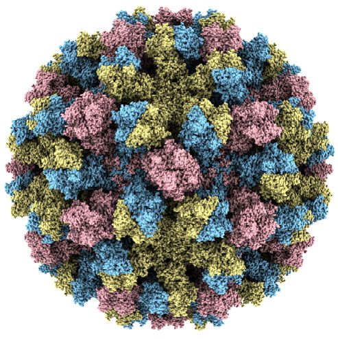
ARTICLE NATURE COMMUNICATIONS | https://doi.org/10.1038/s41467-022-28757-z
a A B A B
180° 90° R476
E63
66° 70°
b C C
C C
180° 90°
78° 84°
c d P1'
P218'
L474 Hinge'
T526 Y525 R476
2.6Å 3.4Å P1 3.2Å
V216'
F524
S519 Y88 3.3Å
T65 N522
E63 G61 P60 I52 L215'
G62
L76 W77
S W51
S'
F64
S
e
A1 P1
A5 A2 R516
V485 R484
R476
R69'
E63 E74'
S134' P72'
S
P1'
A5 A3
S'
B1
f N24
P20 E152'
C1 R149 V19 C2
L151'
S14 L18
B E152 F114'
L151 A16
A C R149'
D12
B3 B2
C3
EDTA treatment increases the binding of NORO-320 Fab to the particles showed a discrete or a continuous heterogeneity using
VLP, indicating that chelation of the metal ion results in a con- the recently developed three-dimensional variability analysis
formational change that allows for increased exposure of the (3DVA) in cryoSPARC30. This analysis shows that upon EDTA
partially occluded epitope for Fab binding (Fig. 6d). treatment, the particle images split into two populations
In addition to the above studies, we analyzed the cryo-EM (Supplementary Fig. 5) in clear contrast to untreated VLPs which
images of the HOV VLP treated with EDTA to examine if the exhibit a homogeneous single distribution of VLP images
6 NATURE COMMUNICATIONS | (2022)13:1241 | https://doi.org/10.1038/s41467-022-28757-z | www.nature.com/naturecommunications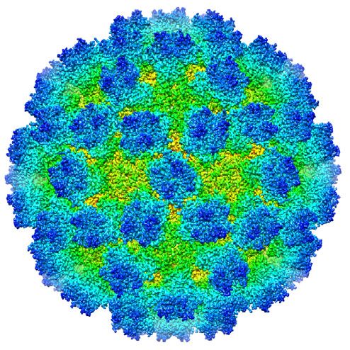
NATURE COMMUNICATIONS | https://doi.org/10.1038/s41467-022-28757-z                                                                             ARTICLE

Fig. 4 Molecular interactions within and between VP1 subunits. a, b Representative A/B and C/C dimer of the crystal structure. The angles formed by the
P dimer and S domain axes are measured using Chimera, showing the bent A/B dimer with smaller angles and the flat C/C dimers with larger angles. The
molecular interactions with and between VP1 are determined using Chimera and shown as blue lines. The S-P1 and S-Hinge interactions are indicated by
gray and magenta boxes, respectively. c Close-up view showing the interactions between the P1 subdomain (green) and the S domain (blue). The
interacting residues are shown as ball-and-stick models. d Close-up view of the interactions between the S domain (blue) and the hinge region of the
neighboring subunit (red). The residues from the adjacent subunit are indicated with prime (′). e Interactions between the A1–A5 subunits at a five-fold
axis. The inset shows the P1 subdomain of A1 interacting with the S domain of the A2 subunit. f The NTA network. Only residues of NTA are shown on the
left panel for clarity. The inset presents how NTAs interact with each other and the S domains at a three-fold axis. The subunits B and C are colored in blue
and pink, respectively.

               a                                        b                                         c

                                                                                                              3.7Å         3.8Å
                                                                                                         H460                        H460'

                                                                                                             Fo-Fc map

                      A/B dimer (crystal)                            C/C dimer (crystal)
               d                                                                  e
                                                                                                                                       L459'
                                                                   L459'
                                                          L459                                                               L459

                                                                                                                              2.8Å     3.0Å
                                                            2.9Å     3.2Å

                                                                                                                       H460
                                                       H460                                                                            H460'
                                                                     H460'
                                                              cryo-EM map                                                     cryo-EM map

                      A/B dimer (cryo-EM)                                                C/C dimer (cryo-EM)

Fig. 5 Ion binding site at the dimeric interface of VP1. a, b Cd2+ metal ion (orange sphere) is bound between the P dimers via a conserved residue
His460. c Simulated annealing omit map of the crystal structure is shown as green mesh and contoured at 3σ level. d, e Cryo-EM density maps of A/B and
C/C dimers reveal density (gray mesh) at the same site. The side chains of Leu459 and His460 are shown as stick models. A water molecule is placed into
the density between these Histidine residues.

(Supplementary Fig. 5a). The visualization of the particle                      Caliciviridae14. In the 8.1 Å cryo-EM structure of the GII.4 VLP
variability in 3D shows that EDTA treatment leads to increased                  expressed to mimic a vaccine strain, the capsid exhibits a
particle diameter with thinning of the density between the P and                T = 3 symmetry12, although the VP1 subunits, as in the
S domains indicating a transition from resting to a raised                      T = 4 structure of the Minerva strain, exhibit a ‘rising’ conforma-
conformation of the VP1 (Supplementary Movies 1 and 2). Taken                   tion, which was not observed previously except in the case of murine
together, these experiments suggest that loss of ion binding has a              NoV. Our studies by providing the structural details at the atomic
profound effect on the capsid structure leading to a raised                     level have allowed insights into the factors that likely influence the
conformation with increased access to antigenic sites.                          conformational dynamics of GII.4 VP1 and how it could impact the
                                                                                symmetry and stability of the capsid.

Discussion
In this study, by determining the structure of a globally dominant              The VP1 flexibility observed in GII.4 capsid is likely an
GII.4 HuNoV using X-ray crystallography and cryo-EM, we have                    intrinsic feature of HuNoV capsids. A remarkable observation
provided the atomic details of how the major capsid protein VP1                 in our 3.0 Å crystal structure of the GII.4 VLP is the extent to
assembles into a native T = 3 icosahedral assembly (Figs. 1 and 3).             which the conformation of capsid protein can deviate within the
Although this is the first crystallographic structure of the GII.4 VLP,          context of an icosahedrally symmetric organization. This obser-
previously, cryo-EM structures of VLPs of two GII.4 variants have               vation was possible because the GII.4 VLP crystallized in the
been reported12,14,20. These cryo-EM structures showed unique                   space group I222, with 45 VP1 subunits in the crystallographic
aspects of the GII.4 capsid structure that were not observed in our             asymmetric unit representing 15 of each of the three quasiequi-
GII.4 structure. In the 4.1 Å cryo-EM structure of the VLP of GII.4             valent subunits in the T = 3 icosahedral lattice (Supplementary
Minerva strain, the capsid displays T = 4 icosahedral organization,             Fig. 1). Our structure determination without imposing icosahe-
which had not been observed before in the members of                            dral symmetry showed that the C subunits varied significantly in

NATURE COMMUNICATIONS | (2022)13:1241 | https://doi.org/10.1038/s41467-022-28757-z | www.nature.com/naturecommunications                                   7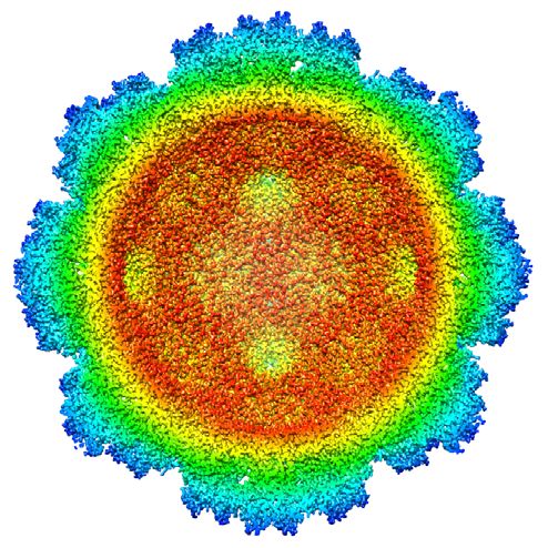
ARTICLE                                                                                            NATURE COMMUNICATIONS | https://doi.org/10.1038/s41467-022-28757-z

    a                                                             b
                                                                                        250                                                                     200
                                                                                                                pH 6.0 + EDTA
      Diameter (nm)

                                                                                        200                                                                     180                    pH 8.0 + EDTA

                                                                   Intensity (A.U.)

                                                                                                                                           Intensity (A.U.)
                                                                                        150                     pH 6.0                                          160

                                                                                        100                                                                     140                    pH 8.0

                                                                                         50                                                                     120

                                                                                          0                                                                     100
                      pH 6.0 pH 6.0   pH 8.0 pH 8.0                                           0   200     400      600      800     1000                              0   200    400     600      800   1000
                        –      +        –      + EDTA                                                   Time (Seconds)                                                          Time (seconds)

    d                                                             c
                                            1 μM + EDTA
                                            0.5 μM + EDTA
                                            0.25 μM + EDTA

                                                                     Intensity (A.U.)

                                                                                                                                             Intensity (A.U.)
                                            1 μM
                                            0.5 μM
                                            0.25 μM

                                                                                                   pH 6.0                pH 6.0                                             pH 8.0              pH 8.0
                                                                                                     –                     + EDTA                                             –                   + EDTA

Fig. 6 Dynamic light scattering, Bis‐ANS, and BLI binding assays. a DLS analysis shows the hydrodynamic radius increase upon removal of metal ions by
chelation with EDTA at pH 6.0 and pH 8.0, indicating the rising of P domain above shell in the absence of ion at the dimeric interface. b Changes in bis-ANS
binding in the absence or presence of EDTA show increased fluorescence intensity when VLPs are incubated with 20 mM EDTA, suggesting more
hydrophobic surfaces of VLP bound with bis-ANS. c Stabilized fluorescence intensities measured during the last minute for each sample were averaged and
presented as a bar graph. d BLI analysis of NORO-320 Fab and GII.4 HOV VLP shows that more VLPs bind to the immobilized Fab of GII.4 mAb NORO-320
in the presence of 20 mM EDTA, suggesting the exposure of mAb-binding epitope with EDTA-treatment. Data presented in each panel are means ± SE
(n = 3 independent study repliates) are shown. Source data are provided.

their conformations compared to the A and B subunits. The                                                caliciviruses and the other HuNoV VLPs. However, in the two
deviations are not in the conformation of the individual domains,                                        recent cryo-EM structures of GII.4 VLPs, irrespective of T = 3 or
which remain mostly invariant between the subunits, but are due                                          T = 4 organization, as noted above, all the three quasiequivalent
to distinct conformational variations in the hinge region, allowing                                      VP1 subunits are in the ‘raised’ conformation with the P domain
the P domain in these subunits to be oriented differently with                                           rotated and significantly elevated from the S domain12. Such a
respect to the S domain (Fig. 2c). These variations in the hinge are                                     raised conformation of the VP1 was first observed in the 8.1 Å
mostly due to the significant changes in the backbone con-                                                cryo-EM structure of the murine norovirus (MNV) with
formational angles of the residue T219 following two proline                                             T = 3 symmetry31. Follow-up cryo-EM studies with MNV VLPs
residues in the hinge region. Compared to T219-E221, other                                               showed VP1 transits to the resting state in the presence of bile
residues (213–218) in the hinge region show minor variations.                                            acids and that the resting state promotes efficient interactions
High conservation of the residues in the hinge region of the                                             with the cellular receptor23. However, in the most recent 3.1 Å
VP1 suggests similar conformational variability is likely an                                             cryo-EM structure of the MNV, the capsid protein is in a resting
intrinsic feature in the capsids of other HuNoVs.                                                        state both in the absence and presence of bile acids22. None-
   Interestingly, such variations are not observed in the cryo-EM                                        theless, these structural studies show that the P domain of the
structure. Despite enforcing icosahedral symmetry, the density for                                       norovirus capsid protein, because of the flexible hinge, can transit
the three quasi-equivalent subunits is well defined. The                                                  from a resting to a rising state. Such a transition appears to
conformations of the A and B subunits are the same as in the                                             depend upon several conditions.
X-ray structure, whereas the C subunit is ‘locked’ into a singular
conformation selected from one of the 15 conformations                                                   Factors that favor ‘resting’ vs. ‘raised’ states of VP1. Compar-
observed in the crystal structure to adhere to the icosahedrally                                         ison of the resting conformation of VP1 as found in our structural
symmetric organization. These observations indicate that under                                           studies with that of the ‘raised’ conformation of the VP1 as
the influence of external forces such as crystal packing, in this                                         observed in the recent 4.1 Å cryo-EM structure of the Minerva
case the capsid protein has certain conformational freedom to                                            GII.4 VLP, we notice several interesting differences despite the
‘breathe’ because of the flexible hinge. It is plausible that such                                        overall structures of the S and the P domain remaining highly
intrinsic conformational flexibility plays a role in optimal                                              similar with a RMSD of ~2.23–2.31 Å and ~2.15–2.30 Å,
engagement with cellular receptors upon contacting the cell                                              respectively, when the individual S or P domains are compared
surface during cell entry processes.                                                                     alone (Fig. 7). The first difference is the loss of all interactions
                                                                                                         between the S and P domain in the ‘raised’ conformation as the P
From ‘resting’ to ‘raised’ conformation of VP1. In both of our                                           domain, with the hinge region fully extended, is rotated and
crystal and cryo-EM structures of the GII.4 VLP, the P domains                                           elevated from the S domain by as much as 24 Å (Fig. 7c, d). The
of all the three quasi-equivalent subunits remain close to the S                                         second difference is the presence of metal ions at the P domain
domain in a ‘resting’ conformation, similar to that observed in the                                      dimeric interface in our structures, which is absent in the ‘raised’
high-resolution T = 3 structures of the several animal                                                   conformation. The residues at the dimeric interface that are close

8                         NATURE COMMUNICATIONS | (2022)13:1241 | https://doi.org/10.1038/s41467-022-28757-z | www.nature.com/naturecommunications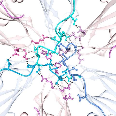
NATURE COMMUNICATIONS | https://doi.org/10.1038/s41467-022-28757-z                                                                          ARTICLE

                      a       A / B dimer in T = 3            e

                                                                                                         L459                 P230'
                                                                                                                    Q463
                                                                                               90°
                                                                                                         H460
                                                                                                                             H460'
                                                                                                                           L459'
                                                                                                          P230 Q463'

                                                                               Hinge

                      b        C / C dimer in T = 3           f

                                                                                                                    Q463
                                                                                                         L459                  P230'

                                                                                               90°       H460
                                                                                                                             H460'
                                                                                                                           L459'
                                                                                                          P230 Q463'
                                                                               Hinge

                      c        A / B dimer in T = 4           g
                                                                                                                  Q463

                                                                                                          Q459                 P230'

                                                                                               90°     H460
                                                                                                                             H460'
                                                                                                                            Q459'
                                                                                                       P230

                                                                                                                   Q463'
                                                                                Hinge

                      d        C / D dimer in T = 4           h
                                                                                                                   Q463
                                                                                                           Q459
                                                                                                                              P230'

                                                                                               90°
                                                                                                       H460                  H460'

                                                                                                       P230                 Q459'
                                                                                                                  Q463'

                                                                               Hinge

Fig. 7 Comparison of different quasiequivalent VP1 dimers in the resting conformation in T = 3 HOV capsid vs the raised conformation in T = 4
Minerva capsid. Comparison of the dimers using the S domain (shown in dashed oval) (left panel, a–d), or the P domain (middle panel, e–h) for structural
alignment. The additional C/D dimer present in the T = 4 capsid is shown in the bottom row (d, h). The inset (right panel) corresponding the the boxed
region in the middle panel shows a close up view of the interactions at the dimeric interface viewed along the two-fold axis of the P dimer. The ion present
only in the T = 3 dimers is shown as a cyan sphere. The A, B, C, and D subunits are labeled, and colored in yellow, blue, purple, and gray respectively (the C
subunit of the T = 4 C/D dimer is shown in lighter shade of purple to distinguish it from the C subunits of C/C dimer)). Residues 213-221 (hinge) and 222-
229 (N-terminus of P domain) are colored red and cyan, respectively.

to the metal ion in the resting conformation move away in the                   with the rest of the protein using the program PISA32. The cal-
rising conformation creating a gap at the dimeric interface                     culated ΔG, binding energy, from the same program, is −8.6 kcal/
(Fig. 7g, h and Supplementary Fig. 4). With the absence of S-P                  mol. In the raised state, these stabilizing interactions are lost.
interactions and the metal ion, it appears that the capsid structure               Although the metal ion observed in our crystal structure is
with the ‘raised’ conformation of the VP1 subunits would be                     likely Cd2+, based on the composition of the crystallization
more labile and less stable compared to the capsid with the                     buffer, the nature of the metal ion in the cryo-EM density is
subunits in the resting conformation. Furthermore, in the resting               uncertain. Postulating that it is likely a divalent metal ion, we
state, the interactions between the P and S domains are con-                    examined the effect of treating the GII.4 VLPs with EDTA using
siderable, with a buried surface area of 2078 Å2, as seen by                    DLS and Bis-ANS assay (Fig. 6). These experiments show that
analyzing the interactions between S (residues 1–213) of the chain              EDTA treatment results in a noticeable increase in the VLP

NATURE COMMUNICATIONS | (2022)13:1241 | https://doi.org/10.1038/s41467-022-28757-z | www.nature.com/naturecommunications                                    9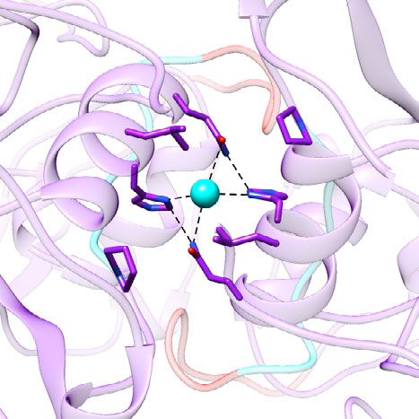
ARTICLE                                                                  NATURE COMMUNICATIONS | https://doi.org/10.1038/s41467-022-28757-z

diameter as well as exposure of new hydrophobic surfaces,                     such a transition may not be of consequence for antibodies that
suggesting that the chelation of the metal ion triggers a possible            neutralize HuNoV infection by blocking the HBGA binding site as
transition from a resting conformation to a raised conformation               these sites are not affected by the transition and remain accessible
as noticed using 3DVA analysis. Interestingly, the EDTA                       in both conformations (Fig. 9c).
treatment coupled with an increased pH, as inferred from the                     In summary, we have provided the first atomic-level descrip-
~8.1 Å resolution cryo-EM studies, has been shown to trigger                  tion of the capsid architecture of a globally dominant GII.4
such a transition in MNV and as well as in the T = 3 capsid                   HuNoV in the native T = 3 icosahedral symmetric state. In both
structure of HuNoV GII.3 VLP21. The authors further showed                    the crystal and solution, the capsid protein VP1 assumes a resting
that transition between resting and rising conformations is                   state stabilized by an elaborate network of NTA linking the capsid
reversible both in MNV and GII.3 VLP and that the infectivity of              subunits and the interactions between the S and P domains, as
MNV is optimal under the conditions favoring the resting                      expected in a T = 3 icosahedral capsid. A novel finding is the
conformation.                                                                 metal ion binding at the dimeric interface of the P domain that
   Our structure suggests a possible mechanism of how the                     further stabilizes the resting conformation. A remarkable feature
transition from a resting conformation can occur in the GII.4                 in our crystal structure is how the flexible hinge allows the metal
capsid. As observed in our GII.4 VLP structure, the metal ion                 ion stabilized P domain to sample an extensive conformational
binding site is close to the hinge region. The removal of the                 space in the resting conformation and allows transition to
bound metal ion may allosterically affect the conformation of the             possibly a more extended rising conformation with the removal
hinge allowing the P domain to transit from a resting to a rising             of the metal ion. Such conformational plasticity perhaps is an
state. The pairs of H460, Q463, L459 from the opposing subunits               inherent feature of the norovirus VP1 akin to the spike proteins
in the P dimer are involved in metal ion binding. Sequence                    of many enveloped and nonenveloped viruses, and maybe a
comparisons show that these residues are well conserved in                    critical factor in optimizing the interactions with the receptor and
HuNoV VP1 sequences, including GII.3 (Fig. 8). However,                       subsequent penetration through the cell membrane and genome
interestingly, in GII.4 Sydney (2012), H460 is replaced by Y460,              release. It is hoped that such a detailed understanding of the GII.4
and a hydrophilic residue Q459 replaces L459 in GII.4 Minerva                 capsid architecture will provide a rational basis for the improved
and GII.4 Sydney (Fig. 8). Whether these sequence changes alter               design and development of VLP-based vaccines to counter global
the metal binding affinity and tilt the preference for either the              HuNoV infections.
resting or the raised state, or whether they alter the transition
kinetics remains unclear.                                                     Methods
                                                                              Expression and purification of GII.4 HOV VLP. The VP1 and VP2 of a GII.4
                                                                              norovirus (Houston/TCH186/2002 or HOV, GenBank no. EU310927) were
Possible implications of ‘resting’ to ‘raised’ transition in anti-            expressed in the baculovirus system using Gibco Sf9 cells as described previously13.
                                                                              Briefly, cells were grown for 10 days and then centrifuged for 15 min at 1120 × g.
body neutralization. Our GII.4 structure also supports the sug-               The supernatant containing VLPs was then clarified by centrifugation at 22,100 × g
gestion by Song et al. that the transition from a more stable resting         for 30 min to remove aggregates and cell debris. Next, the clarified supernatant was
conformation to a more dynamic ‘raised’ conformation of VP1                   transferred to a fresh tube, and a 30% sucrose cushion was gently added to the
may play a critical role during the cell entry processes or the               bottom of the tube. This sample containing the 30% cushion was then centrifuged
disassembly processes. In addition to the cryo-EM structures of               at 120,000 × g for 3 h to pellet the VLPs. VLPs were then suspended by adding 200
                                                                              uL of PBS per tube and incubating at 4 °C overnight. Suspended VLPs were then
MNV and various HuNoVs VLPs, which have captured the resting                  pooled and diluted with PBS containing cesium chloride (1.14 mg/ml) to a final
and the raised states individually, a clear demonstration by Song             concentration of 0.38 mg/ml cesium chloride. The sample was then centrifuged at
et al., that such a transformation is reversible strongly suggests that       150,000 × g for 48 h at 4 °C. Lipid layer was removed, and then a visible blue band
this dynamic aspect is intrinsic to norovirus VP1. Dynamic                    containing VLP was isolated from each gradient tube. The VLP sample was then
                                                                              dialyzed overnight at 4 °C in PBS pH6.0. Dialyzed VLPs were then further purified
structural transitions have been implicated in the cell entry process         using the Sephacryl S500 size exclusion chromatography column. Purified fractions
for several viruses including, for example, the spike proteins of             were then pooled and stored at 4 °C.
rotavirus, influenza virus, and coronavirus18,33–36. If such a tran-
sition is indeed a critical step in the downstream cell entry events          Crystallization of GII.4 HOV VLP. Crystallization trials were carried out using the
of HuNoV, it also may have a consequence in how specific anti-                 hanging-drop vapor diffusion method at room temperature. For each condition,
bodies recognize and neutralize the virus infection as some of the            2 μl of the VLP solution, at a concentration of 3 mg/ml, was mixed with 2 μl of the
antigenic sites may become more accessible in one of these con-               well solution and equilibrated with 0.5 ml of the well solution. Initial screening
                                                                              produced crystals under a few conditions, including the Crystal Screen II condition
formations. This appears the case with the human antibody                     12 (0.1 M Na acetate pH 4.6, 0.1 M cadmium chloride, 30% PEG 400). After
NORO-320 that efficiently neutralizes GII.4 and GII.17 HuNoV                   crystallization condition optimization and on-site synchrotron screening, crystals
infections in the human enteriod system29. As determined by the               of the best diffraction quality were obtained using a well solution containing 0.1 M
crystal structure of the GII.4 P domain in complex with the anti-             Na acetate pH 3.6, 26–27% PEG 400. Cryo-protection of crystals was obtained by
genic binding fragment (Fab) of this antibody, the antigenic site is          soaking the crystals in the mother liquor with increasing glycerol concentration
                                                                              through four steps: 5%, 10%, 15%, and 18%. The equilibration time at each solution
on the sides of the P domain at a region that is conserved in the             was at least 10 min. The crystals were then flash-frozen in liquid nitrogen and
GII genotypes. Mapping this site on our GII.4 capsid structure,               shipped to synchrotron sites for data collection.
with the subunits in the resting conformation, indicates that this
site is occluded to a large extent, limiting the antibody access for          Crystal structure data collection and analysis. The diffraction data from single
efficient binding and neutralization (Fig. 9a). However, when we               crystals were collected under cryo-conditions at APS 19-ID with monochromatic
model the ‘raised’ conformation of the P domain into our struc-               X-rays (wavelength = 0.9795 Å) and a detector to crystal distance of 350 mm on an
ture, the NORO-320 binding site is fully exposed. It allows a                 ADSC 3 × 3 CCD detector using an oscillation angle of 0.3° and an exposure time
                                                                              of 5 sec. Indexing, integration, scaling, post-refinement, and data reduction were
stoichiometrically higher number of antibodies to bind and thus               carried out using the HKL200038. Analysis of the diffraction data indicated that the
increasing the neutralization efficiency (Fig. 9b and Supplementary            HOV VLP crystal belonged to the orthorhombic space group I222 (Supplementary
Fig. 3). Consistent with this hypothesis, the BLI assay shows                 Table 2). The data between 30 and 3.0 Å from 300 frames were scaled with a
increased binding of NORO-320 to VLPs in the presence of EDTA                 Rmerge factor of about 13%. To determine the precise orientation of the virus
                                                                              particle in the unit cell, self-rotation functions were calculated using the program
(Fig. 6d). Similarly, the antigenic site for one of the nanobodies,           GLRF39. These calculations showed all the peaks corresponding to 5-fold (κ = 72°),
although whether it neutralizes virus infection is not determined,            3-fold (κ = 120°), and 2-fold (κ = 180°) axes, which are expected from a particle
becomes more accessible in the ‘raised’ conformation37. However,              with icosahedral symmetry. In the I222 space group, consistent with the unit cell

10                NATURE COMMUNICATIONS | (2022)13:1241 | https://doi.org/10.1038/s41467-022-28757-z | www.nature.com/naturecommunications
NATURE COMMUNICATIONS | https://doi.org/10.1038/s41467-022-28757-z ARTICLE
D12 N24 W51 I52
R69 E74 W77 Y88 F114
S134 R149 E152
L215 P218
H460 L474 R484
R516 S519 T526
S-NTA S-Hinge S-P1 Subunit A-A’ Ion
Fig. 8 Sequence alignment of HuNoV VP1. Multiple sequence alignment was carried using ClustalW and visualized by Jalview50. The genotype | strain |
accession number of each VP1 sequence is given on the left side of the alignment. Residues are color-shaded, corresponding to the schematic in Fig. 1a. The
residues involved in the inter-domain or inter-subunit interactions are indicated using colored boxes.
NATURE COMMUNICATIONS | (2022)13:1241 | https://doi.org/10.1038/s41467-022-28757-z | www.nature.com/naturecommunications 11ARTICLE NATURE COMMUNICATIONS | https://doi.org/10.1038/s41467-022-28757-z
a Resting AB dimers b Raised AB dimers
Resting CC dimers Raised CC dimers
Resting P dimers Raised P dimers
c Antibody escape Antibody neutralization
mAb
P domain
Ion Hinge
S domain
HBGA/Co-receptor
Plasma membrane
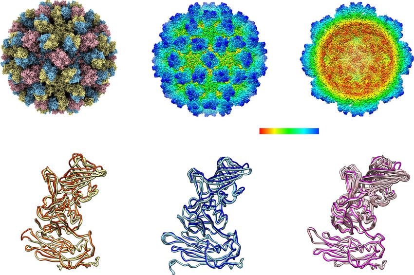
Fig. 9 Model of GII.4 HOV VLP in the ‘resting’ and ‘raised’ conformations. a Superimposition of GII.4 P domain in complex with Fab of NORO-320 onto
the AB and CC dimers of HOV VLP cryo-EM structure at 3.8 Å. The neighboring VP1 is shown as a transparent surface representation. b Modeling of GII.4
HOV VLP in the ‘raised’ conformation using the 8.0 Å GII.4c cryo-EM map in T = 3 symmetry. The P domain/Fab320 complex is superimposed onto the
model as in (a). c Schematic of NoV cell attachment process and human mAb neutralization mechanism.
dimensions and the particle radius, which is estimated to be 200 Å, the particle Cryo-EM sample preparation and data collection. A 3.5 μL aliquot of GII.4 HOV
position is uniquely defined by the intersection of the crystallographic 2-fold axes. VLPs (0.24 mg/ml) in a buffer containing PBS pH 6.0 was applied onto a 200-mesh
In such a setting, an icosahedral particle can assume one of the two possible R2/1 Quantifoil holey carbon grid coated with 0.2 mg/ml Graphene Oxide. The
orientations. This ambiguity in orientation was resolved by the self-rotation grid was blotted and rapidly frozen in liquid ethane using a Vitrobot IV(FEI), with
function calculations using the X-ray diffraction data. Each crystallographic constant temperature and humidity during the process of blotting. Movie stacks
asymmetric unit is composed of 1/4th of the virion with a non-crystallographic 15- (3149) were collected at 300 kV on a JEM-3200FSC electron microscope (JEOL)
fold symmetry. Similar packing of icosahedral particles has been observed in the with an in-column energy filter (50-eV width) equipped with a direct electron
Norwalk virus. detector K2 Summit camera (Gatan). Images were collected semiautomatically by
SerialEM46 in dose fractionation super-resolution counting mode with a binning of
0.5 at 30,000X magnification, corresponding to a pixel size of 1.2 Å. The images
Crystal structure determination and refinement. A properly positioned and were collected with a defocus range from −0.6 to −2.6 μm. The final frame average
oriented Norwalk capsid was placed in an HOV crystal unit cell. One initial model was computed from 25 frames to correct for beam-induced motion during expo-
at 10 Å was calculated from the 15 copies of NV related first by icosahedral 5-fold sure by MotionCor247. The image in each frame was weighted according to
symmetry and then by icosahedral 3-fold symmetry. Another initial model was radiation damage. CTF parameters of the particles in each frame average were
obtained from a cryo-EM reconstruction of HOV at a resolution of 22 Å. The cryo- determined by Relion 3.1 CtfFind and Gctf v1.0648. Two-dimensional (2D)
EM structure was also used to generate a mask. Phase refinement and extension reference-free class averages were computed using RELION 3.1. Data were pro-
were carried out by iterative cycles of real space electron density averaging, solvent cessed for icosahedral reconstruction using CryoSPARC. The 0.143 Fourier shell
flattening, and back transformation with CCP4 program packages40. The phases correlation (FSC) cut-off was used for the resolution determination (Supplemen-
were extended to the final 3 Å resolution, with each step less than one reciprocal tary Fig. 2). Three-dimensional variability analysis (3DVA) implemented in
space point. The averaged 3.0 Å density map is of good quality and readily cryoSPARC30 was performed to understand and visualize the dynamics of GII.4
interpretable in the whole S domain and the P domains of the A and B subunits, HOV VLPs (0.24 mg/ml) in a buffer containing PBS pH 6.0 in the presence of
but not in the P domain of the C subunit. Iterative cycles of refinement and further 20 mM EDTA (Supplementary Movies 1 and 2).
model building were carried out using PHENIX41 and COOT programs42. Non-
crystallographic symmetry (NCS) was applied during refinement using Phe-
nix.refine. In the final refinement, the stereochemistry of the structures was Cryo-EM structure determination and refinement. The crystal structure of GII.4
checked using MolProbity43. Data refinement and statistics are given in Supple- HOV VLP was fitted into the density map using Chimera. Iterative cycles of
mentary Table 2. The interactions within and between VP1 subunits were analyzed refinement and further model building were carried out using PHENIX41,
using Chimera44. Figures were prepared using Chimera and ChimeraX44,45. Rosetta49, and COOT programs42 Water molecules were built using Douse in
12 NATURE COMMUNICATIONS | (2022)13:1241 | https://doi.org/10.1038/s41467-022-28757-z | www.nature.com/naturecommunicationsNATURE COMMUNICATIONS | https://doi.org/10.1038/s41467-022-28757-z ARTICLE
PHENIX and manually verified in COOT. The cryo-EM map was deposited in the 5. Vongpunsawad, S., Venkataram Prasad, B. V. & Estes, M. K. Norwalk virus
Electron Microscopy Databank, and the coordinates of the atomic model were minor capsid protein VP2 associates within the VP1 shell domain. J. Virol. 87,
deposited in the Protein Data Bank. EM density Data collection and processing 4818–4825 (2013).
details are provided in Supplementary Table 3. 6. Chhabra, P. et al. Updated classification of norovirus genogroups and
genotypes. J. Gen. Virol. 100, 1393–1406 (2019).
Dynamic light scattering. The hydrodynamic diameters of GII4 HOV VLPs in the 7. Sherwood, J. et al. Efficacy of an intramuscular bivalent norovirus GI.1/GII.4
absence or presence of the ion chelation reagent EDTA at pH 6.0 or pH 8.0 were virus-like particle vaccine candidate in healthy US adults. Vaccine 38,
measured using dynamic light scattering (DLS) on a ZetaSizer Nano instrument 6442–6449 (2020).
(Malvern Instruments, UK). Samples were diluted to a final concentration of 8. Kim, L. et al. Safety and immunogenicity of an oral tablet norovirus vaccine, a
200 nM for each component in phosphate-buffered saline. Upon addition of phase I randomized, placebo-controlled trial. JCI Insight 3, https://doi.org/
EDTA, samples were incubated on ice for 30 min before measurements were taken. 10.1172/jci.insight.121077 (2018).
Three × 12 measurement runs were performed with standard settings (Refractive 9. Siebenga, J. J. et al. Epochal evolution of GGII.4 norovirus capsid proteins
Index 1.335, viscosity 0.9, temperature 25 °C). The average result was created with from 1995 to 2006. J. Virol. 81, 9932–9941 (2007).
ZetaSizer software. 10. Ettayebi, K. et al. Replication of human noroviruses in stem cell-derived
human enteroids. Science 353, 1387–1393 (2016).
Detection of bis-ANS binding by fluorescence spectroscopy. Purified VLPs (30 11. Ettayebi, K. et al. New insights and enhanced human norovirus cultivation in
μg/ml, 0.5 μM concentration of the VP1) diluted in PBS buffer pH 6.0 or pH 8.0 human intestinal enteroids. mSphere 6, https://doi.org/10.1128/mSphere.01136-20
with or without 20 mM EDTA were incubated at 25 °C for 10 min to allow for (2021).
temperature equilibration. To detect bis-(8-anilinonaphthalene-1-sulfonate) (bis- 12. Devant, J. M. & Hansman, G. S. Structural heterogeneity of a human
ANS) binding to VLP alone or in the presence of EDTA, bis-ANS was added to norovirus vaccine candidate. Virology 553, 23–34 (2021).
each sample for a final concentration of 3 μM immediately before data collection. 13. Prasad, B. V. et al. X-ray crystallographic structure of the Norwalk virus
Bis-ANS was excited at 395 nm, and emission was collected at 495 nm at 30 s capsid. Science 286, 287–290 (1999).
intervals for 15 min on a Flexstation 3 (Molecular Devices, USA). The averaged 14. Jung, J. et al. High-resolution cryo-EM structures of outbreak strain human
results were prepared using PRISM. norovirus shells reveal size variations. Proc. Natl Acad. Sci. USA 116,
12828–12832 (2019).
15. Ossiboff, R. J., Zhou, Y., Lightfoot, P. J., Prasad, B. V. & Parker, J. S.
Modeling of GII.4 HOV VLP in the ‘raised’ conformation. The atomic model of
Conformational changes in the capsid of a calicivirus upon interaction with its
GII.4 VLP P domain and S domain were fit into the 8.1 Å cryo-EM density map of
functional receptor. J. Virol. 84, 5550–5564 (2010).
norovirus GII.4c VLPs with T = 3 icosahedral symmetry using Chimera. The flexible
16. Chen, R., Neill, J. D., Estes, M. K. & Prasad, B. V. X-ray structure of a native
hinge was built using COOT. The axis of P domain dimers was defined using mass-
calicivirus: structural insights into antigenic diversity and host specificity.
weighting in Chimera. We then calculated the crossing angle and distance of the axes
of the P domain in the ‘rising’ and ‘raised’ conformation using Chimera. Proc. Natl Acad. Sci. USA 103, 8048–8053 (2006).
17. Wang, X. et al. Atomic model of rabbit hemorrhagic disease virus by cryo-
electron microscopy and crystallography. PLoS Pathog. 9, e1003132 (2013).
Biolayer interferometry (BLI). Biotinylation of the NORO-320 Fab was carried 18. Conley, M. J. et al. Calicivirus VP2 forms a portal-like assembly following
out using EZ-Link NHC-LC-LC-biotin (catalog no. 21343; Thermo Scientific) receptor engagement. Nature 565, 377–381 (2019).
following the manufacturer’s instructions. The NORO-320 Fab was loaded onto 19. Prasad, B. V. et al. Antiviral targets of human noroviruses. Curr. Opin. Virol.
streptavidin biosensors at a concentration of 1 μg/ml in the BLI running buffer 18, 117–125 (2016).
(20 mM Hepes, 150 mM NaCl, 0.005% surfactant P20, and 2 mg/ml BSA, pH 7.8) 20. Devant, J. M., Hofhaus, G., Bhella, D. & Hansman, G. S. Heterologous
for 600 s, resulting in capture levels of 1.5–2 nm within a row of eight tips. GII.4 expression of human norovirus GII.4 VP1 leads to assembly of T = 4 virus-
HOV VLP was diluted in BLI running buffer with or without the addition of like particles. Antivir. Res 168, 175–182 (2019).
20 mM EDTA to a final concentration of 1000 nM and incubated on ice overnight. 21. Song, C. et al. Dynamic rotation of the protruding domain enhances the
NORO-320 Fab-GII.4 HOV VLP association and dissociation curves were obtained
infectivity of norovirus. PLoS Pathog. 16, e1008619 (2020).
through twofold serial dilutions of GII.4 HOV VLP (1000, 500, 250 nM) plus buffer
22. Snowden, J. S. et al. Dynamics in the murine norovirus capsid revealed by
blanks at 30 °C using the Octet acquisition software. BLI studies were carried out
high-resolution cryo-EM. PLoS Biol. 18, e3000649 (2020).
using an Octet RED96 instrument (FortéBio).
23. Sherman, M. B. et al. Bile salts alter the mouse norovirus capsid conformation:
possible implications for cell attachment and immune evasion. J. Virol. 93,
Reporting summary. Further information on research design is available in the Nature https://doi.org/10.1128/JVI.00970-19 (2019).
Research Reporting Summary linked to this article. 24. Taniguchi, K., Urasawa, S. & Urasawa, T. Further studies of 35–40 nm virus-
like particles associated with outbreaks of acute gastroenteritis. J. Med.
Microbiol. 14, 107–118 (1981).
Data availability
25. Bertolotti-Ciarlet, A., White, L. J., Chen, R., Prasad, B. V. & Estes, M. K.
The data that support this study are available from the corresponding author upon
Structural requirements for the assembly of Norwalk virus-like particles. J.
reasonable request. Atomic coordinates and structure factors for the crystal structure of
Virol. 76, 4044–4055 (2002).
the GII.4 HOV VLP have been deposited in the Protein Data Bank (PDB) with the
26. Sosnovtsev, S. V. & Green, K. Y. Identification and genomic mapping of the
accession code 7K6V [https://doi.org/10.2210/pdb7K6V/pdb]. The cryo-EM structure of
ORF3 and VPg proteins in feline calicivirus virions. Virology 277, 193–203
GII.4 HOV VLP has been deposited in the Protein Data Bank with the accession code
7MRY [https://doi.org/10.2210/pdb7MRY/pdb] and the Electron Microscopy Databank
(2000).
27. Glass, P. J. et al. Norwalk virus open reading frame 3 encodes a minor
with accession code EMD-23960. Source data are provided with this paper.
structural protein. J. Virol. 74, 6581–6591 (2000).
28. Dokmanic, I., Sikic, M. & Tomic, S. Metals in proteins: correlation between
Received: 13 July 2021; Accepted: 8 February 2022; the metal-ion type, coordination number and the amino-acid residues
involved in the coordination. Acta Crystallogr D. Biol. Crystallogr 64, 257–263
(2008).
29. Alvarado, G. et al. Broadly cross-reactive human antibodies that inhibit
genogroup I and II noroviruses. Nat. Commun. 12, 4320 (2021).
30. Punjani, A. & Fleet, D. J. 3D variability analysis: resolving continuous
References flexibility and discrete heterogeneity from single particle cryo-EM. J. Struct.
1. Villabruna, N., Koopmans, M. P. G. & de Graaf, M. Animals as reservoir for Biol. 213, 107702 (2021).
human norovirus. Viruses 11,https://doi.org/10.3390/v11050478 (2019). 31. Katpally, U. et al. High-resolution cryo-electron microscopy structures of
2. Mattison, C. P., Cardemil, C. V. & Hall, A. J. Progress on norovirus vaccine murine norovirus 1 and rabbit hemorrhagic disease virus reveal marked
research: public health considerations and future directions. Expert Rev. flexibility in the receptor binding domains. J. Virol. 84, 5836–5841 (2010).
Vaccines 17, 773–784 (2018). 32. Krissinel, E. & Henrick, K. Inference of macromolecular assemblies from
3. Kirk, M. D. et al. World Health Organization estimates of the global and crystalline state. J. Mol. Biol. 372, 774–797 (2007).
regional disease burden of 22 foodborne bacterial, protozoal, and viral 33. Herrmann, T. et al. Functional refolding of the penetration protein on a non-
diseases, 2010: a data synthesis. PLoS Med 12, e1001921 (2015). enveloped virus. Nature 590, 666–670 (2021).
4. GBD 2015 Mortality and Causes of Death Collaborators. Global, regional, and 34. Russell, C. J. Hemagglutinin stability and its impact on influenza A virus
national life expectancy, all-cause mortality, and cause-specific mortality for infectivity, pathogenicity, and transmissibility in avians, mice, swine, seals,
249 causes of death, 1980–2015: a systematic analysis for the Global Burden of ferrets, and humans. Viruses 13, https://doi.org/10.3390/v13050746
Disease Study 2015. Lancet 388, 1459–1544 (2016). (2021).
NATURE COMMUNICATIONS | (2022)13:1241 | https://doi.org/10.1038/s41467-022-28757-z | www.nature.com/naturecommunications 13You can also read