BESTSEL: WEBSERVER FOR SECONDARY STRUCTURE AND FOLD PREDICTION FOR PROTEIN CD SPECTROSCOPY
←
→
Page content transcription
If your browser does not render page correctly, please read the page content below
W90–W98 Nucleic Acids Research, 2022, Vol. 50, Web Server issue Published online 11 May 2022
https://doi.org/10.1093/nar/gkac345
BeStSel: webserver for secondary structure and fold
prediction for protein CD spectroscopy
András Micsonai1 , Éva Moussong1 , Frank Wien2 , Eszter Boros3 , Henrietta Vadászi1 ,
Nikoletta Murvai3,4 , Young-Ho Lee5,6,7 , Tamás Molnár1 , Matthieu Réfrégiers2,8 , Yuji Goto9 ,
Ágnes Tantos4 and József Kardos 1,*
1
ELTE NAP Neuroimmunology Research Group, Department of Biochemistry, Institute of Biology, ELTE Eötvös
Loránd University, Budapest H-1117, Hungary, 2 Synchrotron SOLEIL, Gif-sur-Yvette 91192, France, 3 Department of
Biochemistry, Institute of Biology, ELTE Eötvös Loránd University, Budapest H-1117, Hungary, 4 Institute of
Downloaded from https://academic.oup.com/nar/article/50/W1/W90/6584425 by guest on 01 August 2022
Enzymology, Research Centre for Natural Sciences, Budapest H-1117, Hungary, 5 Research Center of
Bioconvergence Analysis, Korea Basic Science Institute (KBSI), Ochang 28119, Republic of Korea, 6 Bio-Analytical
Science, University of Science and Technology (UST), Daejeon 34113, Republic of Korea, 7 Graduate School of
Analytical Science and Technology (GRAST), Chungnam National University (CNU), Daejeon 34134, Republic of
Korea, 8 Centre de Biophysique Moléculaire, CNRS UPR4301, Orléans, France and 9 Global Center for Medical
Engineering and Informatics, Osaka University, Osaka 565-0871, Japan
Received March 28, 2022; Revised April 18, 2022; Editorial Decision April 22, 2022; Accepted May 09, 2022
ABSTRACT GRAPHICAL ABSTRACT
Circular dichroism (CD) spectroscopy is widely used
to characterize the secondary structure composition
of proteins. To derive accurate and detailed struc-
tural information from the CD spectra, we have devel-
oped the Beta Structure Selection (BeStSel) method
(PNAS, 112, E3095), which can handle the spectral
diversity of -structured proteins. The BeStSel web-
server provides this method with useful accessories
to the community with the main goal to analyze sin-
gle or multiple protein CD spectra. Uniquely, BeSt-
Sel provides information on eight secondary struc-
ture components including parallel -structure and
antiparallel -sheets with three different groups of
twist. It overperforms any available method in ac-
curacy and information content, moreover, it is ca-
pable of predicting the protein fold down to the INTRODUCTION
topology/homology level of the CATH classification.
The far-UV circular dichroism (CD) spectrum of a protein
A new module of the webserver helps to distinguish
is characteristic to the secondary structure composition and
intrinsically disordered proteins by their CD spec- is widely used to investigate protein structure. Although it
trum. Secondary structure calculation for uploaded does not provide site-specific structural information, CD
PDB files will help the experimental verification of spectroscopy is useful when a fast, inexpensive technique
protein MD and in silico modelling using CD spec- is needed or the application of high-resolution techniques
troscopy. The server also calculates extinction coeffi- (X-ray or NMR) is problematic. Applications cover all ar-
cients from the primary sequence for CD users to de- eas of protein science. CD spectroscopy can be used to ver-
termine the accurate protein concentrations which is ify the correct fold of recombinant proteins, study the ef-
a prerequisite for reliable secondary structure deter- fect of environmental conditions (pH, ionic strength, addi-
mination. The BeStSel server can be freely accessed tives, crowding) and protein modifications (e.g. mutations
at https://bestsel.elte.hu. and post-translational modifications) on the structure and
* To whom correspondence should be addressed. Tel: +36 1 372 2500/1795; Fax: +36 1 381 2172; Email: kardos@elte.hu.
C The Author(s) 2022. Published by Oxford University Press on behalf of Nucleic Acids Research.
This is an Open Access article distributed under the terms of the Creative Commons Attribution-NonCommercial License
(http://creativecommons.org/licenses/by-nc/4.0/), which permits non-commercial re-use, distribution, and reproduction in any medium, provided the original work
is properly cited. For commercial re-use, please contact journals.permissions@oup.com
Nucleic Acids Research, 2022, Vol. 50, Web Server issue W91
stability. Moreover, CD spectroscopy is a suitable technique offered from 175–250 nm to 200–250 nm at 5 nm steps. For
for the experimental verification of the structural informa- each range, there are eight sets of the eight basis spectra, and
tion predicted by bioinformatics tools that make use of the each set is optimized to be the most accurate for one of the
ever-growing protein databases. secondary structure components. The fraction of that com-
The instrumentation of CD spectroscopy is well devel- ponent is taken from the fitting with the linear combination
oped and benchtop instruments are routinely used. Syn- of the corresponding eight spectra to the CD spectrum of
chrotron radiation (SR) CD is also available with a broad the unknown protein and thus eight fittings will provide the
wavelength range and can be used for high quality and fractions of the eight secondary structure components.
special applications (1). A central question in protein CD
spectroscopy is the spectral contribution of the various sec-
Protein fold prediction by BeStSel
ondary structural elements. Numerous algorithms were de-
veloped in the last decades to gain information on the sec- Protein folds can be characterized by specific secondary
ondary structure from the CD spectra, however, accurate structure patterns. The eight secondary structure compo-
Downloaded from https://academic.oup.com/nar/article/50/W1/W90/6584425 by guest on 01 August 2022
structure estimation was mostly limited to ␣-helical pro- nents of BeStSel provide sufficient structural information
teins because of the large spectral diversity of -structured to predict the protein fold. The -sheet composition, includ-
proteins (2,3). We have shown that the parallel-antiparallel ing the parallel-antiparallel -sheets and the level of twist in
orientation and the twist of the -sheets account for this the antiparallel -sheets adequately characterize the diverse
spectral diversity and developed the Beta Structure Selec- folds with -structures, while the two ␣-helix components
tion (BeStSel) method for the accurate secondary structure reveal the number and average length of ␣-helices in the pro-
estimation from protein CD spectra (4). BeStSel provides teins (4). In the BeStSel package, the CATH protein fold
detailed structural information distinguishing eight struc- classification is used (8) distinguishing hierarchical levels of
tural components and overperforms any other method in protein fold from Class to Architecture, Topology, Homol-
accuracy. Moreover, it is capable of fold prediction down to ogy (superfamily) and further levels. The advantage of using
the topology/homology level of CATH protein fold clas- CATH is that most of the protein domains in the PDB are
sification (5). The BeStSel webserver provides free access classified and the database is continuously maintained. Ev-
to the method for the scientific community and enables the ery single protein structure of the PDB can be represented as
fast, easy and accurate analysis of CD spectra. Here, we in- a point in the eight-dimensional secondary structural space
troduce the recent developments and present status of the of BeStSel and, in turn, the result of the BeStSel analysis
BeStSel webserver. of the CD spectrum can also be projected to this space. To
find the fold of a protein, we search for points representing
MATERIALS AND METHODS (WEBSERVER DE- PDB structures that have similar secondary structure com-
SCRIPTION) position based on their Euclidean distance and then we de-
termine their fold classification. However, the regions that
Secondary structure components of BeStSel and the twist of different protein folds occupy in the eight-dimensional sec-
-sheets ondary structure space can be overlapping and it might be
Eight secondary structure components are defined in BeSt- challenging to find the correct fold. The BeStSel package
Sel based on the Dictionary of Secondary Structure of Pro- offers various different methods for fold recognition. A sim-
teins (DSSP) (6,7). Residues assigned to ␣-helix by DSSP ple method performs a search on the entire PDB database
are divided into two groups, regular, and distorted, as the for 20 structures closest to the target structure in Euclidean
middle part of ␣-helices (Helix1) and two-two residues at distance in the eight dimensional secondary structure space.
the ends of ␣-helices (Helix2), respectively. Residues as- The CATH classification of the corresponding structures
signed to -strands by DSSP are considered for the four are listed. In the case of multidomain proteins, it might be
-sheet groups of BeStSel, (i) parallel -sheet and antipar- difficult to pinpoint the correct fold. To predict the fold of
allel -sheet of three different twists: (ii) left-hand twisted single domain proteins, we use a reference database contain-
(Anti1), (iii) relaxed (slightly right-hand twisted, Anti2), ing a non-redundant (95% maximal sequence identity) sin-
and (iiii) right-hand twisted (Anti3) (4). Turn is defined gle domain reference subset of CATH 4.3 (5). The corre-
identically to that in DSSP (7). All other elements includ- sponding secondary structure compositions are calculated
ing missing residues are assigned to ‘Others’. from the PDB structures of the domains.
This subset contains 61 932 single domains covering the
five fold classes, 43 architectures, 1467 topologies and 6540
Optimization of BeStSel basis spectra
homologies. Three prediction methods were constructed. (i)
BeStSel uses precalculated, fixed basis spectra correspond- Search for the closest structures in the Euclidean space. This
ing to the eight structural components to determine the sec- is useful for structures lying in a rarely populated part of
ondary structure composition of proteins. These basis spec- the fold space. (ii) Search for all the chains that lie within
tra are optimized using a reference CD spectrum set of 73 the expectable error of BeStSel secondary structure deter-
proteins with known 3D-structures (4). An independent set mination, more exactly, within a distance of 1.5 × RMSD
of -structure rich proteins or proteins with rare structural of BeStSel’s average performance on SP175 reference set.
composition was also used as test. The optimization pro- The hits are sorted out for classes, architectures and topolo-
cedure to get the basis spectra sets is described in detail by gies. The result table shows the frequencies and percentages
Micsonai et al. (4). The entire optimization process was ex- of the different groups in the CATH categories. In dense
ecuted separately for the different wavelength ranges now regions, hundreds of structures can be found within the ex-
W92 Nucleic Acids Research, 2022, Vol. 50, Web Server issue
pected error of BeStSel, and the closest ones are not nec- results can be saved as .txt or .csv files for further data pro-
essarily the correct ones (4). Usually, this method provides cessing or figure preparation. Secondary structure contents
the highest reliability. (iii) The weighted k nearest neigh- can be recalculated by rescaling the spectrum with a chosen
bors’ (WKNN) (9) method predicts the Class, Architecture, factor. The ‘Best factor’ function makes multiple recalcula-
Topology and Homology of the protein. In each layer, the tions with scaling factors between 0.5 and 2. This might help
predicted categories are ordered by WKNN score (10). to examine the dependence of the fitting results on the CD
amplitude and might help to find possible errors in concen-
tration determination or normalization. However, the fac-
Disordered-ordered binary classification
tor with the lowest NRMSD should not be taken as correc-
262 ordered or disordered protein CD spectra were col- tion for the normalized spectrum when used in the 190–250
lected from the PCDDB (11), were the results of our or 200–250 nm range. The correct concentration determi-
own measurements, or were collected from the literature nation is essential for accurate analysis.
(12). The classification method uses the k nearest neigh- Protein fold prediction based on the secondary structure
Downloaded from https://academic.oup.com/nar/article/50/W1/W90/6584425 by guest on 01 August 2022
bor model with cosine distance function (13) using CD data content can be initiated by one click. Four different types
at three wavelengths (197-206-233nm, or 212-217-225 nm). of analyses are carried out as described in Materials and
Disordered-ordered classification is based on the analysis of Methods. The Fold recognition module can be used sepa-
the 10 nearest neighbors in the reference set. rately from CD spectrum analysis to predict the protein fold
by manually entering the eight secondary structure contents
and the chain length. The output of fold prediction is a list
The operation of the BeStSel web server
of the highest ranked 1, 5, 10 and 15 CATH classes, architec-
The BeStSel web server is freely accessible. A detailed guide tures, topologies and homologies, respectively. We recom-
is provided in the tutorial file, which can be downloaded mend the WKNN method for structural studies to discover
from the website in pdf format. The homepage also pro- the fold of model structures or structures originated from
vides short explanations and tips for users. Error messages the PDB.
explain if input data format is not suitable. Warning mes- Multiple spectra analysis will analyze a series of CD spec-
sages draw the attention to possible problems like abnormal tra simultaneously which might be helpful when multiple
spectral amplitudes, which can be a result of improper data spectra are collected as a function of ligand or denaturant
normalization or CD unit choice. concentration, temperature, time, etc. Data table can be
The server provides 8 program modules: Single spectrum copied to the text window or can be opened from a text
analysis, Multiple spectra analysis, Fold recognition, Sec- file. Input units are the same as in Single spectrum analysis.
ondary structure decomposition for 3D-structures, Calcu- After ‘Data examination’ secondary structure contents are
lation of extinction coefficients from the primary sequence, calculated by a single click and presented either as a graph-
Disordered-ordered binary classification, a searchable col- ical output or can be saved in .txt or .csv files for the conve-
lection of publications using CD spectroscopy with BeStSel nience of the users. Wavelength range and scaling factor can
analysis, and a Guide to CD spectroscopy and data analysis be set for recalculation the same way as in Single spectrum
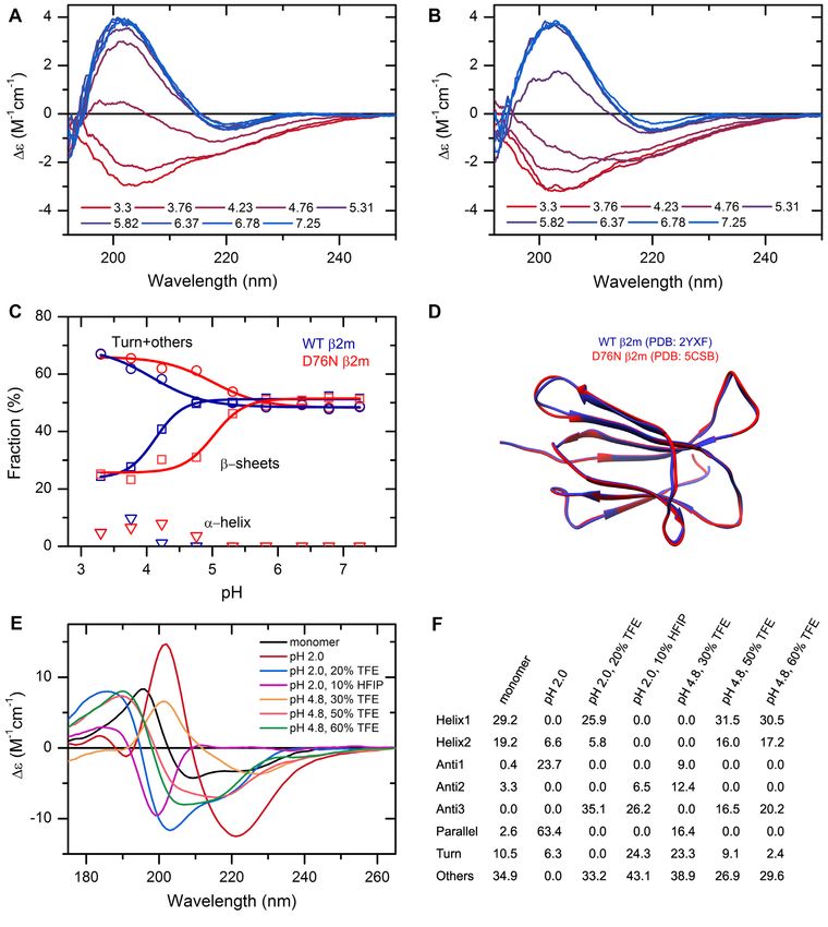
helping CD users. A schematic diagram shows the modules analysis.
and function of the web server in Figure 1. The Secondary structure from PDB files module calcu-
In Single spectrum analysis, a CD spectrum can be up- lates the eight BeStSel components and for comparison,
loaded and analyzed by the BeStSel method for secondary DSSP (7) and SELCON3 (14) composition for 3D struc-
structure content. Data can be copied to the text window tures. For structures deposited in the PDB the input is
in the form of two columns or can be uploaded from txt the four-letter PDB ID and the program will provide the
files. The program automatically recognizes the file headers CATH information as well, if it exists. Structural files in
and in case of data pitch different from 1 nm, sorts out and PDB format (max. 20 MB) can also be uploaded to the
uses integer nm data for analysis. Measurement files of var- server and the calculation will be carried out automatically.
ious instruments saved in text format are handled properly Both graphical and text outputs are available. This module
by the server. Input units can be ε (M–1 cm–1 ), [] (mean is especially useful for experimental verification of MD re-
residue ellipticity in deg cm2 dmol–1 ) or measured elliptic- sults or in silico models by CD spectroscopy, making the
ity in mdeg units. In the latter case, concentration, residue structural information comparable (see in Results and Dis-
number and pathlength data should be given and the server cussion).
will normalize the data to ε. After clicking on the submit The Extinction coefficient calculation module provides
button, a Data examination window appears to verify that the extinction coefficients of proteins and peptides at 214
the data was uploaded correctly. With one more click, the nm (15) and 205 nm (16), based on their primary sequence
secondary structure contents are calculated using the eight and number of disulfide bridges. The amino acid sequence
secondary structure components and presented in the form should be entered or copied to the text window. The extinc-
of a graphical output together with the spectral fitting with tion coefficients can be used for concentration determina-
RMSD and normalized RMSD (NRMSD) data. At first, tion directly on the CD sample.
fitting is carried out for the widest wavelength range. The The Disordered-ordered classification module analyses
user then can change the lower wavelength limit at 5 nm in- far-UV CD data to identify disordered structures. The clas-
crements and recalculate the secondary structures. Details sification is based on CD data at three wavelengths (197-
of the output image can be configured at the bottom of the 206-233 nm or 212-217-225 nm triplet). Data can be copied
page and the image can be redrawn. Alternatively, fitting into the text window. The first column contains the wave-Nucleic Acids Research, 2022, Vol. 50, Web Server issue W93
Downloaded from https://academic.oup.com/nar/article/50/W1/W90/6584425 by guest on 01 August 2022
Figure 1. Schematic representation of the BeStSel server. Block diagram shows the modules and function of the BeStSel package. Arrows indicate the input
and output data. From a single CD spectrum the secondary structure contents are estimated and then, based on these, the protein fold can be predicted.
A series of CD spectra as input can be evaluated at once to get the secondary structure contents. Users can provide arbitrary secondary structure contents
and carry out the fold prediction for that secondary structure composition. Users can also enter PDB IDs or upload structure files in PDB format as input
to find the corresponding secondary structure contents and fold classification. Based on the CD data, a binary disordered-ordered classification can be
carried out. To aid correct concentration determination, extinction coefficients at 205 and 214 nm can be calculated from the primary sequence of the
protein (15,16).
length values and the other columns contain the corre- tural information buried in the CD spectra. By develop-
sponding spectral data. Entire spectrum, series of spectra, ing the BeStSel method, we could solve a general problem
or CD data only at the necessary wavelengths, all will be of structure determination by CD spectroscopy: the diver-
accepted and handled properly. The output is a table con- sity of -structures. By distinguishing parallel and antipar-
taining the CD data at the wavelength triplet used for the allel -structures and three different twists of antiparallel
classification and the predicted results. -sheets and including two ␣-helix components, the over-
The Guide to CD and data analysis opens a separate win- all eight components of BeStSel provide a more accurate
dow with practical and important considerations for CD structural estimation for any secondary structure compo-
spectroscopy measurements. nent than any previous method (4). A great advantage of the
Cited by. . . opens a separate window and provides a method is that it can be used for a reliable structure estima-
database of scientific articles that used CD spectroscopy tion of -sheet-rich proteins, including membrane proteins,
with BeStSel analysis. Article identifiers and keywords are protein aggregates and amyloid fibrils. Moreover, BeStSel
provided, and the collection is searchable showing useful ex- provides extra structural information, which is sufficient
amples of using CD spectroscopy and BeStSel for the con- for protein fold prediction down to the topology/homology
venience of the users. levels of CATH fold classification. In the present upgrade,
BeStSel basis spectra were re-optimized by using the DSSP
3.0 algorithm to assign the major secondary structure com-
RESULTS AND DISCUSSION ponents in the reference database. In the earlier version of
Performance DSSP, residues in -helices might have been erroneously as-
signed to ␣-helix. Moreover, the BeStSel basis spectra were
Whereas the instrumentation of CD spectroscopy is well de- calculated for new wavelength ranges, now available at 5 nm
veloped, there is a great need to efficiently extract the struc- increments starting from 175 nm. Supplementary Table S1W94 Nucleic Acids Research, 2022, Vol. 50, Web Server issue
shows the performance compared to the previous version ence of gold nanoparticles than fibrils alone. They investi-
of BeStSel on the reference database. Overall, the accuracy gated the conformational changes of fibrils caused by gold
of the method is improved, e.g. in the 190–250 nm wave- nanoparticles. Brito et al. (20) showed that stability and gain
length range, the RMSD for ␣-helix estimation decreased of specific activity of bromelain protein complexes can be
from 0.052 to 0.042 (Supplementary Table S1). Performance improved by immobilizing bromelain on gold nanoparti-
was also tested on an independent set of -sheet rich or rare cles. By applying CD spectroscopy, the authors observed
protein structures and compared to other available methods structural changes in proteins upon binding to nanoparti-
for secondary structure estimation (Supplementary Table cles. Barbir et al. (21) studied the effects of silver nanopar-
S2). Calculated to a common basis of helix, antiparallel , ticles on the structure of plasma transport proteins by
parallel , overall -sheet and ‘turn + others’ structures, the CD.
RMSDs for secondary structure estimation were proved to There are several cases where BeStSel was applied in
be 0.034, 0.049, 0.037, 0.035 and 0.038 for BeStSel, while the SARS-CoV-2-related experiments. Mycroft-West et al. (22)
other methods provided RMSDs in the ranges 0.083–0.26, found that heparin alters the conformation of the SARS-
Downloaded from https://academic.oup.com/nar/article/50/W1/W90/6584425 by guest on 01 August 2022
0.12–0.214, 0.076–0.198, 0.068–0.23 and 0.074–0.232, re- CoV-2 spike protein and inhibits infection. Van Oosten et
spectively. None of the previous methods performed evenly al. (23) developed a virus-like particle (VLP)-based vaccine
for the different secondary structure components. for SARS-CoV-2 using the baculovirus––insect cell expres-
The Fold prediction module was upgraded from CATH sion system. They used secondary structure analysis to com-
4.2 (17) to using CATH 4.3 (5) data, resulting in a significant pare the recombinant and the wild-type spike protein. A
increase in the number of protein folds at all levels of classi- number of further studies involve the application of BeStSel
fication. Supplementary Table S3 shows the theoretical reli- for demonstrating that recombinant proteins have the cor-
ability of fold prediction on the domains of CATH 4.3 (5) as rect secondary structure (24–27). Another purpose BeStSel
secondary structure inputs in a 5-fold cross-validated man- is often used for is investigating the structure of antibodies
ner. (28–30).
A new function at the webserver is the Disordered-ordered Numerous works addressed the structural changes and
binary classification of proteins based on their CD spectra -sheet conversion or formation upon protein aggregation
(12). Such classifier, using experimental data has not been and amyloid formation. Kazman et al. (31) studied anti-
available yet and is highly needed by the community study- body light chain amyloid formation and explored the pro-
ing intrinsically disordered proteins (IDPs). It can be used cess of -sheet transition from antiparallel to parallel in
for simple and fast experimental verification for a variety oligomers. Kaur et al. (32) revealed that the CarD transcrip-
of bioinformatics tools identifying IDPs and can also facil- tion regulator from M. tuberculosis has a tendency to form
itate the growth of experimental data in IDP databases, such amyloid-like fibrils and undergoes reversible thermal fold-
as DisProt (18). The method uses the k nearest neighbors ing in solution. Do et al. (33) followed the aggregation of
mathematical model with cosine-distance function on CD the functional amyloid CRES (cystatin-related epididymal
data at three wavelengths (13). Using the 197-206-233 nm spermatogenic) and pointed out that its amyloid form is rich
wavelength triplet, the estimation error is 4.7, 1.7 and 3.9% in antiparallel -sheets instead of the more common paral-
on ordered, disordered proteins and in overall error, respec- lel -sheets. Amodeo et al. (34) discovered that the c subunit
tively, on the dataset of CD spectra with 190 nm wavelength of the ATP synthase is amyloidogenic and spontaneously
cut-off (12). For a cut-off at 200 nm, using the 212–217–225 folds into -sheets.
nm wavelength triplet, the error of classification is 3.3, 7.5 There are also studies about characterizing individual
and 4.6%, respectively. proteins of interest: Bowen et al. (35) microbially produced
The functionality of the BeStSel webserver is compared high-performance titin polymers and used CD to examine
to the other available online tools in Table 1. Besides its the secondary structure and fold of the purified polymers.
superior accuracy and the more detailed secondary struc- Balacescu et al. (36) investigated the structural behavior
ture information, the BeStSel webserver has an intelligent of apomyoglobin under different denaturing conditions. Ji
interface and provides useful functions for CD spectroscopy et al. (37) designed a self-assembling drug delivery system
users. and examined its target protein by CD spectroscopy.
Recently we predicted the structure of ␣-synuclein, an
IDP associated with Parkinson’s disease, by AlphaFold2
Applications
(38), which predicted 64% ␣-helix content. Experimen-
Application of CD spectroscopy in combination with BeSt- tal verification by CD spectroscopy and BeStSel analysis
Sel analysis covers all areas of protein science. BeStSel has showed no ␣-helix content under physiological conditions.
been used in over 1,000 scientific studies since its first publi- However, in the presence of 30% TFE, 47% ␣-helix was ob-
cation in 2015 (4). We made a searchable database of these served (12).
works at the webserver, providing valuable examples for
users. The broad applicability of BeStSel is represented by
Case studies
the vast variation of studies conducted using the algorithm.
Some notable examples are presented below. 2 -Microglobulin (2m) is the light chain of the major
An increasing number of users are applying the method histocompatibility complex I. Dissociating from the com-
to investigate the effects of nanoparticle–protein interac- plex, the protein circulates in monomeric form in the blood
tions on protein structure. Barbalinardo et al. (19) report and is associated with dialysis related amyloidosis in long-
that lysozyme amyloid fibrils are less cytotoxic in the pres- term haemodialysis patients. In 2012, a variant of the pro-Nucleic Acids Research, 2022, Vol. 50, Web Server issue W95
Table 1. Comparison of the functionality of the BeStSel webserver to other available online services for secondary structure estimation from the CD
spectra of proteins
BeStSel Dichroweb(46) CAPITO(47) K2D2(48) K2D3(49)
Access without registration • • • •
Text input • • • •
File input • • •
Input unit selection • • •
Auto normalization from mdeg • •
Change wavelength range without resubmission •
Best factor •
Download fitted spectrum • •a • •
Different reference sets •
Different algorithms •b
• •
Downloaded from https://academic.oup.com/nar/article/50/W1/W90/6584425 by guest on 01 August 2022
Multiple spectra analysis
Decompose parallel/antiparallel -sheet and antiparallel -sheet •
twist
Fold recognition •
Disordered-ordered classification • •c
PDB file analysis • •d
Extinction coefficient calculation •
Similarity analysis •
a Not for VARSLC; b For comparison of the performance of various algorithms to BeStSel, see Micsonai et al. (4) and Supplementary Table S1 and S2;
c Plot only; d Via 2Struct server (50).
tein carrying a D76N point mutation was discovered caus- analysis are now available at 5 nm increments. Fold pre-
ing a hereditary systemic amyloidosis with pathophysiol- diction was further enhanced by processing the CATH 4.3
ogy strikingly different from that of the wild-type protein data. The background databases are up-to-date, 184 307
(39,40). The first investigations found that the mutant ex- PDB structures are now recognized for secondary structure
hibits a high-resolution structure almost identical to that of analysis and fold classification. The updated single domain
the wild-type protein (Figure 2D) and its unique behavior dataset used as a basis for fold prediction contains 61932
can be discovered by using a rather complex methodology. single domains based on CATH 4.3 covering 43 architec-
Here, using CD spectroscopy and BeStSel analysis we show tures, 1467 topologies and 6540 homologies. A recent addi-
that the mutant protein exhibits increased sensitivity to a tion is the binary classification of ordered-disordered struc-
decrease in pH and below pH 6 it tends to unfold and loose tures based on the CD spectra, which helps the experimen-
its -structured native state, which might facilitate its amy- tal identification of intrinsically disordered proteins (IDPs)
loid aggregation. The wild-type protein is more resistant to and the verification of the results of bioinformatics tools.
low pH as shown in Figure 2A–C. One of the new features is that users can upload any
CD spectroscopy has a great advantage in characterizing 3D structures in PDB format and have the eight secondary
the conformation of proteins as a function of environmen- structure components of BeStSel calculated along with
tal conditions. High-resolution techniques cannot handle DSSP and SELCON components for comparison. This is
large number of samples, while in silico methods often can- crucial for the experimental verification of MD simulation
not address environmental parameters properly. We stud- results or in silico models, such as AlphaFold2 (42) struc-
ied the structure of human insulin at different pH values tures, making the structural comparison with the results of
in the presence of additives. As revealed by the CD spec- CD spectrum analysis possible. Another useful accessory is
tra, under native conditions insulin exhibits ␣-helical struc- the extinction coefficient calculator from the amino acid se-
ture. At low pH and in the presence of TFE or HFIP, insulin quence based on the works of Kuipers et al. (15) and An-
forms oligomers and amyloid fibrils with various different this et al. (16) for direct concentration determination of CD
secondary structure composition (Figure 2E, F and (41)). samples.
The detailed, downloadable tutorial has been updated
and further improved. Information and help is provided
NEW FEATURES throughout the use of the webserver.
After the first release in 2015 (4), the next larger update of
BeStSel was introduced in 2018 when fold prediction was
LIMITATIONS AND FURTHER DEVELOPMENTS
improved on the basis of the CATH 4.2 and the WKNN
search engine was built-in (10). The current version of the The eight secondary structure components of BeStSel do
webserver uses updated background databases. The DSSP not account for polyproline-II helix which is characteristic
algorithm was replaced by the 3.0 version, which solved of collagen-like structures, different type of turns that are
issues with assigning residues in -helices as ␣-helix. The often the main structural components of short peptides, 310 -
BeStSel basis spectra were re-optimized on the new assig- helices, which appear in higher amounts in some globular
nations resulting in improved accuracy (Supplementary Ta- proteins, and thus, analysis for such structures is not ade-
bles S1-S2). Moreover, wavelength ranges for CD spectrum quate. BeStSel does not treat aromatic contributions (otherW96 Nucleic Acids Research, 2022, Vol. 50, Web Server issue
Downloaded from https://academic.oup.com/nar/article/50/W1/W90/6584425 by guest on 01 August 2022
Figure 2. The effect of environmental conditions on the protein structure studied by CD spectroscopy and analyzed by the BeStSel web server. (A, B) CD
spectra of wild type (A) and D76N mutant 2m (B) were recorded at various pH values in 10 mM Na-citrate buffer at 37◦ C. (C) Secondary structure contents
provided by BeStSel were added up as ␣-helix, -sheet and turn + others. The mutant protein (red) is more sensitive to pH drop than the wild-type one (blue)
and starts to loose its -structure below pH 6.0. CD measurements were carried out on a benchtop Jasco J-1500 spectropolarimeter (1 mm pathlength, 50
nm/min scan rate, 2 sec response time, 1 nm bandwidth, accumulation: 6). (D) The two 2m variants exhibit very similar high-resolution native structure
making difficult to explain the difference in the pathology. (E) Insulin can exhibit various conformations depending on the solution conditions. Its native,
monomeric state is ␣-helical. Under nonnative conditions, such as low pH and the presence of alcohols, it forms -structured aggregates with different
-sheet compositions as shown in (F). At pH 2.0, it forms amyloid fibrils with characteristic parallel -sheets. These spectra were collected by SRCD at
DISCO beamline in SOLEIL Synchrotron, France.Nucleic Acids Research, 2022, Vol. 50, Web Server issue W97
algorithms neither do), which might affect the results in the DATA AVAILABILITY
case of high number of aromatic residues.
The datasets generated for this study are available on re-
For highly disordered proteins, some part of the disor-
quest to the corresponding author. The BeStSel web server
dered structure is counted as highly right-twisted antiparal-
is freely accessible at https://bestsel.elte.hu.
lel -sheet (Anti3) because of the spectral similarities (4,10).
When a protein or peptide is not expected to have globular
structure, and the secondary structure estimation provides SUPPLEMENTARY DATA
high Anti3 component with no or very low Anti2 content,
Anti3 might be considered as disordered and added to the Supplementary Data are available at NAR Online.
‘Others’ component.
BeStSel has an advantage over the previously available FUNDING
methods that it is capable of estimating the -sheet-rich
structure of protein aggregates and amyloid fibrils (4). How- National Research, Development and Innovation Fund
Downloaded from https://academic.oup.com/nar/article/50/W1/W90/6584425 by guest on 01 August 2022
ever, in case of such samples, spectral artifacts caused by dif- of Hungary [K120391, K138937, K125340, PD135510,
ferential light scattering, precipitation, or linear dichroism 2017-1.2.1-NKP-2017-00002]; International Collabora-
might affect or obstruct the accurate secondary structure tion [2019-2.1.11-TÉT-2019-00079, 2018-2.1.17-TÉT-
analysis. Therefore, it is essential to make sure that the sam- KR-2018-00008, 2019-2.1.6-NEMZ KI-2019-00012,
ple measured is a transparent, homogenous solution with- 2019-2.1.11-TÉT-2020-00101]; SOLEIL Synchrotron,
out large insoluble precipitates (4,43). An indication of light France [20181890, 20191810, 20200751]; Institute for
scattering might be when after a proper baseline subtrac- Protein Research, Osaka University; Japan Society for
tion there is a substantial remaining signal in the 250–260 the Promotion of Science, Core-to-Core Program A
nm wavelength region (be sure it is not nucleic acid contam- (Advanced Research Networks to Y.G.). Funding for
ination). Light scattering effects can be decreased by mak- open access charge: National Research, Development and
ing the size of the aggregates or amyloid fibrils smaller by Innovation Fund of Hungary [K120391, K138937, PD
applying a slight ultrasonication on the sample and plac- 135510].
ing the cuvette close to the detector. Precipitation makes the Conflict of interest statement. None declared.
sample inhomogeneous and results in absorption flattening
(distortion and shrinking of the CD signal) (4,43,44), which
makes the quantitative structure analysis impossible. Amy- REFERENCES
loid fibrils might become oriented in the cell causing linear 1. Wallace,B.A. (2000) Synchrotron radiation circular-dichroism
dichroism effects (45), which can be detected by rotating the spectroscopy as a tool for investigating protein structures. J.
Synchrotron Radiat., 7, 289–295.
cell in the instrument. 2. Greenfield,N.J. (2006) Using circular dichroism spectra to estimate
One of the main goals for the future is to significantly in- protein secondary structure. Nat. Protoc., 1, 2876–2890.
crease the number of reference proteins and further improve 3. Khrapunov,S. (2009) Circular dichroism spectroscopy has intrinsic
the accuracy on -structured proteins and IDPs. limitations for protein secondary structure analysis. Anal. Biochem.,
389, 174–176.
4. Micsonai,A., Wien,F., Kernya,L., Lee,Y.H., Goto,Y., Refregiers,M.
and Kardos,J. (2015) Accurate secondary structure prediction and
fold recognition for circular dichroism spectroscopy. Proc. Natl.
CONCLUSIONS Acad. Sci. U.S.A., 112, E3095–E3103.
The BeStSel web server provides the BeStSel method for 5. Sillitoe,I., Bordin,N., Dawson,N., Waman,V.P., Ashford,P.,
Scholes,H.M., Pang,C.S.M., Woodridge,L., Rauer,C., Sen,N. et al.
the community to analyze protein CD spectra for sec- (2021) CATH: increased structural coverage of functional space.
ondary structure composition and protein fold prediction. Nucleic Acids Res., 49, D266–D273.
The eight secondary structure components give detailed 6. Cooley,R.B., Arp,D.J. and Karplus,P.A. (2010) Evolutionary origin
structural information from the CD spectra including the - of a secondary structure: pi-helices as cryptic but widespread
structure composition (orientation and twist). The method insertional variations of alpha-helices that enhance protein
functionality. J. Mol. Biol., 404, 232–246.
has an accuracy superior to any previously available method 7. Kabsch,W. and Sander,C. (1983) Dictionary of protein secondary
on any type of secondary structures. It is especially usable structure: pattern recognition of hydrogen-bonded and geometrical
for -sheet-rich proteins, protein aggregates and membrane features. Biopolymers, 22, 2577–2637.
proteins. Single and multiple CD spectra can be analyzed 8. Orengo,C.A., Michie,A.D., Jones,S., Jones,D.T., Swindells,M.B. and
Thornton,J.M. (1997) CATH–a hierarchic classification of protein
and the protein fold can be predicted with a few clicks. Ad- domain structures. Structure, 5, 1093–1108.
justable wavelength ranges, scaling of spectra, links to corre- 9. Dudani,S.A. (1976) The distance-weighted k-nearest-neighbor rule.
sponding PDB structures make the site a swiss-knife for CD IEEE Trans. Syst. Man Cybern., SMC-6, 325–327.
users. A new module of the webserver helps to distinguish 10. Micsonai,A., Wien,F., Bulyaki,E., Kun,J., Moussong,E., Lee,Y.H.,
intrinsically disordered proteins by their CD spectrum. Sec- Goto,Y., Refregiers,M. and Kardos,J. (2018) BeStSel: a web server for
accurate protein secondary structure prediction and fold recognition
ondary structure calculation for uploaded PDB files will from the circular dichroism spectra. Nucleic Acids Res., 46,
help the experimental verification of protein MD and in sil- W315–W322.
ico modelling using CD spectroscopy. The server is capa- 11. Whitmore,L., Woollett,B., Miles,A.J., Klose,D.P., Janes,R.W. and
ble of high-throughput calculations and makes the method- Wallace,B.A. (2011) PCDDB: the protein circular dichroism data
bank, a repository for circular dichroism spectral and metadata.
ology of protein CD spectroscopy complete with accurate Nucleic Acids Res., 39, D480–D486.
analyses at any field of protein science, structural biochem- 12. Micsonai,A., Moussong,E., Murvai,N., Tantos,A., Toke,O.,
istry, biotechnology and pharmaceutical industry. Réfrégiers,M., Wien,F. and Kardos,J. (2022) Disordered-orderedW98 Nucleic Acids Research, 2022, Vol. 50, Web Server issue
protein binary classification by circular dichroism spectroscopy. displaying HIV-1 env V1V2 loop in a native-like trimeric
Front. Mol. Biosci., 863141. conformation as vaccine antigen. Nanomedicine, 16, 206–216.
13. Manning,C.D., Raghavan,P. and Schütze,H. (2008) Introduction to 31. Kazman,P., Absmeier,R.M., Engelhardt,H. and Buchner,J. (2021)
Information Retrieval. Cambridge, Cambridge University Press. Dissection of the amyloid formation pathway in AL amyloidosis.
14. Sreerama,N., Venyaminov,S.Y. and Woody,R.W. (1999) Estimation of Nat. Commun., 12, 6516.
the number of alpha-helical and beta-strand segments in proteins 32. Kaur,G., Kaundal,S., Kapoor,S., Grimes,J.M., Huiskonen,J.T. and
using circular dichroism spectroscopy. Protein Sci., 8, 370–380. Thakur,K.G. (2018) Mycobacterium tuberculosis CarD, an essential
15. Kuipers,B.J. and Gruppen,H. (2007) Prediction of molar extinction global transcriptional regulator forms amyloid-like fibrils. Sci. Rep.,
coefficients of proteins and peptides using UV absorption of the 8, 10124.
constituent amino acids at 214 nm to enable quantitative reverse 33. Do,H.Q., Hewetson,A., Myers,C., Khan,N.H., Hastert,M.C.,
phase high-performance liquid chromatography-mass spectrometry F,M.H., Latham,M.P., Wylie,B.J., Sutton,R.B. and Cornwall,G.A.
analysis. J. Agric. Food Chem., 55, 5445–5451. (2019) The functional mammalian CRES (Cystatin-Related
16. Anthis,N.J. and Clore,G.M. (2013) Sequence-specific determination epididymal spermatogenic) amyloid is antiparallel beta-Sheet rich
of protein and peptide concentrations by absorbance at 205 nm. and forms a metastable oligomer during assembly. Sci. Rep., 9, 9210.
Protein Sci., 22, 851–858. 34. Amodeo,G.F., Lee,B.Y., Krilyuk,N., Filice,C.T., Valyuk,D.,
17. Sillitoe,I., Lewis,T.E., Cuff,A., Das,S., Ashford,P., Dawson,N.L., Otzen,D.E., Noskov,S., Leonenko,Z. and Pavlov,E.V. (2021) C
Downloaded from https://academic.oup.com/nar/article/50/W1/W90/6584425 by guest on 01 August 2022
Furnham,N., Laskowski,R.A., Lee,D., Lees,J.G. et al. (2015) CATH: subunit of the ATP synthase is an amyloidogenic calcium dependent
comprehensive structural and functional annotations for genome channel-forming peptide with possible implications in mitochondrial
sequences. Nucleic Acids Res., 43, D376–D381. permeability transition. Sci. Rep., 11, 8744.
18. Quaglia,F., Meszaros,B., Salladini,E., Hatos,A., Pancsa,R., 35. Bowen,C.H., Sargent,C.J., Wang,A., Zhu,Y., Chang,X., Li,J., Mu,X.,
Chemes,L.B., Pajkos,M., Lazar,T., Pena-Diaz,S., Santos,J. et al. Galazka,J.M., Jun,Y.S., Keten,S. et al. (2021) Microbial production
(2021) DisProt in 2022: improved quality and accessibility of protein of megadalton titin yields fibers with advantageous mechanical
intrinsic disorder annotation. Nucleic Acids Res., 50, D480–D487. properties. Nat. Commun., 12, 5182.
19. Barbalinardo,M., Antosova,A., Gambucci,M., Bednarikova,Z., 36. Balacescu,L., Schrader,T.E., Radulescu,A., Zolnierczuk,P.,
Albonetti,C., Valle,F., Sassi,P., Latterini,L., Gazova,Z. and Holderer,O., Pasini,S., Fitter,J. and Stadler,A.M. (2020) Transition
Bystrenova,E. (2020) Effect of metallic nanoparticles on amyloid between protein-like and polymer-like dynamic behavior: internal
fibrils and their influence to neural cell toxicity. Nano Res., 13, friction in unfolded apomyoglobin depends on denaturing conditions.
1081–1089. Sci. Rep., 10, 1570.
20. Brito,A.M.M., Oliveira,V., Icimoto,M.Y. and Nantes-Cardoso,I.L. 37. Ji,T., Li,Y., Deng,X., Rwei,A.Y., Offen,A., Hall,S., Zhang,W.,
(2021) Collagenase activity of bromelain immobilized at gold Zhao,C., Mehta,M. and Kohane,D.S. (2021) Delivery of local
nanoparticle interfaces for therapeutic applications. Pharmaceutics, anaesthetics by a self-assembled supramolecular system mimicking
11, 810. their interactions with a sodium channel. Nat. Biomed. Eng., 5,
21. Barbir,R., Capjak,I., Crnkovic,T., Debeljak,Z., Domazet Jurasin,D., 1099–1109.
Curlin,M., Sinko,G., Weitner,T. and Vinkovic Vrcek,I. (2021) 38. Jumper,J., Evans,R., Pritzel,A., Green,T., Figurnov,M.,
Interaction of silver nanoparticles with plasma transport proteins: a Ronneberger,O., Tunyasuvunakool,K., Bates,R., Zidek,A.,
systematic study on impacts of particle size, shape and surface Potapenko,A. et al. (2021) Highly accurate protein structure
functionalization. Chem. Biol. Interact., 335, 109364. prediction with alphafold. Nature, 596, 583–589.
22. Mycroft-West,C.J., Su,D., Pagani,I., Rudd,T.R., Elli,S., Gandhi,N.S., 39. Bulyaki,E., Kun,J., Molnar,T., Papp,A., Micsonai,A., Vadaszi,H.,
Guimond,S.E., Miller,G.J., Meneghetti,M.C.Z., Nader,H.B. et al. Marialigeti,B., Kovacs,A.I., Gellen,G., Yamaguchi,K. et al. (2021)
(2020) Heparin inhibits cellular invasion by SARS-CoV-2: structural Pathogenic D76N variant of beta2-Microglobulin: synergy of diverse
dependence of the interaction of the spike S1 receptor-binding effects in both the native and amyloid states. Biology (Basel), 10,
domain with heparin. Thromb. Haemost., 120, 1700–1715. 1197.
23. van Oosten,L., Altenburg,J.J., Fougeroux,C., Geertsema,C., van den 40. Valleix,S., Gillmore,J.D., Bridoux,F., Mangione,P.P., Dogan,A.,
End,F., Evers,W.A.C., Westphal,A.H., Lindhoud,S., van den Berg,W., Nedelec,B., Boimard,M., Touchard,G., Goujon,J.M., Lacombe,C.
Swarts,D.C. et al. (2021) Two-Component nanoparticle vaccine et al. (2012) Hereditary systemic amyloidosis due to asp76asn variant
displaying glycosylated spike S1 domain induces neutralizing beta2-microglobulin. N. Engl. J. Med., 366, 2276–2283.
antibody response against SARS-CoV-2 variants. mBio, 12, e0181321. 41. Muta,H., Lee,Y.H., Kardos,J., Lin,Y., Yagi,H. and Goto,Y. (2014)
24. Kibria,M.G., Fukutani,A., Akazawa-Ogawa,Y., Hagihara,Y. and Supersaturation-limited amyloid fibrillation of insulin revealed by
Kuroda,Y. (2021) Anti-EGFR VHH antibody under thermal stress is ultrasonication. J. Biol. Chem., 289, 18228–18238.
better solubilized with a lysine than with an arginine SEP tag. 42. Tunyasuvunakool,K., Adler,J., Wu,Z., Green,T., Zielinski,M.,
Biomolecules, 11, 810. Zidek,A., Bridgland,A., Cowie,A., Meyer,C., Laydon,A. et al. (2021)
25. Brindha,S., Kibria,M.G., Saotome,T., Unzai,S. and Kuroda,Y. (2021) Highly accurate protein structure prediction for the human proteome.
EGFR extracellular domain III expressed in escherichia coli with Nature, 596, 590–596.
SEP tag shows improved biophysical and functional properties and 43. Micsonai,A., Bulyaki,E. and Kardos,J. (2021) BeStSel: from
generate anti-sera inhibiting cancer cell growth. Biochem. Biophys. secondary structure analysis to protein fold prediction by circular
Res. Commun., 555, 121–127. dichroism spectroscopy. Methods Mol. Biol., 2199, 175–189.
26. Bortnov,V., Tonelli,M., Lee,W., Lin,Z., Annis,D.S., Demerdash,O.N., 44. Wallace,B.A. and Teeters,C.L. (1987) Differential absorption
Bateman,A., Mitchell,J.C., Ge,Y., Markley,J.L. et al. (2019) Solution flattening optical effects are significant in the circular dichroism
structure of human myeloid-derived growth factor suggests a spectra of large membrane fragments. Biochemistry, 26, 65–70.
conserved function in the endoplasmic reticulum. Nat. Commun., 10, 45. Wallace,B.A. (2009) Protein characterisation by synchrotron
5612. radiation circular dichroism spectroscopy. Q. Rev. Biophys., 42,
27. Anathy,V., Lahue,K.G., Chapman,D.G., Chia,S.B., Casey,D.T., 317–370.
Aboushousha,R., van der Velden,J.L.J., Elko,E., Hoffman,S.M., 46. Lobley,A., Whitmore,L. and Wallace,B.A. (2002) DICHROWEB: an
McMillan,D.H. et al. (2018) Reducing protein oxidation reverses lung interactive website for the analysis of protein secondary structure
fibrosis. Nat. Med., 24, 1128–1135. from circular dichroism spectra. Bioinformatics, 18, 211–212.
28. Eliseev,I.E., Ukrainskaya,V.M., Yudenko,A.N., Mikushina,A.D., 47. Wiedemann,C., Bellstedt,P. and Gorlach,M. (2013) CAPITO–a web
Shmakov,S.V., Afremova,A.I., Ekimova,V.M., Vronskaia,A.A., server-based analysis and plotting tool for circular dichroism data.
Knyazev,N.A. and Shamova,O.V. (2021) Targeting erbb3 receptor in Bioinformatics, 29, 1750–1757.
cancer with inhibitory antibodies from llama. Biomedicines, 9, 1106. 48. Perez-Iratxeta,C. and Andrade-Navarro,M.A. (2008) K2D2:
29. Dash,R. and Rathore,A.S. (2021) Freeze thaw and lyophilization estimation of protein secondary structure from circular dichroism
induced alteration in mAb therapeutics: trastuzumab as a case study. spectra. BMC Struct. Biol., 8, 25.
J. Pharm. Biomed. Anal., 201, 114122. 49. Louis-Jeune,C., Andrade-Navarro,M.A. and Perez-Iratxeta,C. (2012)
30. Karch,C.P., Bai,H., Torres,O.B., Tucker,C.A., Michael,N.L., Prediction of protein secondary structure from circular dichroism
Matyas,G.R., Rolland,M., Burkhard,P. and Beck,Z. (2019) Design using theoretically derived spectra. Proteins, 80, 374–381.
and characterization of a self-assembling protein nanoparticle 50. Klose,D.P., Wallace,B.A. and Janes,R.W. (2010) 2Struc: the
secondary structure server. Bioinformatics, 26, 2624–2625.You can also read