XCELLigence RTCA DP Instrument Flexible Real-Time Cell Monitoring - For life science research only. Not for use in diagnostic procedures.
←
→
Page content transcription
If your browser does not render page correctly, please read the page content below
xCELLigence RTCA DP Instrument
           Flexible Real-Time Cell Monitoring

For life science research only.
Not for use in diagnostic procedures.
The xCELLigence RTCA DP Instrument
Flexible Real-Time Cell Monitoring
The RTCA DP Instrument expands the throughput and application options of the xCELLigence
Real-Time Cell Analyzer (RTCA) portfolio. Featuring a dual-plate (DP) format, the instrument
measures impedance-based signals in both cellular and cell invasion/migration (CIM)
assays – without the use of exogenous labels. With outstanding application flexibility, the RTCA
DP Instrument supports multiple users performing short-term and long-term experiments.
The xCELLigence System continuously and non-invasively
Explore the wide range of applications detects cell responses throughout an experiment, without the use
of exogenous labels that can disrupt the natural cell environment.
Cell invasion and migration assays
Obtain complete, continuous data profiles from cell
Compound- and cell-mediated cytotoxicity responses generated during in vitro experiments (Figure 1).
Cell adhesion and cell spreading T ake advantage of real-time data to identify optimal time
Cell proliferation and cell differentiation points for downstream assays.
Receptor-mediated signaling Combine real-time monitoring of cellular responses with
complementary functional endpoint assays, and maximize
Virus-mediated cytopathogenicity data quality before, during, and after your experiment.
Continuous quality control of cells
Cytotoxicity Analysis in E-Plates
RTCA Control Unit RTCA DP Analyzer
Compact. Convenient. Versatile.
The RTCA DP Instrument consists of two components: the RTCA
Control Unit and the RTCA DP Analyzer with three integrated
stations for measuring cell responses in parallel or independently.
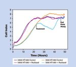
Figure 1: Reveal cytotoxic effects through continuous
Choose from three types of impedance-based 16-well plates:
monitoring. HT1080 cells were seeded in an E-Plate at two — E-Plate 16 and E-Plate VIEW 16 for cellular assays
different densities (5,000 and 10,000 cells) and treated 24 hours — CIM-PLATE 16 for cell invasion/migration assays
later with 12.5 nM Paclitaxel, or DMSO as a control. As shown
Use all three different plate types in any combination.
by the Cell Index profile, which reflects cell adherence, the
antimitotic effect of Paclitaxel was observed in HT1080 cells that Easily achieve optimal cell culture conditions by placing the
were proliferating, whereas confluent cells showed no response. RTCA DP Analyzer and plates into standard CO2 incubators.
2E-Plates for the RTCA DP Instrument
More Flexibility. More Data. More Insight.
Obtain detailed information about your cells with the versatile RTCA DP Instrument, which supports
up to three plates of any type – E-Plate 16, E-Plate VIEW 16, or CIM-Plate 16 – in any combination.
For example, cell invasion/migration assays and cytotoxicity assays or short- and long-term assays
may be run simultaneously.
E-Plate 16 and E-Plate VIEW 16:
Cellular Assays in a 16-Well Format
Quantitatively monitor changes in cell number, cell adhesion,
cell viability, and cell morphology.
E-Plate 16
Easily add compounds during an experiment.
Assess short- and long-term cellular effects.
1.9 Control Antimycin A
With the E-Plate VIEW 16, observe measured changes using 5 µM administration
100 µM
Normalized Cell Index
microscopes. 300 µM
E-Plate 16 E-Plate VIEW 16
0.9
- 0.1
9 18 27 36 45 54
Time (Hours)
4h 22 h
Control
300 µM
Antimycin A
500 µm
Figure 2: Easily visualize cells while measuring cell response with
xCELLigence System E-Plate VIEW technology. A modified version
of the standard E-Plate 16, the E-Plate VIEW 16 enables image acqui-
sition using microscopes or automated cell-imaging systems. For the Figure 3: Continuously monitor cells and determine optimal time
modification, four rows of microelectrode sensors were removed in each points for assessing cytotoxicity. Cell proliferation and cell death
well to create a window for visualizing cells. Approximately 70% of each were continuously monitored using the xCELLigence RTCA DP Instrument.
well bottom is covered by the microelectrodes, providing cell impedance The optimal time points for visual inspection of HeLa cells were
measurements nearly identical to those obtained with the standard determined and images taken 4 and 22 hours after compound treatment
E-Plate 16. Both plate types can be used in parallel. using a Z16 Apo Microscope with light base (Leica Microsystems).
3CIM-Plate 16:
Quantitative Cell Invasion/Migration Analysis
Monitor cell invasion and migration continuously in real time
over the entire time course of an experiment.
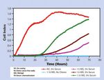
Eliminate time-consuming manual detection (Figure 4).
CIM-Plate 16
Perform CIM analysis in a convenient one-well system (Figure 5).
Invasion/Migration Analysis in CIM-Plates
Lid
Cells
Upper Chamber
Gel Layer
(user provided)
Microporous Membrane
Microelectrodes
Figure 4: Quantitatively measure the rate and onset of invasion
Adherent Cell
while concurrently assessing migration. HT1080 cells (2 x 10 4 ) were Lower Chamber
seeded in the upper chamber of CIM-Plate wells coated with varying dilutions Chemoattractant
of Matrigel, or in wells with no coating. Serum was added to the lower
chamber of selected wells as a chemoattractant. Invasion was observed and
migration monitored continuously over a 70-hour period. All serum-starved Figure 5: Analyze invasion/migration in real time with the
samples resulted in base-line Cell Index levels, indicating the absence of CIM-Plate 16. The plate features two separable sections for ease
invasion/migration, while those wells with chemoattractant induced migration. of experimental setup. Cells seeded in the upper chamber move
through the microporous membrane into the lower chamber that
contains a chemoattractant. Cells adhering to the microelectrode
sensors lead to an increase in impedance, which is measured in
real time by the RTCA DP Instrument.
4E-Plate Insert 16:
Co-Culture in Real-Time
Continuously monitor indirect cell-cell interactions.
Assess short- and long-term cell response without
labor-intensive labeling and microscopy.
Co-culture different cell types under physiological conditions
for a broad range of applications, including:
Cancer Research: Assess paracrine stimulation of
cancer cell proliferation by fibroblasts.
Immunology: Investigate immune cell interactions.
Figure 6. Real-time monitoring of co-culture-induced proliferation
Stem Cell Research: Monitor proliferation and stimulation and its inhibition using the E-Plate Insert. Intercellular
differentiation in the presence of stimulation cells. interactions play an important role in normal cell development and
tumorigenesis. Results show that the proliferation of hormone-responsive
Toxicology: Determine cytotoxicity of agents and tumor cells is likely mediated by hormones and growth factors exchanged
assess effects of cytokine release. between the two cell populations separated by the E-Plate Insert.
Elevated T47D cell proliferation on the E-Plate (green trace ) was
induced by hormone secretion of H295R cells in the insert, and inhibited
by the hormone synthesis inhibitor Prochloraz (blue trace ). Incubation
of T47D cells with only the E-Plate Insert did not affect proliferation (red
trace ).
Lid
E-Plate Insert
E-Plate 16
5Selected Publications for the RTCA DP Instrument
1. Cell Invasion and Migration
MicroRNA-200c Represses Migration and Invasion of Breast Cancer Cells by Targeting Actin-Regulatory
Proteins FHOD1 and PPM1Ferences.
Jurmeister S, Baumann M, Balwierz A, Keklikoglou I, Ward A, Uhlmann S, Zhang JD, Wiemann S, Sahin O.
Mol Cell Biol. 2012; 32(3):633–651.
c-Myb regulates matrix metalloproteinases 1/9, and cathepsin D: implications for matrix-dependent bre-
ast cancer cell invasion and metastasis.
Knopfová L, Beneš P, Pekarčíková L, Hermanová M, Masařík M, Pernicová Z, Souček K, Smarda J.
Mol Cancer. 2012; 11:15.
Comparative Analysis of Dynamic Cell Viability, Migration and Invasion Assessments by Novel Real-
Time Technology and Classic Endpoint Assays.
Limame R, Wouters A, Pauwels B, Fransen E, Peeters M, Lardon F, De Wever O, Pauwels P.
PLoS One. 2012; 7(10): e46536.
2. Compound-mediated Cytotoxicity/Apoptosis
Screening and identification of small molecule compounds perturbing mitosis using time-dependent
cellular response profiles.
Ke N, Xi B, Ye P, Xu W, Zheng M, Mao L, Wu MJ, Zhu J, Wu J, Zhang W, Zhang J, Irelan J, Wang X, Xu
X, Abassi YA.
Anal Chem. 2010; 82(15):6495-503.
Kinetic cell-based morphological screening: prediction of mechanism of compound action and off-target
effects.
Abassi YA, Xi B, Zhang W, Ye P, Kirstein SL, Gaylord MR, Feinstein SC, Wang X, Xu X.
Chem Biol. 2009; 16(7):712-23.
3. Cell-mediated Cytotoxicity
Real-time profiling of NK cell killing of human astrocytes using xCELLigence technology.
Moodley K, Angel CE, Glass M, Graham ES.
J Neurosci Methods. 2011; 200(2): 173-180.
Unique functional status of natural killer cells in metastatic stage IV melanoma patients and its modula-
tion by chemotherapy.
Fregni G, Perier A, Pittari G, Jacobelli S, Sastre X, Gervois N, Allard M, Bercovici N, Avril MF, Caignard A.
Clin Cancer Res. 2011; 17(9): 2628–37.
4. Cell Adhesion and Cell Spreading
A role for adhesion and degranulation-promoting adapter protein in collagen-induced platelet activati-
on mediated via integrin a2b1.
Jarvis GE, Bihan D, Hamaia S, Pugh N, Ghevaert CJ, Pearce AC, Hughes CE, Watson SP, Ware J, Rudd
CE, Farndale RW.
Journal of Thromb Haemost. 2012; 10(2): 268–277.
Dynamic monitoring of cell adhesion and spreading on microelectronic sensor arrays.
Atienza JM, Zhu J, Wang X, Xu X, Abassi Y.
J Biomol Screen. 2005; 10(8): 795-805.
6Selected Publications continued
5. Receptor-mediated Signaling
Impedance responses reveal b2-adrenergic receptor signaling pluridimensionality and allow classification of
ligands with distinct signaling profiles.
Stallaert W, Dorn JF, van der Westhuizen E, Audet M, Bouvier M.
PLoS One. 2012; 7(1): e29420.
Label-free impedance responses of endogenous and synthetic chemokine receptor CXCR3 agonists correlate
with Gi-protein pathway activation.
Watts AO, Scholten DJ, Heitman LH, Vischer HF, Leurs R.
Biochem Biophys Res Commun. 2012; 419(2):412-8.
Impedance measurement: A new method to detect ligand-biased receptor signaling.
Kammermann M, Denelavas A, Imbach A, Grether U, Dehmlow H, Apfel CM, Hertel C.
Biochem Biophys Res Commun. 2011; 412(3): 419-424.
6. Virus-mediated Cytopathogenicity
Novel, real-time cell analysis for measuring viral cytopathogenesis and the efficacy of neutralizing antibodies to
the 2009 influenza A (H1N1) virus.
Tian D, Zhang W, He J, Liu Y, Song Z, Zhou Z, Zheng M, Hu Y.
PloS One. 2012; 7(2):e31965.
Real-time monitoring of flavivirus induced cytopathogenesis using cell electric impedance technology.
Fang Y, Ye P, Wang X, Xu X, Reisen W.
J Virol Methods. 2011; 173(2):251–8.
7. Quality of Control of Cells
Rapid and quantitative assessment of cell quality, identity, and functionality for cell-based assays using real-time
cellular analysis.
Irelan JT, Wu MJ, Morgan J, Ke N, Xi B, Wang X, Xu X, Abassi YA.
J Biomol Screen. 2011; 16(3):313-22.
Live cell quality control and utility of real-time cell electronic sensing for assay development.
Kirstein SL, Atienza JM, Xi B, Zhu J, Yu N, Wang X, Xu X, Abassi YA.
Assay Drug Dev Technol. 2006; 4(5):545-53.
8. Endothelial Barrier Function
An inverted blood-brain barrier model that permits interactions between glia and inflammatory stimuli.
Sansing HA, Renner NA, MacLean AG.
J Neurosci Methods. 2012; 207(1):91–6.
A dynamic real-time method for monitoring epithelial barrier function in vitro.
Sun M, Fu H, Cheng H, Cao Q, Zhao Y, Mou X, Zhang X, Liu X, Ke Y.
Anal Biochem. 2012; 425(2):96–103.
9. Cell-Cell Interactions: Co-Culture
Dynamic assessment of cell viability, proliferation and migration using real time cell analyzer system (RTCA).
Roshan Moniri M, Young A, Reinheimer K, Rayat J, Dai LJ, Warnock GL.
Cytotechnology. 2014 Jan 19. [Epub ahead of print]
Using real-time impedance based assays to monitor the effects of fibroblast derived media on the adhesion,
proliferation, migration and invasion of colon cancer cells.
Dowling CM, Herranz Ors C, Kiely PA.
Biosci Rep. 2014 Jun 17. [Epub ahead of print]
7Ordering Information for xCELLigence RTCA DP System
Product Cat. No. Pack Size
xCELLigence RTCA DP Instrument 00380601050 1 Bundled Package
RTCA DP Analyzer 05469759001 1 Instrument
RTCA Control Unit 05454417001 1 Notebook PC
E-Plate 16 05469830001 6 Plates
05469813001 6 x 6 Plates
E-Plate VIEW 16 06324738001 6 Plates
06324746001 6 x 6 Plates
E-Plate VIEW 16 PET (Polyethylene Terephthalate) 00300600890 6 Plates
00300600880 6 x 6 Plates
E-Plate Insert 16 06465382001 1 x 6 Devices (6 16-Well Inserts)
CIM-Plate 16 05665817001 6 Plates
05665825001 6 x 6 Plates
CIM-Plate 16, Assembly Tool 05665841001 1 Assembly Tool
Learn more about the enabling technology of the xCELLigence System and its broad range of
applications at www.aceabio.com
Published by
ACEA Biosciences, Inc.
For life science research only. 6779 Mesa Ridge Road Ste. 100
Not for use in diagnostic procedures. San Diego, CA 92121
U.S.A.
XCELLIGENCE, E-PLATE, CIM-PLATE, and ACEA BIOSCIENCES are registered trademarks of ACEA Biosciences, Inc. in the US
and other countries. www.aceabio.com
All other product names and trademarks are the property of their respective owners.
© 2014 ACEA Biosciences, Inc.
All rights reserved.You can also read


