What a stranded whale with scoliosis can teach us about human idiopathic scoliosis
←
→
Page content transcription
If your browser does not render page correctly, please read the page content below
www.nature.com/scientificreports
OPEN What a stranded whale
with scoliosis can teach us
about human idiopathic scoliosis
Steven de Reuver1, Lonneke L. IJsseldijk2*, Jelle F. Homans1, Dorien S. Willems3,
Stefanie Veraa3, Marijn van Stralen4, Marja J. L. Kik2, Moyo C. Kruyt1, Andrea Gröne2 &
René M. Castelein1
Scoliosis is a deformation of the spine that may have several known causes, but humans are the only
mammal known to develop scoliosis without any obvious underlying cause. This is called ‘idiopathic’
scoliosis and is the most common type. Recent observations showed that human scoliosis, regardless
of its cause, has a relatively uniform three-dimensional anatomy. We hypothesize that scoliosis is
a universal compensatory mechanism of the spine, independent of cause and/or species. We had
the opportunity to study the rare occurrence of scoliosis in a whale (Balaenoptera acutorostrata)
that stranded in July 2019 in the Netherlands. A multidisciplinary team of biologists, pathologists,
veterinarians, taxidermists, radiologists and orthopaedic surgeons conducted necropsy and
imaging analysis. Blunt traumatic injury to two vertebrae caused an acute lateral deviation of the
spine, which had initiated the development of compensatory curves in regions of the spine without
anatomical abnormalities. Three-dimensional analysis of these compensatory curves showed strong
resemblance with different types of human scoliosis, amongst which idiopathic. This suggests that any
decompensation of spinal equilibrium can lead to a rather uniform response. The unique biomechanics
of the upright human spine, with significantly decreased rotational stability, may explain why only in
humans this mechanism can be induced relatively easily, without an obvious cause, and is therefore
still called ‘idiopathic’.
Scoliosis is a three-dimensional (3D) deformity of the spine and trunk, in which rotation of the vertebral column
in the horizontal plane together with extension in the sagittal plane plays a consistent role, that may be caused by
traumatic injury, syndromic conditions, congenital malformations or neuromuscular disease1. In mammals, the
development of scoliosis without an obvious underlying cause is exclusively observed in humans, this is called
‘idiopathic’ scoliosis and is the most frequently observed type1–3. The condition occurs with a prevalence of
1–4% in otherwise healthy individuals, most commonly adolescent f emales1. Treatment is currently focused on
limiting progression of the spinal curve until skeletal maturity, which can necessitate bracing therapy or spinal
fusion surgery1. Many theories have been brought forward in search of the aetiology of idiopathic s coliosis1,4–12.
Upright spinal biomechanics, that implies a reduction of stability in the horizontal plane, was shown to play an
important role13–19. And while the shape of the scoliotic spine has been described for over a century10,20, 21, recent
observations have shown that the 3D anatomy is very uniform across human scoliosis with different aetiology,
including vertebral rotation into the curve convexity and anterior lengthening of the intervertebral d iscs22–26.
We hypothesize that scoliosis is a universal compensatory mechanism of the spine, that consists of vertebral
rotation into the convexity of the curve, accompanied by anterior lengthening of the intervertebral discs, that
can be caused by different primary challenges to spinal equilibrium. One of these challenges, and a possible
explanation of idiopathic scoliosis in humans is the unique upright posture with the centre of weight balanced
straight above the pelvis, resulting in a unique biomechanical loading of the t runk13–17,27. This specific sagittal
plane configuration of the human spine was shown to lead to decreased rotational stability, making it more prone
than other spines in nature to decompensate into s coliosis18,19,28,29. Scoliosis is found rarely in other vertebrates
than humans and is usually caused by anatomic abnormalities30,31.
1
Department of Orthopaedic Surgery, University Medical Center Utrecht, Utrecht, The Netherlands. 2Division of
Pathology, Department of Biomolecular Health Sciences, Faculty of Veterinary Medicine, Utrecht University,
Utrecht, The Netherlands. 3Division of Diagnostic Imaging, Department of Clinical Sciences, Faculty of Veterinary
Medicine, Utrecht University, Utrecht, The Netherlands. 4Imaging Division, University Medical Center Utrecht,
Utrecht, The Netherlands. *email: l.l.ijsseldijk@uu.nl
Scientific Reports | (2021) 11:7218 | https://doi.org/10.1038/s41598-021-86709-x 1
Vol.:(0123456789)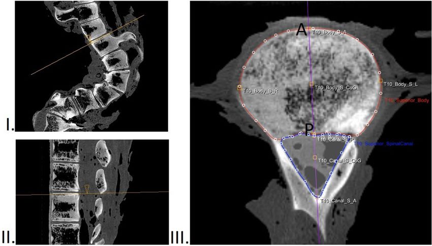
www.nature.com/scientificreports/
Figure 1. Photograph of the common minke whale (Balaenoptera acutorostrata) that washed ashore on the 8th
of July 2019 at Texel, the Netherlands. Photograph by Pierre Bonnet (Ecomare, Texel).
Recently, we had the opportunity to study the compensatory curves in an anatomically normal area of the
spine of a whale with a post-traumatic scoliosis. Whales are sea mammals that are not known to develop scoliosis
spontaneously: several reports on cetaceans with scoliosis exist, however all cases have a clear cause which is
mostly of traumatic origin, e.g. following ship c ollision32–34. In the current study we examined a young common
minke whale (Balaenoptera acutorostrata), which was found stranded in July 2019 in the Netherlands with an
obvious spine trauma and subsequent severe local post-traumatic scoliosis (Fig. 1). This post-traumatic scoliosis
initiated compensatory 3D curves in the area of the spine that was not affected by the trauma, apparently in
an attempt of the animal to re-align its trunk. We were interested in these compensatory curves, as they could
provide insights into the more general, intrinsic mechanisms that govern alignment of the mammalian spine.
A multidisciplinary team of biologists, pathologists, veterinarians, taxidermists, radiologists and orthopaedic
surgeons studied the whale and conducted a necropsy and 3D imaging analysis of the spine and compared the
findings to non-scoliotic whales. The aim of the study was to assess whether scoliosis is a universal compensatory
mechanism that occurs independent of cause and/or species. The hypothesis tested in the current study was that
the injured whale would re-align its trunk by creating compensatory curves in the essentially normal spine and
that these curves show a similar 3D configuration as is observed in human scoliosis.
Results
Post mortem findings. The common minke whale was a 403 cm long, 530 kg female juvenile, with an
estimated age between 0.5 and 4 years35. Besides the clear lateral post-traumatic curvature of the spine, other
important findings of external examination were multifocal areas of deep haemorrhage and oedema that were
present in the subcutis and longissimus dorsi muscle, as well as the presence of blood tinge liquid in the spinal
canal and congestion and haemorrhage of the brain. The animal had a poor nutritional condition (blubber layers
of 20–25 mm) despite recent feeding36. Histology of the fractured vertebrae demonstrated fibrin deposits, some
eosinophilic granulocytes, and necrosis, indicative of chronic changes that were still ongoing. Therefore, the
most likely cause of death was considered to be acute recent blunt trauma.
Furthermore, there was clear evidence that earlier trauma had resulted in the fractures and other deformations
of the lumbar vertebrae, which had led to a localized, post-traumatic deformity of the spine. Visual inspection
showed that the deformity was mostly in the coronal plane with no significant lordosis or kyphosis at that region.
This was further investigated after removing all of the soft tissues of the entire vertebral column. Visual inspec-
tion showed an epiphysiolysis at the left-side of the lower endplate of vertebra L3, a burst upper endplate at the
right-side of vertebra L4 and fractured/missing spinous processes at multiple levels (Fig. 2).
Furthermore, spinal curvatures were observed in the adjacent, anatomically normal parts of the spine, outside
the traumatically affected area (Fig. 3). Therefore, the thoraco-lumbar area was the suspected site of an acute
(dorso-)lateral blunt traumatic injury, which subsequently initiated a double compensatory curve cranially and
a single compensatory curve caudally in areas of the spine unaffected by the trauma (Fig. 4). We analysed these
compensatory curves in 3D and compared the morphology with the non-scoliotic spine of 10 control whales.
The levels T11/12 (severe wedging) and L3/L4 (traumatic injury) were excluded before CT-scan analysis of the
compensatory curvatures.
CT‑measurements. The 3D analysis of the compensatory curves showed a rotation of the vertebral bodies
in the transverse plane into the convexity of the curve (Fig. 5). The mean anterior–posterior length discrepancy
(AP%) of the total compensatory curvature was + 9.4% in the whale. This means that the anterior (ventral) length
of the compensatory scoliotic curvature was 9.4% greater than the posterior (dorsal) length, indicating a regional
lordosis. This is significantly different from the kyphosis in the same part of the spine in the non-scoliotic control
group, with a total AP% of − 2.1 ± 0.4%, meaning that the anterior length of the spine was 2.1% shorter than
the posterior length (p < 0.001). On the contrary, the bony morphology of the vertebral bodies was similar to
the controls; the vertebral body AP% of the whale was − 2.5%, which was comparable to the kyphotic shape of
the vertebral bodies in controls with − 1.8 ± 0.8% (p = 0.429). Almost all anterior lengthening took place in the
intervertebral discs, as the disc AP% in the compensatory curvature of the whale was + 99.5%, which meant a lor-
Scientific Reports | (2021) 11:7218 | https://doi.org/10.1038/s41598-021-86709-x 2
Vol:.(1234567890)
www.nature.com/scientificreports/
Figure 2. Photographs of the post-traumatic primary curve after removing the soft tissues. The dorsal overview
on the right-hand side shows the post-traumatic primary, abrupt coronal curve at level L3/L4. Close-up
inspection reveals an epiphysiolysis at the left-side of the lower endplate of vertebra L3, and a burst upper
endplate at the right-side of vertebra L4. Furthermore, fractured spinous processes at multiple levels are present.
There are multiple post mortem marks following tissue selection for histopathology, and also centre holes and
screws that were drilled through the endplates in the process of framing the complete skeleton for museum
display. These artefacts did not influence the presented post-traumatic features.
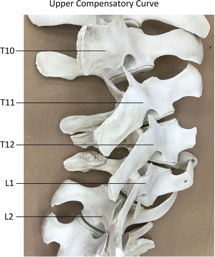
Figure 3. Close-up photograph of the apex of the upper compensatory curve, directly cranial of the post-
traumatic primary curve, after removing the soft tissues. This compensatory curvature occurred in an (initially)
anatomically normal part of the spine, unaffected by the trauma.
dotic shape of the intervertebral discs with an anterior length almost twice the posterior length. This is in sharp
contrast to the kyphosis in the discs of controls with − 4.6 ± 5.0% (p < 0.001). The AP% for the separate vertebral
bodies and intervertebral discs at every level is shown in Fig. 6.
Discussion
Idiopathic scoliosis is a 3D decompensation of a spine with no anatomical abnormalities, in an individual without
underlying manifest d isease1. In the search for its aetiology, many theories involving just as many of the body’s
organ systems have been suggested to play a role1,4–12. The usually present lengthening of the anterior side of the
thoracic spine in idiopathic s coliosis5,6,37, was suggested to be the result of a generalized bony overgrowth disor-
der (relative anterior spinal overgrowth; RASO), possibly as a compensation for a disturbance of synchronized
Scientific Reports | (2021) 11:7218 | https://doi.org/10.1038/s41598-021-86709-x 3
Vol.:(0123456789)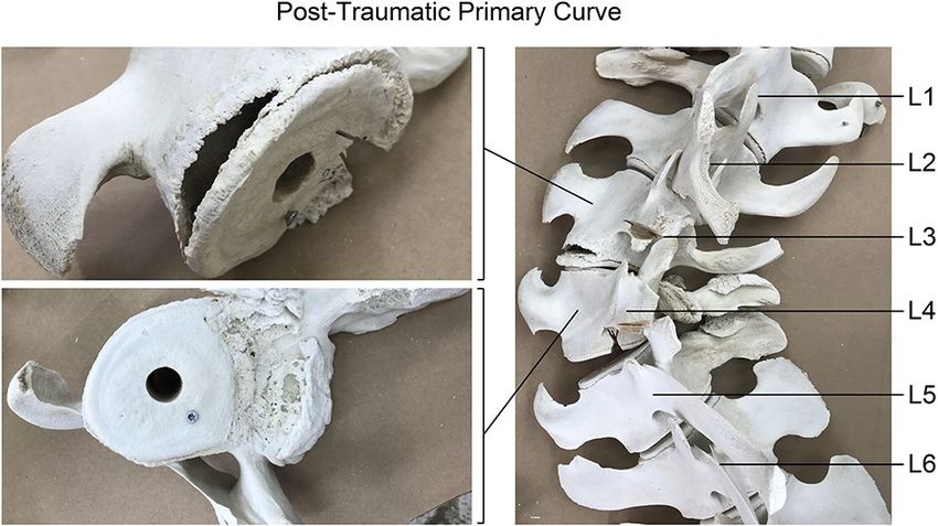
www.nature.com/scientificreports/
Figure 4. Dorsal view with the cranial side upwards from the CT-scan of level C1 to L7. The site of the blunt
traumatic injury at level L3/L4 (indicated with an asterisk) initiated a double compensatory curve cranially and
a single compensatory curve caudally.
growth between the neural and the osseous e lements7–9,38. Recent observations have shown that this anterior
lengthening occurs predominantly in the discs and is not restricted to idiopathic s coliosis22–26.
We propose that scoliosis is a rather universal compensatory mechanism that can occur as a response to a
(perceived) disturbance of spinal equilibrium. The crucial difference between the human spine and that of other
mammals is not in its anatomy, but in the way it is biomechanically loaded, not by the fact that man is bipedal
(many species are) but by the fact that humans carry their center of mass more posteriorly than any other
species13–17,27. This leads to a sagittal profile that makes the human spine, in comparison with any other spine in
nature, quadrupedal and bipedal alike, a rotationally less stable s tructure18,19,39. This means that, whereas in other
species often draconic measures are necessary to induce a s coliosis3, in humans much less is needed to initiate
this mechanism. We propose that the possible value of scoliosis research in experimental animals is not in the
primary, artificially induced curve, but in the response that follows in the untouched area of the spine, i.e. the
compensatory curve. This is supported by the observation that compensatory curves in congenital scoliosis in
humans show a very similar 3D morphology to idiopathic scoliosis25, and that in porcine tether induced scolio-
sis the compensatory curves outside the instrumented spinal segment showed a similar rotational d eformity40.
The objective of the current study was to investigate the mechanism through which a scoliosis developed in
the normal area of the spine, in an animal that is not known to develop a scoliosis spontaneously. We studied
Scientific Reports | (2021) 11:7218 | https://doi.org/10.1038/s41598-021-86709-x 4
Vol:.(1234567890)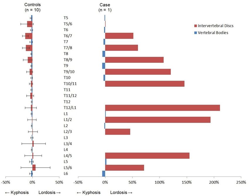
www.nature.com/scientificreports/
Figure 5. Of the whale with scoliosis, the rotation of the vertebral bodies in the transverse plane is shown in
degrees. Positive values indicate that the anterior part of the vertebral body is pointing towards the right. All
rotation of the vertebral bodies is into the convexity of the curve.
the 3D morphology of the compensatory curves that evolved around a traumatically induced coronal plane
deformity in a whale. A (dorso-)lateral blunt traumatic injury caused a localized, acute, predominantly lateral
deviation of the spine, that subsequently initiated 3D compensatory scoliotic curves in the anatomically normal
areas of the spine. These compensatory curves showed rotation of the vertebral bodies into the convexity of the
curve, and an apical lordosis (+ 9.4%), which differed significantly from the kyphosis in the spine of the control
group (− 2.1%). Although, since the animal was still growing, some wedging of vertebrae occurred, the shape of
the vertebral bodies in the sagittal plane showed no difference with the normal, kyphotic shape of the vertebrae
in the control group (− 1.8%). The observed lordosis in the compensatory curves of the whale was exclusively
located in the intervertebral discs, they showed severe anterior lengthening (+ 99.5%) which was in stark contrast
with the kyphotic discs of controls (− 4.6%). This 3D morphology is very similar to what is found in humans
with idiopathic scoliosis, but also in human neuromuscular scoliosis as well as in the compensatory curve in
human congenital s coliosis22–25.
The acute primary scoliosis caused by the traumatic accident, resulted in the head and tail of the whale being
out of line and inhibiting proper locomotion and swimming manoeuvres. As many mammals have a vestibular
Scientific Reports | (2021) 11:7218 | https://doi.org/10.1038/s41598-021-86709-x 5
Vol.:(0123456789)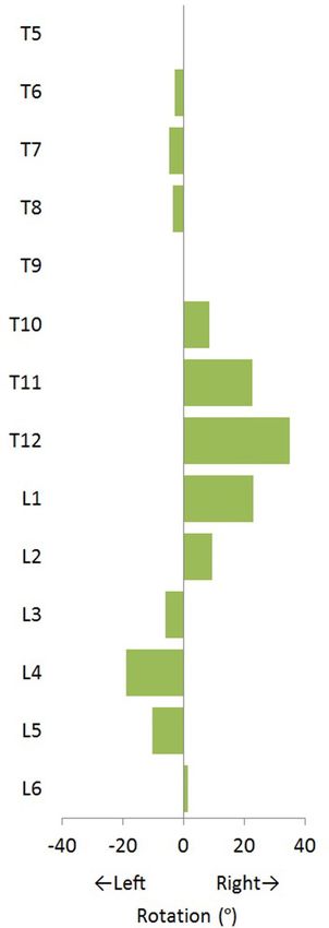
www.nature.com/scientificreports/
Figure 6. The mean anterior–posterior length discrepancy (AP%) with the standard deviation is shown for the
intervertebral discs in red and the vertebral bodies in blue, for both the non-scoliotic controls and the whale
with scoliosis. Endplates severely affected by trauma (L3/L4) and wedging (T11/T12) were excluded due to
impossibility of proper CT-scan analysis. Positive AP% indicates a larger anterior length than posterior length.
reflex of self-righting41,42, the whale most likely compensated this trunk imbalance in an attempt to re align its
head to its tail, inducing compensatory curves in the anatomically normal areas of the spine with a 3D morphol-
ogy that strongly resembles human scoliosis. Whereas most spines in nature require substantial effort to start a
permanent rotational deformity due to the stabilizing action of gravity in combination with the trunk’s muscles
(i.e. the follower load)39,43, the human spine is much less rotationally stable due to its unique sagittal profile with
the body’s centre of gravity straight above, rather than in front of the p elvis13–17,27. This reduces the stabilizing
anterior shear loading and even induces posteriorly directed shear loads that render the involved spinal segments
unstable in the horizontal plane1,18,19,28,29,39,44.
This rare occurrence of scoliosis in a species that is not known to develop a spinal curvature spontaneously,
provided a unique chance to study scoliosis in a completely different model. A limitation of this study was that
the common minke whale was not compared to non-scoliotic controls of the exact same species. This is due
to the low frequency of stranded common minke whales in the Netherlands, in combination with their large
size and weight exceeding the capacity of most CT-scanning facilities. However, the smaller harbour porpoise
(Phocoena phocoena) share strong commonalities in spinal anatomy and were therefore used as controls in the
current study45,46. Furthermore, the fractured and extensively deformed vertebrae were excluded since proper
recognition of the anatomical planes was impossible during CT-scan analysis. However, visual inspection showed
wedging mainly in the coronal plane and overview images in the sagittal plane of the CT-scan did not show a
significant kyphosis at the site of traumatic injury. Therefore, we could exclude a post-traumatic kyphosis as the
initiator of the apical lordosis observed in this study. Also, the observation of a lordosis or kyphosis could be influ-
enced by the fact that the scoliotic whale, nor the controls were alive during CT-scanning and were positioned
prone outside of their naturally aquatic habitat. We know from human scoliosis that kyphosis and lordosis are
underestimated during prone or supine imaging compared to upright, but there is no difference between prone
or supine47. Gravity obviously plays an important role in humans, but not in submerged mammals, therefore we
feel that the influence of positioning on our results is limited.
The aim of this study was to analyse whether scoliosis can be considered a more generalized compensatory
mechanism that occurs independent of cause and/or species. In line with our hypothesis, we observed that the
compensatory curves that developed in the normal area of the spine of a whale, that suffered severe but localized
Scientific Reports | (2021) 11:7218 | https://doi.org/10.1038/s41598-021-86709-x 6
Vol:.(1234567890)www.nature.com/scientificreports/
trauma to the spine, show strong similarities in 3D configuration with different types of human scoliosis. This
suggests a shared and rather uniform mechanical basis, implying that any perceived decompensation of spinal
equilibrium can lead to a uniform response, with uniform 3D morphology. The unique biomechanics of the
upright human s pine13–17,27, with significantly decreased rotational s tability1,18,19,39,44, may explain why only in
humans this mechanism can be induced relatively easily, without an obvious cause, and is therefore still called
‘idiopathic’.
Methods
Post mortem examination. Since 2008, cetaceans that stranded dead or died shortly after stranding on
the Dutch coast are subjected to post mortem examination, which is conducted at the division of pathology
of the Faculty of Veterinary Medicine (Utrecht University). The animals described in the current study were
not used for scientific or commercial testing. All were free-living whales which died of natural causes or were
euthanized on welfare grounds and not for the purpose of this, or other studies. Therefore, since there was no
handling of live animals in the current study, according to institutional guidelines, no consent from the Animal
Use Committee was required, and animal ethics committee approval was not applicable to this work. On the 8th
of July 2019, a young common minke whale washed up on the North Sea beach of Texel, the Netherlands (Fig. 1),
and subsequently underwent post mortem investigation aiming to determine the cause of its death. A necropsy
and tissue sampling procedure was conducted following internationally standardized guidelines48. This included
the collection of the following measures: total length (measured from the tip of the rostrum to the fluke notch, in
a straight line next to the body, in cm), weight (kg) and blubber thickness. The latter was measured immediately
anterior to the dorsal fin at three locations (dorsal, lateral and ventral, in mm). Age class was determined based
on total length and gross examination of reproductive organs. Tissue samples from various organs, as well as the
vertebral bone, were fixed in 4% phosphate-buffered formalin, embedded in paraffin, cut into 4 µm sections, and
stained with haematoxylin and eosin. Samples from vertebra were decalcified prior to paraffin imbedding and
staining procedures.
Diagnostic imaging. Upon gross examination of the whale, the spinal malformation was noted. The entire
vertebral column was therefore wrapped in plastic sheets and submitted for computed tomography (CT)-scan-
ning. The spine was positioned in ventral recumbency on the table of a 64-slice sliding gantry CT scanner
(Somatom Definition AS, Siemens AG, München, Germany).
Control group. As common minke whale strandings infrequently occur on the Dutch coast, a control group
of the same species was not possible to acquire. Therefore, a control group was assembled of the harbour por-
poise; a smaller member of the cetacean family and the most abundant whale species in the North Sea. Har-
bour porpoises are regularly subjected to post mortem examination and in a previous study focusing on their
ecropsies49. Ten cases which did not present
anatomy, animals were subjected for full-body CT-scan prior to the n
spinal abnormalities and were positioned straight during CT-scanning were selected from this database and
used as a control group in this study.
CT measurements. The orientation of the scanned whales in this study was defined the same way as in
humans, with anterior indicating the ventral side and posterior indicating the dorsal side, and furthermore cra-
nial, caudal, left and right as standard. The CT-scans of the whale and control group was measured with dedicated
software (ScoliosisAnalysis 4.1; Image Sciences Institute, Utrecht, The Netherlands, developed with MeVisLab,
MeVis Medical Solutions AG, Bremen, Germany) to measure the direction and amount of rotation, anterior and
posterior height of vertebral bodies and vertebral discs in the exact mid-sagittal plane, corrected for deformity
in all three planes. This software is in-house developed and validated with excellent intra- and interobserver
reliability50. This semi-automated method is used and extensively described in multiple earlier studies23,25,50, 51.
For all upper and lower endplates in the included part of the spine, the observer adjusted the plane of view for
coronal and sagittal tilt. In this true transverse plane, the vertebral body and spinal canal were manually seg-
mented by the observer, whereafter the software automatically determined the 3D coordinates of the anterior
and posterior point of the endplate, adjusted for rotation and deformity in all planes. The distances between
these points were calculated to obtain the anterior and posterior heights of the vertebral bodies and interver-
tebral discs (Fig. 7). This was done for all the compensatory curves (Cobb-to-Cobb). The corresponding levels
of the spine analysed in the whale were also measured in controls. After measurements, the anterior–posterior
length discrepancy (AP%) was calculated as [(anterior length – posterior length)/(posterior length)] × 100%, for
the total compensatory curved spine, and for the vertebral bodies and the intervertebral discs separately. End-
plates severely affected by trauma or wedging were excluded, as proper segmentation was not possible. Positive
AP% values indicated that the anterior (ventral) side was longer than the posterior (dorsal) side.
Statistical analysis. The mean AP% results for the total curve, the vertebral bodies and the intervertebral
bodies were determined for the minke whale and for the non-scoliotic harbour porpoise control group given
with ± standard deviation. The differences in mean AP% between the scoliotic whale and non-scoliotic controls
were tested with an independent samples T-test. The data for Figs. 5, 6 were provided separately (Supplementary
Information). Statistical analysis was performed in SPSS 25.0 for Windows (IBM, Armonk, NY, USA). The level
of statistical significance was set at p ≤ 0.05.
Scientific Reports | (2021) 11:7218 | https://doi.org/10.1038/s41598-021-86709-x 7
Vol.:(0123456789)www.nature.com/scientificreports/
Figure 7. Method of 3D measurements on CT-scans in this study. For all upper and lower endplates, the
observer adjusted the plane of view for coronal (I) and sagittal (II) tilt. In the true transverse plane, the vertebral
body and spinal canal were manually segmented (III), whereafter the software automatically determined the 3D
coordinates of the anterior (A) and posterior (P) point of the endplate, adjusted for rotation and deformity in
all planes. The distances between these points were calculated to obtain the anterior and posterior heights of the
vertebral bodies and intervertebral discs.
Animal ethics committee approval. The animals described in the current study were not used for sci-
entific or commercial testing. All were free-living whales which died of natural causes or were euthanized on
welfare grounds and not for the purpose of this, or other studies. Therefore, since there was no handling of live
animals in the current study, according to institutional guidelines, no consent from the Animal Use Committee
was required, and animal ethics committee approval was not applicable to this work.
Data availability
All generated or analysed data in this study are included in this article or the supplementary information files.
Received: 15 December 2020; Accepted: 19 March 2021
References
1. Cheng, J. C. et al. Adolescent idiopathic scoliosis. Nat. Rev. Dis. Prim. 1, 15–30 (2015).
2. Kouwenhoven, J.-W.M. & Castelein, R. M. The pathogenesis of adolescent idiopathic scoliosis: Review of the literature. Spine 33,
2898–2908 (2008).
3. Janssen, M. M. A., de Wilde, R. F., Kouwenhoven, J.-W.M. & Castelein, R. M. Experimental animal models in scoliosis research:
A review of the literature. Spine J. 11, 347–358 (2011).
4. Schlösser, T. P. C., van der Heijden, G. J. M. G., Versteeg, A. L. & Castelein, R. M. How ‘idiopathic’ is adolescent idiopathic scoliosis?
A systematic review on associated abnormalities. PLoS ONE 9, e97461 (2014).
5. Somerville, E. W. Rotational lordosis; the development of single curve. J. Bone Joint Surg. Br. 34-B, 421–427 (1952).
6. Dickson, R. A., Lawton, J. O., Archer, I. A. & Butt, W. P. The pathogenesis of idiopathic scoliosis. Biplanar spinal asymmetry. J.
Bone Joint Surg. Br. 66, 8–15 (1984).
7. Guo, X., Chau, W. W., Chan, Y. L. & Cheng, J. C. Y. Relative anterior spinal overgrowth in adolescent idiopathic scoliosis. Results
of disproportionate endochondral-membranous bone growth. J. Bone Joint Surg. Br. 85, 1026–1031 (2003).
8. Newell, N. et al. Quantifying progressive anterior overgrowth in the thoracic vertebrae of adolescent idiopathic scoliosis patients:
A sequential magnetic resonance imaging study. Spine 41, E382–E387 (2016).
9. Stokes, I. A. F., Burwell, R. G. & Dangerfield, P. H. Biomechanical spinal growth modulation and progressive adolescent scoliosis—A
test of the ‘vicious cycle’ pathogenetic hypothesis: Summary of an electronic focus group debate of the IBSE. Scoliosis 1, 16 (2006).
10. Nicoladoni, C. Anatomie und Mechanismus der Skoliose. Kocher, König, von Mikulicz, eds. Bibl. Medica. Stuttgart Verlag von Erwin
Nagele (1904).
11. Roth, M. Idiopathic scoliosis and Scheuermann’s disease: Essentially identical manifestations of neuro-vertebral growth dispropor-
tion. Radiol. Diagn. 22, 380–391 (1981).
12. Porter, R. W. Can a short spinal cord produce scoliosis?. Eur. Spine J. 10, 2–9 (2001).
13. Abitbol, M. M. Evolution of the ischial spine and of the pelvic floor in the Hominoidea. Am. J. Phys. Anthropol. 75, 53–67 (1988).
14. Voutsinas, S. A. & MacEwen, G. D. Sagittal profiles of the spine. Clin. Orthop. Relat. Res. 210, 235–242 (1986).
15. Washburn, S. L. The analysis of primate evolution with particular reference to the origin of man. Cold Spring Harb. Symp. Quant.
Biol. 15, 67–78 (1950).
Scientific Reports | (2021) 11:7218 | https://doi.org/10.1038/s41598-021-86709-x 8
Vol:.(1234567890)www.nature.com/scientificreports/
16. Alexander, R. M. Bipedal animals, and their differences from humans. J. Anat. 204, 321–330 (2004).
17. Payne, R. C. et al. Morphological analysis of the hindlimb in apes and humans. II. Moment arms. J. Anat. 208, 725–742 (2006).
18. Kouwenhoven, J.-W.M. et al. Effects of dorsal versus ventral shear loads on the rotational stability of the thoracic spine: A biome-
chanical porcine and human cadaveric study. Spine 32, 2545–2550 (2007).
19. Homminga, J. et al. Posteriorly directed shear loads and disc degeneration affect the torsional stiffness of spinal motion segments:
A biomechanical modeling study. Spine 38, E1313–E1319 (2013).
20. Lovett, R. W. A contribution to the study of the mechanics of the spine. Am. J. Anat. 2, 457–462 (1903).
21. Lovett, R. W. The mechanism of the normal spine and its relation to scoliosis. Bost. Med. Surg. J. 153, 349–358 (1905).
22. Schlösser, T. P. C. et al. Anterior overgrowth in primary curves, compensatory curves and junctional segments in adolescent idi-
opathic scoliosis. PLoS ONE 11, e0160267 (2016).
23. Brink, R. C. et al. Anterior-posterior length discrepancy of the spinal column in adolescent idiopathic scoliosis-a 3D CT study.
Spine J. 18, 2259–2265 (2018).
24. Brink, R. C. et al. Anterior spinal overgrowth is the result of the scoliotic mechanism and is located in the disc. Spine 42, 818–822
(2017).
25. de Reuver, S. et al. Anterior lengthening in scoliosis occurs only in the disc and is similar in different types of scoliosis. Spine J.
20, 1653–1658 (2020).
26. Will, R. E., Stokes, I. A., Qiu, X., Walker, M. R. & Sanders, J. O. Cobb angle progression in adolescent scoliosis begins at the
intervertebral disc. Spine 34, 2782–2786 (2009).
27. Bernhardt, M. & Bridwell, K. H. Segmental analysis of the sagittal plane alignment of the normal thoracic and lumbar spines and
thoracolumbar junction. Spine 14, 717–721 (1989).
28. Pasha, S. 3D deformation patterns of S shaped elastic rods as a pathogenesis model for spinal deformity in adolescent idiopathic
scoliosis. Sci. Rep. 9, 16485 (2019).
29. Castelein, R. M., Pasha, S., Cheng, J. C. & Dubousset, J. Idiopathic scoliosis as a rotatory decompensation of the spine. J. Bone
Miner. Res. 35, 1850–1857 (2020).
30. Arkin, A. M. The mechanism of the structural changes in scoliosis. J. Bone Joint Surg. Am. 31A, 519–528 (1949).
31. Naique, S. B. et al. Scoliosis in an orangutan. Spine 28, E143–E145 (2003).
32. Sharp, S. M. et al. Gross and histopathologic diagnoses from North Atlantic right whale Eubalaena glacialis mortalities between
2003 and 2018. Dis. Aquat. Organ. 135, 1–31 (2019).
33. Weir, C. R. & Wang, J. Y. Vertebral column anomalies in Indo-Pacific and Atlantic humpback dolphins Sousa spp. Dis. Aquat.
Organ. 120, 179–187 (2016).
34. Bertulli, C. G. et al. Vertebral column deformities in white-beaked dolphins from the eastern North Atlantic. Dis. Aquat. Organ.
116, 59–67 (2015).
35. Nielsen, N. H., Víkingsson, G. A., Hansen, S. H., Ditlevsen, S. & Heide-Jørgensen, M. P. Two techniques of age estimation in
cetaceans: GLGs in teeth and earplugs, and measuring the AAR rate in eye lens nucleus. NAMMCO Sci. Publ. 10, (2017).
36. Christiansen, F., Víkingsson, G. A., Rasmussen, M. H. & Lusseau, D. Minke whales maximise energy storage on their feeding
grounds. J. Exp. Biol. 216, 427–436 (2013).
37. Roaf, R. The basic anatomy of scoliosis. J. Bone Joint Surg. Br. 48, 786–792 (1966).
38. Chu, W. C. W. et al. Relative shortening and functional tethering of spinal cord in adolescent idiopathic scoliosis? Study with
multiplanar reformat magnetic resonance imaging and somatosensory evoked potential. Spine 31, E19-25 (2006).
39. Castelein, R. M., van Dieën, J. H. & Smit, T. H. The role of dorsal shear forces in the pathogenesis of adolescent idiopathic scoliosis—
A hypothesis. Med. Hypotheses 65, 501–508 (2005).
40. Barrios, C. et al. Novel porcine experimental model of severe progressive thoracic scoliosis with compensatory curves induced by
interpedicular bent rigid temporary tethering. J. Orthop. Res. 36, 174–182 (2017).
41. Spoor, F., Bajpai, S., Hussain, S. T., Kumar, K. & Thewissen, J. G. M. Vestibular evidence for the evolution of aquatic behaviour in
early cetaceans. Nature 417, 163–166 (2002).
42. Spoor, F. et al. The primate semicircular canal system and locomotion. Proc. Natl. Acad. Sci. USA. 104, 10808–10812 (2007).
43. Patwardhan, A. G., Meade, K. P. & Lee, B. A frontal plane model of the lumbar spine subjected to a follower load: Implications for
the role of muscles. J. Biomech. Eng. 123, 212–217 (2001).
44. Janssen, M. M., Kouwenhoven, J. W. & Castelein, R. M. The role of posteriorly directed shear loads acting on a pre-rotated growing
spine: A hypothesis on the pathogenesis of idiopathic scoliosis. Stud. Health Technol. Inform. 158, 112–117 (2010).
45. Thewissen, J. G. M., Cooper, L. N., George, J. C. & Bajpai, S. From land to water: The origin of whales, dolphins, and porpoises.
Evol. Educ. Outreach 2, 272 (2009).
46. Fordyce, R. E. Cetacean Evolution. Encyclopedia of Marine Mammals (Elsevier, 2018).
47. Brink, R. C. et al. Upright, prone, and supine spinal morphology and alignment in adolescent idiopathic scoliosis. Scoliosis Spinal
Disord. 12, 6 (2017).
48. IJsseldijk, L. L., Brownlow, A. C. & Mazzariol, S. (eds). Best practice on cetacean post mortem investigation and tissue sampling.
Joint ASCOBANS/ACCOBAMS document. (2019).
49. Willems, D., IJsseldijk, L. & Veraa, S. Vertebral pattern variation in the North Sea Phocoena phocoena by computed tomography.
Anat. Rec. Epub ahead of print (2020).
50. Schlösser, T. P. C. et al. Three-dimensional characterization of torsion and asymmetry of the intervertebral discs versus vertebral
bodies in adolescent idiopathic scoliosis. Spine 39, E1159–E1166 (2014).
51. Brink, R. C. et al. A reliability and validity study for different coronal angles using ultrasound imaging in adolescent idiopathic
scoliosis. Spine J. 18, 979–985 (2018).
Acknowledgements
We are grateful for the help of all organizations and volunteers included in the Dutch Stranding Network, for their
help in reporting and collecting of stranded marine mammals. In particular, the authors want to acknowledge
staff of Ecomare Texel, especially Mariette Smit and Pierre Bonnet, for their help with collecting the common
minke whale, and Chris Walen for facilitating the visual inspection of the cleaned vertebral column. In addition,
the authors thanks the staff of the Division of Diagnostic Imaging for their help in scanning marine mammals,
and the staff of the division of pathology for their assistance during the post mortem investigations. Furthermore,
we want to thank the Fondation Yves Cotrel for their support to our research on the role of the intervertebral
disc in the aetiology human idiopathic scoliosis. Post mortem research in the Netherlands is commissioned by
the Dutch Ministry of Agriculture, Nature and Food Quality (harbour porpoises under project reference number
WOT-04-009-045 and the minke whale under project reference number 1400010530). The skeleton of the whale
was included in the collection of Ecomare Texel and visible in their exposition in the future.
Scientific Reports | (2021) 11:7218 | https://doi.org/10.1038/s41598-021-86709-x 9
Vol.:(0123456789)www.nature.com/scientificreports/
Author contributions
The necropsy was performed by L.L.IJ., M.J.L.K. and A.G. Diagnostic imaging was performed by D.S.W. and S.V.
The software for three-dimensional analysis was developed by M.v.S. CT-scan measurements were performed
by S.d.R. and analysis of these measurements were performed by S.d.R., J.F.H., M.C.K. and R.M.C. S.d.R. and
L.L.IJ. wrote the main manuscript text and all authors reviewed and edited the manuscript.
Competing interests
The authors declare no competing interests.
Additional information
Supplementary Information The online version contains supplementary material available at https://doi.org/
10.1038/s41598-021-86709-x.
Correspondence and requests for materials should be addressed to L.L.I.
Reprints and permissions information is available at www.nature.com/reprints.
Publisher’s note Springer Nature remains neutral with regard to jurisdictional claims in published maps and
institutional affiliations.
Open Access This article is licensed under a Creative Commons Attribution 4.0 International
License, which permits use, sharing, adaptation, distribution and reproduction in any medium or
format, as long as you give appropriate credit to the original author(s) and the source, provide a link to the
Creative Commons licence, and indicate if changes were made. The images or other third party material in this
article are included in the article’s Creative Commons licence, unless indicated otherwise in a credit line to the
material. If material is not included in the article’s Creative Commons licence and your intended use is not
permitted by statutory regulation or exceeds the permitted use, you will need to obtain permission directly from
the copyright holder. To view a copy of this licence, visit http://creativecommons.org/licenses/by/4.0/.
© The Author(s) 2021
Scientific Reports | (2021) 11:7218 | https://doi.org/10.1038/s41598-021-86709-x 10
Vol:.(1234567890)You can also read