Ulnar longitudinal deficiency: a rare case report and review
←
→
Page content transcription
If your browser does not render page correctly, please read the page content below
International Journal of Research in Orthopaedics
Lamba A et al. Int J Res Orthop. 2021 Jan;7(1):159-161
http://www.ijoro.org

                                                 DOI: https://dx.doi.org/10.18203/issn.2455-4510.IntJResOrthop20205581
Case Report

            Ulnar longitudinal deficiency: a rare case report and review
               Akshay Lamba1*, Naresh Kumar1, Chaitanya Krishna2, Sargam Chhabra3

  1
    Department of Orthopaedics, B. P. S. Government Medical College, Haryana, India
  2
    Consultant, Healthworld Hospital, Durgapur, West Bengal, India
  3
    Department of Ophthalmology, Pt. B. D. S. PGIMS, Rohtak, Haryana, India

  Received: 22 September 2020
  Revised: 11 November 2020
  Accepted: 12 November 2020

  *Correspondence:
  Dr. Akshay Lamba,
  E-mail: aslamba@gmail.com

  Copyright: © the author(s), publisher and licensee Medip Academy. This is an open-access article distributed under
  the terms of the Creative Commons Attribution Non-Commercial License, which permits unrestricted non-commercial
  use, distribution, and reproduction in any medium, provided the original work is properly cited.

   ABSTRACT

   Ulnar hemimelia is a rare postaxial partial or complete longitudinal deficiency of ulna. It has an estimated incidence of
   1/100,000-150,000 live births, with a male to female ratio of 3:2. There is usually ulnar deviation of hand and shortening
   of forearm. Radial head subluxation and fixed flexion deformity of the hand may be associated with it. Complex carpal,
   metacarpal, and digital abnormalities including absence of triquetrum, capitate and three fingered hand (tridactyly) are
   additional findings commonly found in association. Here, we present a case of a 17-year-old female with left sided
   ulnar club hand due to isolated partial ulnar aplasia.

   Keywords: Aplasia, Ectromelia, Hemimelia, Ulna, Upper extremity deformities, Congenital

INTRODUCTION                                                           more absence of ulna causes more severe flexion
                                                                       deformity. Usually there is associated radial head
Ulnar hemimelia is a rare congenital upper limb anomaly                subluxation.1-3 There may be some associated skeletal
characterized by complete or partial absence of the ulna               anomalies like syndactyly.
bone. Isidore Geoffroy Saint-Hilaire coined the term
“hemimelia” in the early 19th century, while in 1951,                  Here, we present a report of a patient with left sided ulnar
O'Rahilly suggested the term "paraxial hemimelia" for the              club hand who was managed conservatively with good
longitudinal variety, because either the preaxial or                   functional outcome.
postaxial side of the limb is involved.1 Incidence is
estimated at 1/100,000-150,000 live births, with a male to             CASE REPORT
female ratio of 3:2. Ulnar hemimelia is rarer than its radial
counterpart. It occurs in about 1 in 1.5 million population.           A 17-year-old female patient presented to us with
Ulnar ray longitudinal deficiency has been                             deformed left upper limb. There was no history of intake
embryologically shown to be due to a deficiency of the                 of any teratologic drug by mother during the antenatal
Sonic Hedgehog that is the main controller of the                      period. The patient was born through normal vaginal
anteroposterior axis of limb development.4 There is                    delivery at full term. There was no history of a congenital
usually shortening of forearm and ulnar sided deviation of             skeletal anomaly in parents or any of the siblings. On
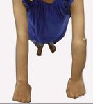
the hand, which leads to its another name ulnar clubhand.              examination, there was shortening of forearm on the
Usually found unilaterally. Such rarity in its occurrence              effected side (Figure 1). Radial head was grossly
leads to controversy and dilemma in proper way of                      subluxated and palpable as a rounded bony mass
management of these patients. Ulnar hemimelia patients                 continuous with radius. There was no restriction of motion
usually have fixed flexion deformity of elbow joint. The               at elbow but range of motion was restricted at wrist with

                                 International Journal of Research in Orthopaedics | January-February 2021 | Vol 7 | Issue 1   Page 159
Lamba A et al. Int J Res Orthop. 2021 Jan;7(1):159-161
decreased wrist flexion. There was no associated hand Treatment
anomaly.
A regular and well-defined physiotherapy plan was
developed to maximize the use of the limb and prevent
development of contractures but no surgery was planned.
DISCUSSION
Ulnar hemimelia is a postaxial complete or partial
longitudinal deficiency of ulna. It may be an isolated
finding or it may be associated with complex carpal,
metacarpal, and digital abnormalities. Various
classification systems have been used, depending on the
deformities in elbow joint, ulna, carpal, metacarpal and
digits. Bayne classification which was later on extended by
Havenhill and Goldfarb gives 6 different types from type
0 to type 6. Type 1 with dysplastic ulna is the most
Figure 1: Forearm is shortened on the left and radial common followed by type 0 with normal ulna and
head is grossly subluxated. involvement of carpus or hand only. Type 2 is the one with
partial aplasia of ulna which was present in our case
(Figure 1). Type 3 patients have complete ulnar aplasia.
Type 4 has radiohumeral synostosis and type 5 comprises
of phocomelic deficiency.9-12
The triquetrum and the capitate are frequently absent in
these patients. Radiohumeral joint fusion is also a frequent
association. Postaxial (ulnar-sided) deficiency of
metacarpals and digits is a common finding in these
patients. Three-fingered hand (tridactyly) is the most
common hand anomaly followed closely by mono-digital
hand.1 Cases with digital anomalies, camptodactyly and
postaxial syndactyly have also been reported in
literature.13 In general, the severity of ulnar dysplasia
Figure 2: Range of motion at the elbow is not correlates with the degree of hand and wrist anomalies.
restricted.
Rarely, ulnar hemimelia may also be associated with
Radiological imaging revealed a deformed misshapen ulna syndromes unlike the radial clubbed hand that has been
with absence of its distal end, ulnar deviation of hand and much more frequently associated with congenital
radial bowing. Radial head was subluxated and proximally syndromes. It may be a part of Poland syndrome, Klippel-
migrated (Figure 3). Feil syndrome, Goltz-Gorlin syndrome, Cornelia De
Lange syndrome, or Femur Fibula Ulna syndrome.5,7 The
radius is typically longer than the ulna in patients with
achondroplasia, and in patients with
mucopolysaccharidosis, the distal ulna and radius can be
hypoplastic and may slope toward each other.6,8 In
syndromic cases the clinical course and the prognosis
depends on the severity of the syndrome.
CONCLUSION
Ulnar hemimelia is a rare postaxial partial or complete
longitudinal deficiency of ulna. It presents with congenital
shortening of forearm, ulnar sided deviation of the hand,
fixed flexion deformity of elbow joint and radial head
subluxation. There may also be some associated skeletal
anomalies of the hand. In the extended Bayne
classification, isolated dysplastic ulna is the most common
Figure 3: Radiograph showing deformed forearm, type. Early recognition is essential to maximize the range
longitudinal ulnar deficiency distally, subluxated of motion at elbow and wrist by preventing contractures
radial head and ulnar deviation of hand. and surgical correction in appropriate cases.
International Journal of Research in Orthopaedics | January-February 2021 | Vol 7 | Issue 1 Page 160Lamba A et al. Int J Res Orthop. 2021 Jan;7(1):159-161
Funding: No funding sources 8. Aucourt J, Budzik JF, Manouvrier S, Mezel A,
Conflict of interest: None declared Cagneaux M, Boutry N, et al. Congenital upper limb
Ethical approval: Not required malformations: pictorial review. ECR 2011/C-2085.
9. Al-Qattan MM, Al-Sahabi A, Al-Arfaj N. Ulnar ray
REFERENCES deficiency: a review of the classification systems, the
clinical features in 72 cases, and related
1. Frantz CH, O'Rahilly R. Ulnar Hemimelia. Artificial developmental biology. J Hand Surg Eur.
Limbs. 1971;15:25-35. 2010;35:699-707.
2. Drachman DB, Sokoloff. The role of movement in 10. Bayne LG. Ulnar club hand (ulnar deficiencies). In:
embryonic joint development. Develop Biol. Green DP (Ed.) Operative hand surgery. New York:
1966;14:401-20. Churchill Livingstone. 1982;245-57.
3. Duken J. Uber der Beziehungen zwischen 11. Goldfarb CA, Manske PR, Busa R, Mill J, Carter P,
Assimilationshypophalangie und Aplasie der Ezaki M. Upper-extremity phocomelia reexamined:
Interphalangealgelenke. Virchows Arch Path Anat a longitudinal dysplasia. J Bone Joint Surg Am.
Physiol. 1921;233:204-25. 2005;87:2639-48.
4. Al-Qattan MM, Al-Thunyan A. Ulnar Deficiencies. 12. Havenhill TG, Manske PR, Patel A, Goldfarb CA.
In: Abzug J, Kozin S, Zlotolow D. (eds). The Type ‘0’ ulnar longitudinal deficiency. J Hand Surg
Pediatric Upper Extremity. Springer, New York, NY. Am. 2005;30:1288-93.
2015. 13. Özdemir M, Turan A, Kavak RP. Ulnar hemimelia: a
5. Agrawal AK, Kosada D, Patel S, Patel J, Desai S, report of four cases. Skeletal Radiol. 2019;48:1137-
Patwa JJ. Ulnar hemimelia in deformed left forearm 43.
treated with Ilizarov fixator. Int J Orthop Sci.
2017;3:1013-6.
6. Panda A, Gamanagatti S, Jana M, Gupta AK. Skeletal
dysplasias: a radiographic approach and review of
common non-lethal skeletal dysplasias. World J
Radiol. 2014;6:808-25. Cite this article as: Lamba A, Kumar N, Krishna C,
7. Palmucci S, Attinà G, Lanza ML. Imaging findings Chhabra S. Ulnar longitudinal deficiency: a rare case
of mucopolysaccharidoses: a pictorial review. report and review. Int J Res Orthop 2021;7:159-61.
Insights Imaging. 2013;4:443-59.
International Journal of Research in Orthopaedics | January-February 2021 | Vol 7 | Issue 1 Page 161You can also read


