Treatment of a Radio-Ulnar Atrophic Pseudoarthrosis in a Toy Poodle Using an Autologous Coccygeal Vertebrae Transfer and Plate Fixation
←
→
Page content transcription
If your browser does not render page correctly, please read the page content below
Animal and Veterinary Sciences
2022; 10(6): 170-173
http://www.sciencepublishinggroup.com/j/avs
doi: 10.11648/j.avs.20221006.11
ISSN: 2328-5842 (Print); ISSN: 2328-5850 (Online)

Treatment of a Radio-Ulnar Atrophic Pseudoarthrosis in a
Toy Poodle Using an Autologous Coccygeal Vertebrae
Transfer and Plate Fixation
Giuseppe Bartoletta1, *, Franco Pizzirani1, Stefano Pizzirani2
1
    Clinica Veterinaria Europa, Firenze, Italy
2
    Cumming School of Veterinary, Medicine Tuft University, Boston, USA

Email address:
*
    Corresponding author

To cite this article:
Giuseppe Bartoletta, Franco Pizzirani, Stefano Pizzirani. Treatment of a Radio-Ulnar Atrophic Pseudoarthrosis in a Toy Poodle Using an
Autologous Coccygeal Vertebrae Transfer and Plate Fixation. Animal and Veterinary Sciences. Vol. 110, No. 6, 2022, pp. 170-173.
doi: 10.11648/j.avs.20221006.11

Received: July 26, 2022; Accepted: August 16, 2022; Published: January 10, 2023

Abstract: Description of the treatment of a radio-ulnar atrophic pseudo-arthrosis in a one-and-a-half-year-old female toy
tramp dog. Due to a domestic trauma the patient suffred a radio-ulnar fracture. Unfortunately at the time of the first visit we
were not in possession of the patient’s medical record and for this reason we did not know the evolution of the previous
surgical revision. The owner reports that at another centre, the subject underwent three osteosynthesis procedures
(osteosynthesis with radio-ulnar intramedullary nails, plate fixation and external circular fixator), with negative results. After a
long consultation with the owner it was decided to perform a surgery to restore the bone radius and ensure good mobility. Our
procedure included debridement and canalisation of the bone stumps, harvesting and grafting of the coccygeal VII vertebra,
autologous spongy bone grafting, osteosynthesis with VCP1.5/2.0 mm plate and 1.5- and 2.0-mm cortical screws. Clinical and
radiographic evaluation were carried out regularly, during which we decided to remove some screw in order to achieve an
implant dynamization. At weeks 7 and 16 four screws were removed. At week 60 the plate was removed and further controls at
weeks 64 and 90 confirmed anatomical and functional healing. No wound or bone healing complication were reported.

Keywords: Atrophic Pseudoarthrosis, Vertebrae Transfer, Radio-Ulnar Fracture, Plate Osteosynthesis

                                                                       callus include stability between cells, absence of
1. Introduction                                                        micromovements, and adequate vascularisation [5]. These
   The term pseudo-arthrosis defines the failure of bone               environmental conditions are interdependent and, in
healing of a fracture, with interposition of fibrous or                particular, decreased oxygen supply to the fracture site can
cartilaginous tissue in the fracture site or the osteotomy [1].        interfere with bone formation and instead favour the
Bone healing is spontaneously regulated by physiological               deposition of cartilage tissue, leading to pseudo-arthrosis [5-
(intrinsic) factors that may vary depending on the patient's           7]. The detailed classification of pseudo-arthrosis is still a
age and/or general health status. Under certain conditions,            source of debate [8]; the one most widely used is the one
usually related to stump instability, surgical procedures              proposed by Weber and Cech [9] which differentiates
(extrinsic factors) must be associated, whose primary                  pseudo-arthrosis into vital and non-vital. In vital
objective is to stabilise the fracture stumps to restore               pseudoarthrosis, the failure to consolidate the bone callus is
anatomical function [2]. Bone healing of a fracture, including         characterised by a proliferative bone reaction, with the
the importance of cytokine stimulation, respect for vascular           interposition of cartilage and fibrous tissue that can be seen
microcirculation and the fundamental need for stump                    both     radiographically      and     histologically.    Vital
stability has been described in detail in articles and reference       pseudoarthrosis, depending on the activity of the bone callus,
texts [3, 4]. Fundamental concepts for a stable and functional         is distinguished        into   three types:      hypertrophic,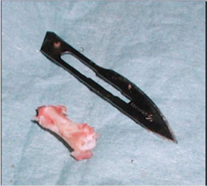
Animal and Veterinary Sciences 2022; 10(6): 170-173 171
normothrophic, and oligotrophic. Regarding non-vital L/min). After aseptically preparing the limb, a cranio-medial
pseudo-arthrosis, they are further subdivided into dystrophic, access to the radial diaphysis was performed according to
necrotic, deficient and atrophic. According to a study standard procedure [15]. Once the fracture site was reached,
conducted on 2825 cases of pseudoarthrosis [10], they were the apex of both stumps was debrided with a bone forceps,
found in the radio-ulnar segment in 40.6% of cases, in the removing the necrotic material and fibrous tissue surrounding
femoral in 39.5% of cases, in the humeral in 12.5% of cases, the ends. Osteostixis was then performed by inserting a 1.0
and in the tibial in 4.2% of cases. While hypertrophic mm Kirshner wire mounted on a drill (Synthes air compact
pseudo-arthroses are generally easier to treat [11], atrophic drive II) into the respective bony canals, performing
pseudo-arthroses present a more complex challenge as they continuous washings with saline to avoid thermal necrosis. A
are poorly vascularised. In toy breed dogs they are found surgical access was then made to the terminal portion of the
more in the distal radial-ulnar segment due to the deficient tail, which had previously been sterile prepared and isolated.
arborization of the metaphyseal vasculature [12]. The seventh coccygeal vertebra was harvested, and subjected
The purpose of this report is to describe the treatment of a to skeletonization, removal of the articular cartilaginous
chronic atrophic pseudo-arthrosis in a toy breed dog, using an surfaces and canalisation using a 0.8 mm Kirshner wire
autologous cortical implant obtained from a coccygeal (Figure 2). To obtain a bone segment of adequate size for the
vertebra, already described for a different bone segment [13], recipient site prior to the surgery, the length of the
as an alternative to other techniques (bank bone, synthetic contralateral radius and the space between the bone stumps
bone, distraction osteogenesis) [14]. of the fractured radius were measured, resulting 10.2 cm and
1.6 cm (16% of the entire length of the radius), respectively.
2. Clinical Case During the surgery, with the help of a Castroviejo Caliper, the
numerical figure was reported on the vertebral body and an
A twenty-month-old whole female toy poodle weighing additional apical ostectomy was performed using an
three kilograms was referred to our clinic for an orthopaedic oscillating saw (Synthes air 70 compact Drive II"). Once the
consultation. Nine months before, the patient had suffered a vertebral graft was inserted between the stumps, the fracture
radioulnar fracture that was initially treated with an was realigned and stabilised by applying a cuttable plate
osteosynthesis using intramedullary Kirshner wires. The first (Veterinary instrumentation) 1.5/2.0 mm - nine holes, placing
surgery was revised using a plate. After forty days, the plate three 1.5 mm screws in the proximal stump, three 2.0 mm
was replaced by a circular external fixator. About thirty days screws in the distal stump and two 1.5- and 2.0-mm screws in
after surgery, the implant was removed due to infection and the vertebral graft (Figure 3).
the limb was immobilised with a rigid cast. At the time of the
examination, after the bandage was removed, swelling, pain
on palpation and bone instability of the fracture site were
appreciated. An X-ray study under sedation, using medio-
lateral and antero-posterior projections of the left forearm,
showed an atrophic pseudo-arthrosis of the diaphysis of the
radius and ulna with thinning of the bone stumps, loss of
substance and lack of osteogenic activity (Figure 1). One of
the concerns raised during the pre-surgery planning was the
lack of bone tissue to restore the correct length of the bone
radius. After analysing the advantages and disadvantages of
a b
viable options, including limb amputation, the client opted
for an autologous bone graft. The patient underwent general Figure 1. Mid-lateral projection (a): Antero-posterior projection of the
anaesthesia using sedation with methadone (0.2 mg/kg) and affected limb (b). The two projections were taken at the time of the first
examination from which a picture of atrophic pseudoarthrosis is evident.
dexmedetomidine (2 µg/kg) intramuscularly. Induction was Note the thinning of the bone stumps, the loss of substance and the lack of
performed with propofol (2mg/kg) intravenously. General osteogenic activity. The holes for the screws and nails used in the previous
anaesthesia was maintained with isoflurane in oxygen (1 treatments are also evident.
a b c d
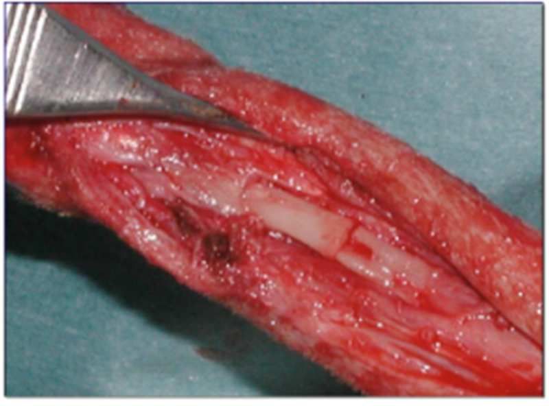
Figure 2. (a) Intra-surgery image of the coccygeal vertebra after removal from the donor site. (b) Intra-surgery image of the coccygeal vertebra after
skeletisation, removal of the apical cartilages and osteostixis. (c) Intra-surgery image of the vertebra at the recipient site. (d) Intra-surgery image after
osteosynthesis and subsequent autologous spongy bone grafting.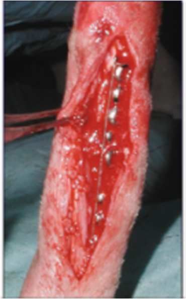
172                      Giuseppe Bartoletta et al.: Treatment of a Radio-Ulnar Atrophic Pseudoarthrosis in a Toy Poodle
                                     Using an Autologous Coccygeal Vertebrae Transfer and Plate Fixation

   The spongy graft was obtained from the right and left                            sixteen weeks after surgery, two more screws in positions 3-
humeral greater tubercle by transcortical access with a 1.8                         7, in the proximal-distal direction, were removed (Figure 3).
mm drill bit and harvesting the spongy tissue with a                                The plate with the remaining four screws was removed at
Volkmann's spoon. It was then inserted around the transplant                        sixty weeks and a rigid cast was applied to the limb for thirty
site. Post-surgery radiographs confirmed proper alignment of                        days, after which a further radiographic check was made to
the bone radius and correct positioning of the synthesis                            assess the state of bone healing in correspondence of the
media. After the surgery, the limb was immobilised with a                           screw holes. Sufficient osteogenic activity was observed and
modified Robert-Jones cast for eight days, replacing it every                       therefore the bandage was removed, advising the owner to
three days. At follow-up control performed twelve days after                        limit the dog's physical activity for further two weeks.
surgery, Grade I lameness was observed but pain on                                  Further clinical and radiographic controls at ninety and one-
palpation was absent, and the wound was in good condition.                          hundred-and-twenty weeks after surgery, respectively,
At follow-up control performed twenty-five days after                               showed a good anatomical and functional result, without
surgery, there was no lameness. Seven weeks after surgery,                          complications. Clinical and radiographic controls have been
two screws in position 5 and 6 (in the proximal-distal                              performed annually since then. The radiographic images of
direction) were removed after radiographic control. At                              the last check-up at fifteen years can be seen in figure 4.

                                        a                    b                  c                   d                   e
Figure 3. (a) Post-surgery X-ray, mid-lateral projection. (b) Antero-posterior projection after partial screw removal at seven weeks. (c) Mid-lateral projection
after partial removal of screws at seven weeks. (d) Mid-lateral projection after partial screw removal at sixteen weeks. (e) Antero-posterior projection after
partial screw removal at sixteen weeks.

                                                                                    canal and osteosynthesis with compression of the fracture site,
                                                                                    followed by a spongy bone graft [11], in our case the large
                                                                                    bone defect would have led to a decrease in radius of more
                                                                                    than 20% and would have favoured the onset of mechanical
                                                                                    lameness [14]. The therapeutic options to restore symmetry
                                                                                    with the contralateral limb are different (synthetic BMP [16],
                                                                                    titanium mesh integration [17], autologous graft, homologous
                                                                                    graft [18], distraction osteogenesis [14]). The choice of using a
                                                                                    cortical-spongiosa graft was motivated by its structural
                                                                                    characteristics, by all the essential components for bone
                                                                                    formation including osteoprogenitor cells, matrix and bone
                                                                                    morphogenetic proteins and by osteoinduction and
           a                        b                         c                     osteoconduction activity [5, 12, 18]. Furthermore, autografts
Figure 4. (a) Mid-lateral projection after plaque removal. (b) Antero-              have less ability to activate the antigenic response decreasing
posterior projection of follow-up ninety weeks after surgery. (c) Mid-lateral       the risk of rejection [13, 18]. The application of this technique
projection of follow-up fifteen years after surgery.                                in the radio-ulnar segment of a toy breed dog must take into
                                                                                    account not only the small size of the bone segment but also
3. Discussion                                                                       the poor metaphyseal vascularity [12]. Furthermore, in our
                                                                                    case, some of the holes drilled in previous operations
   The surgical treatment of atrophic pseudo-arthrosis is part of                   conflicted with the screws of the plate. To remedy this
orthopaedic practice, albeit to a lesser extent than other routine                  problem, screws with a diameter of 2 mm were used, while the
procedures, and often presents a challenge to the surgeon.                          length of some of the screws, which also affected the ulnar
Although the standard surgical treatment involves debridement                       cranial cortical bone, and the deposition of autologous
of the extremity of the two stumps, osteo-fixation of the spinal                    spongiosa bone further favoured the development of the radio-
Animal and Veterinary Sciences 2022; 10(6): 170-173                                        173

ulnar synostosis (already visible at seven weeks after surgery).        [7]   Greenbaum MA, Kanat IO (1993). Current concepts in bone
Some authors have described autologous adipose grafting                       healing. Review of the literature. Journal of the American
                                                                              Pediatric Medical Association. 83 (3): 123-9.
between radius and ulna to prevent the development of
synostosis, the cause of decreased or absent pronation and              [8]   Calori GM, Albisetti W, Agus A, et al (2007).. Risk factors
supination movements and thus lameness [19]. Numerous                         contributing to fracture non-unions. Injury International
radiographic and clinical controls were conducted both to                     Journal Care Injured, 38S, 11-18.
observe the possible development of infection of the surgical           [9]   Weber BG, Cech O. Pseudoarthrosis (1976). Stuttgart, Vienna,
wound and the tail end tract and to monitor the state of bone                 Hans Huber Bern.
healing, paying particular attention to the development of
                                                                        [10] Atilola MAO, Summer–Smith G: Nonunion fractures in dogs
infection at the implants. The removal of the screws conducted               (1984). Journal Veterinary Orthopedics 3: 21-24.
at seven and sixteen weeks was motivated by the intention to
increase the load on the bone without reducing its protection.          [11] Bennett D. Complications of Fracture Healing (1998). In:
In both cases and even after the removal of the plate, a rigid               Manual of Small Animal Fracture Repair and Management.
                                                                             Ed A Coughlan, A Miller. Cheltenham, BSAVA. pp 329-340.
bandage was applied post-surgery for fifteen days, to increase
the protection on the bone to avoid refracturing in                     [12] Welch JA, Boudieau RJ, DeJardin LM, Spodnick GJ (1997).
correspondence of the screw holes.                                           The Intraosseus Blood Supply of the Canine Radius:
                                                                             Implications for Healing of distal Fractures in Small Dogs.
                                                                             Veterinary Surgery 26: 57-61.
4. Conclusion
                                                                        [13] Yeh LS, You SM. Repair of a mandibular defect with a free
   Finally, although our surgical choice led to a satisfactory               vascularized coccygeal vertebra transfer in a dog (1994).
result from an anatomical and functional point of view,                      Veterinary Surgery July-August.
having only one case available it is difficult to interpret our         [14] (Robert D. Welch, DVM, PhD, and Daniel D. Lewis, DVM;
data to compare them with other techniques described in the                  Distraction osteogenesis (1999). Veterinary Clinics of North
literature.                                                                  America: Small animal practice: volume 29 number 5
                                                                             september.

                                                                        [15] Piermattei, D, Jhonson A (2004). An Atlas of surgical
References                                                                   approaches to the bones and joint of dog and cat, IV edition:
                                                                             Elsevier. pp 196-198.
[1]   Permattei DL, Flo GL, DeCamp CE (2006). Handbook of
      small animal orthopedics and fracture repair. 4th ed. Missouri:   [16] James AW, LaChaud G, Shen J, et al. A Review of the Clinical
      Saunders Elsevier. pp 170-202.                                         Side Effect of Bone Morphogenic Protein-2 (2016). Tissue
                                                                             Engineering Part. B Reviews: 22 (4): 284-297, August.
[2]   Tobias KM, Johnston SA (2012). Delayed Unions, Nonunions,
      and Malunions. In: Veterinary Surgery Small Animal Vol I:         [17] Zoi SI, Papadimitriou SA, Galatos AD, et al. Influence of a
      Elsevier S. aunders. pp 647.                                           titanium mesh on the management of segmental long bone
                                                                             defects (2015). Veterinary and Comparative Orthopaedics and
[3]   Tobias KM, Johnston SA (2021). Delayed Unions, Nonunions,              Traumatology: 28 (06): 417-424.
      and Malunions. In: Veterinary Surgery Small Animal Vol I:
      Elsevier Saunders. pp 647- 655.                                   [18] G. Vertenten, F. Gasthuys, M. Cornelissen et al. Enhancing
                                                                             bone healing and regeneration (April 2010): present and future
[4]   Newton CD, Nunamaker DM. Texbook of small Animal                       perspectives in veterinary orthopaedics. Veterinary and
      Orthopaedics (1985): Lippincott. pp 35-41.                             Comparative Orthopaedics and Traumatology.
[5]   Rhinelander FW, Philips RS, Steel WM, et al (1962).               [19] Ali Said Durmus, Emine Unsaldi. Treatment of distal
      Microangiography and bone healing: Undisplaced closed                  radioulnar synostosis and growth deformity in a dog (2008).
      fractured. Journal of Bone and Joint Surgery 44A: 1273.                Olgu Sunumu; 22 (5): 299-301.
[6]   Marsell R, Einhorn TA (June 2011). The biology of fracture
      healing. Injury International Journal Care Injured. 42 (6) pp
      551-555.
You can also read