Recurrent Cutaneous Pyogenic Granuloma of the Nose Following Rhinoplasty: A Case Report - Cureus
←
→
Page content transcription
If your browser does not render page correctly, please read the page content below
Open Access Case
                               Report                                                DOI: 10.7759/cureus.21849

                                               Recurrent Cutaneous Pyogenic Granuloma of the
                                               Nose Following Rhinoplasty: A Case Report
Review began 01/16/2022
                                               Fareeda S. Alghamdi Jr. 1 , Ghassan Barnawi 1 , Awadh M. Alamri 2 , Hussam Alqarni 1 , Sameer Kutbi 1
Review ended 02/01/2022
Published 02/02/2022                           1. Dermatology, King Saud Bin Abdulaziz University for Health Sciences College of Medicine, Jeddah, SAU 2.
© Copyright 2022                               Dermatology, King Abdulaziz Medical City, King Saud Bin Abdulaziz University for Health Sciences College of Medicine,
Alghamdi Jr. et al. This is an open access     Jeddah, SAU
article distributed under the terms of the
Creative Commons Attribution License CC-
                                               Corresponding author: Ghassan Barnawi, gn-al@hotmail.com
BY 4.0., which permits unrestricted use,
distribution, and reproduction in any
medium, provided the original author and
source are credited.
                                               Abstract
                                               Pyogenic granuloma (PG) is a vascular tumor originating from the skin and mucosal membranes. The most
                                               common sites include the oral and nasal cavities. It appears as a solitary erythematous lesion that bleeds
                                               easily. Various triggers were present in the literature such as pregnancy, drugs, and trauma. Trauma to the
                                               nose such as nose piercing was reported several times. However, there has been only one case study that has
                                               reported rhinoplasty as a potential trigger for the development of PG. Here, we report a case of recurrent PG
                                               following rhinoplasty in a 45-year old female.

                                               Categories: Dermatology
                                               Keywords: cosmetics, dermatology, rhinoplasty, granuloma, pyogenic

                                               Introduction
                                               Pyogenic granuloma (PG) or lobular capillary hemangioma refers to a benign vascular tumor arising from the
                                               skin or mucosal membranes of the head and neck, and it commonly affects the oral and nasal cavities [1].
                                               Clinically, it appears as a red to pink to purple solitary, lobulated, friable, and highly vascularized lesion with
                                               a smooth or exophytic surface [2]. Historically, PG was thought to be due to an infectious etiology hence the
                                               term “pyogenic” was given. However, the etiology of PG is not clearly understood and is still unknown to
                                               this day. Several factors were implicated in the pathogenesis of PG, including infections, trauma, pregnancy,
                                               and drugs [3]. Moreover, hormonal factors are suggested as predisposing factors to PG, as it is seen
                                               commonly during pregnancy and has a peak incidence at the childbearing age, especially when a positive
                                               history of hormonal contraceptive use is present [1]. The likely explanation behind trauma as a cause for PG
                                               is that insults may result in a disproportion between pro-angiogenic and anti-angiogenic factors leading to
                                               the proliferation of capillaries in a rapid fashion, consequently creating a friable and lobulated
                                               neovascularization [1].

                                               A commonly reported trauma to the nose that led to PG formation was nose piercing [4]. While trauma to the
                                               nose plays a role in PG development, the literature has only reported one case of an infraorbital PG
                                               following rhinoplasty [5]. In rhinoplasty, referred to as a “nose job” by patients, the manipulation of nasal
                                               tissues creates some type of trauma to the nose. This procedure can be classified as primary and secondary,
                                               which is a rhinoplasty following an operation by another surgeon. Also, it can be classified according to the
                                               type of surgery performed such as closed and open rhinoplasty, osteotomies, septoplasty, cephalic trim, and
                                               altering tip projection [6].

                                               Complications following rhinoplasty are not uncommon, and they range from systemic complications like
                                               deep venous thrombosis/pulmonary embolism to local complications to the nose including hematoma,
                                               infection, scarring, and olfactory changes [7]. Nevertheless, PG may occur as a rare complication of
                                               rhinoplasty. In the present case report, we report a novel case of cutaneous PG of the nose following
                                               rhinoplasty in a middle-aged woman.

                                               Case Presentation
                                               A 45-year-old woman presented to our clinic in September 2019 with a red solitary friable papule at the tip
                                               of the nose that started to grow three months earlier (Figure 1). The lesion initially appeared in mid-June of
                                               2019 as a pimple measuring 2-3 mm and evolved into a larger papule (5-6 mm) with occasional watery and
                                               bloody discharge. The patient had self-medicated with several over-the-shelf and generic topical
                                               treatments, including honey and antimicrobial ointments, but showed no improvement. The lesion
                                               continued to enlarge in size over time despite the use of the topicals by the patient. It was associated with
                                               discomfort and mild pain especially when pressure was applied to the area, leading to interference with the
                                               patient’s quality of life. Upon history, our patient reported undergoing a nasal augmentation (rhinoplasty)
                                               in 2018 with the use of expanded polytetrafluoroethylene (Gore-Tex) implants. No history of another trauma
                                               to the area or other family history of the same presentation. The clinical features of the lesion were typical
                                               for a pyogenic granuloma, and treatment was aimed to remove the lesion for definitive treatment and
                                               histopathological assessment. Due to the site of the lesion and possible cosmetic concerns, the patient

                                How to cite this article
                                Alghamdi Jr. F S, Barnawi G, Alamri A M, et al. (February 02, 2022) Recurrent Cutaneous Pyogenic Granuloma of the Nose Following Rhinoplasty:
                                A Case Report. Cureus 14(2): e21849. DOI 10.7759/cureus.21849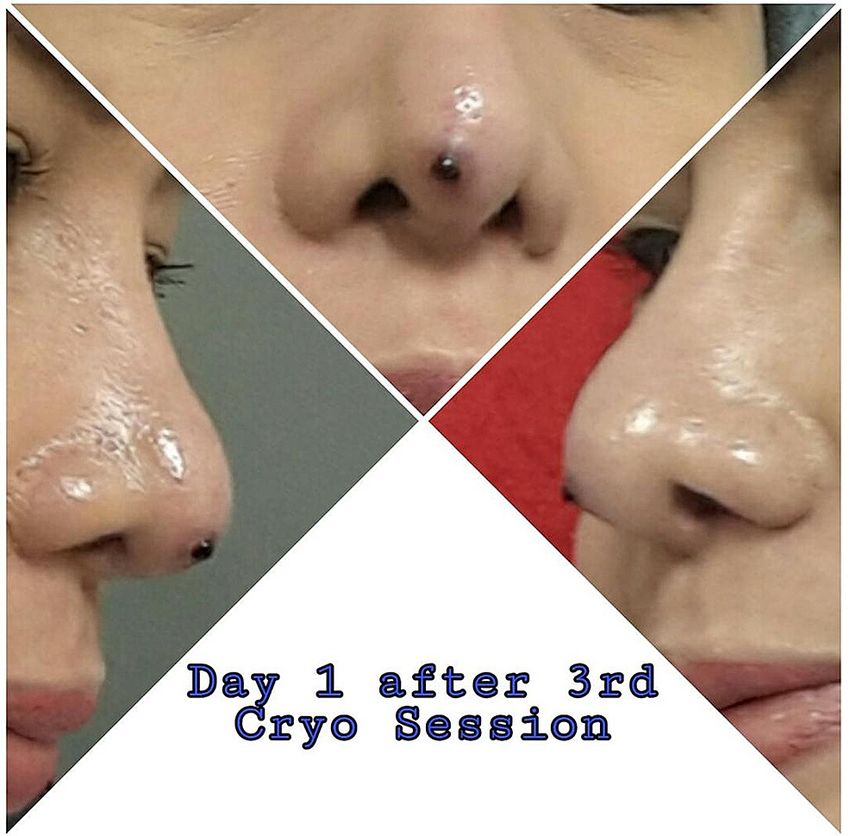
underwent three sessions of cryotherapy on three consecutive days to achieve hemostasis and possible
                                                  reduction in size in preparation for a shave excision with base cauterization (Figure 2).

                                                    FIGURE 1: First Presentation

2022 Alghamdi Jr. et al. Cureus 14(2): e21849. DOI 10.7759/cureus.21849                                                                                  2 of 7
FIGURE 2: Day 1 After the Third Cryotherapy Session

                                                  Shave excision with electrocauterization of the base of the lesion was performed, and microscopic analysis
                                                  of the lesion showed a granulation tissue-like tumor with prominent vascular spaces and endothelial cells
                                                  without cellular atypia, which was consistent with the diagnosis of a pyogenic granuloma. The lesion
                                                  appeared to heal appropriately throughout the following days but started regrowing rapidly into a round red
                                                  papule at the same site within a few weeks. The patient returned six weeks later with a cutaneous pink itchy
                                                  papule at the nasal tip (Figure 3). The diagnosis was made clinically at the second time, as the lesion
                                                  appeared quite similar to the patient's first presentation and histopathological assessment was not required.
                                                  A different line of treatment was used for management at that time, and the patient was treated with three
                                                  separate sessions of silver nitrate (AgNO3) cautery. The lesion demonstrated rapid regression in size after
                                                  each treatment session and continued to heal after the last session with no signs of recurrence for the
                                                  following three weeks (Figure 4).

2022 Alghamdi Jr. et al. Cureus 14(2): e21849. DOI 10.7759/cureus.21849                                                                                           3 of 7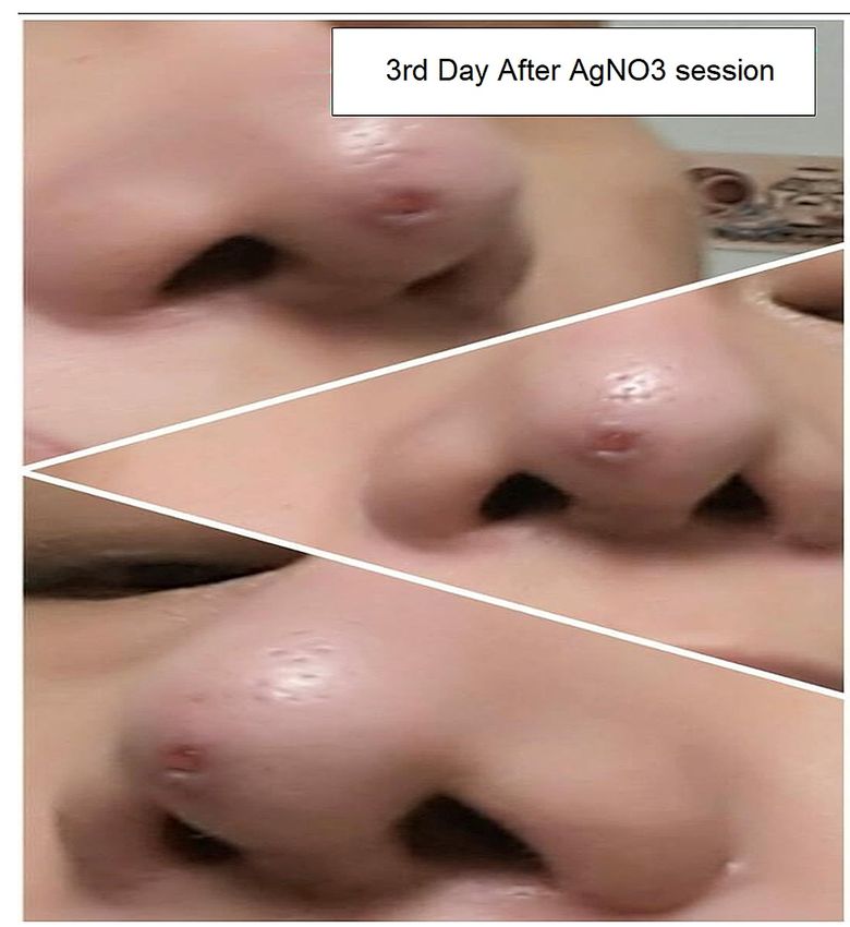
FIGURE 3: First Recurrence

2022 Alghamdi Jr. et al. Cureus 14(2): e21849. DOI 10.7759/cureus.21849          4 of 7
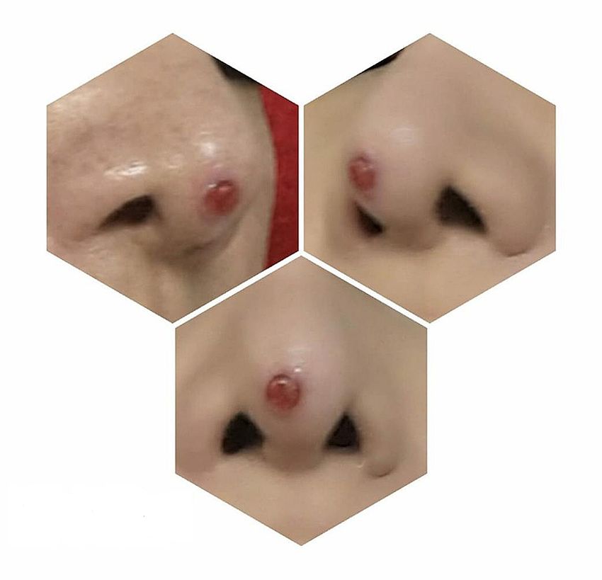
FIGURE 4: Healing After Silver Nitrate (AgNO3)

                                                  Eight months later, however, the patient contracted coronavirus disease 2019 (COVID-19) infection and
                                                  started to notice a small wound or ulcer-like lesion developing at the site of the previously treated pyogenic
                                                  granuloma. The lesion started to grow very slowly again into a papular lesion over months. The patient was
                                                  referred to a plastic surgeon for further assessment of the implant because local and systemic infections are
                                                  considered one of the factors that may lead to the development of PG. In addition, an infected Gore-Tex
                                                  implant is a rare complication of this type of implant, which necessitates implant removal. Our patient
                                                  was assessed for the viability of the implant and to rule out any active association of the procedure with the
                                                  patient’s recurrent presentations. The patient also underwent wide local excision of the lesion with primary
                                                  closure. The procedure allowed for direct visualization of the implant and the area beneath the lesion, which
                                                  showed no signs of infection. However, a deep extension of the tumor base into the subcutaneous
                                                  tissue possibly due to initial incomplete excision, which eventually contributed to our patient's disease
                                                  recurrence, was noticed. The site of the lesion has been healing since the procedure with no signs of
                                                  recurrence so far.

                                                  Discussion
                                                  Pyogenic granuloma is a benign vascular lesion of unknown etiology that affects the skin and mucous
                                                  membranes. This vascular lesion tends to grow rapidly and reaches its final size within a few weeks. It can
                                                  grow on the skin surface or other mucous membranes like the nasal cavity and usually presents as a painless
                                                  pink or red solitary papule that bleeds easily upon trauma or irritation [1]. Despite its idiopathic nature,
                                                  pyogenic granuloma has been associated most commonly with hormonal changes, physical trauma that
                                                  disrupts the skin integrity, and systemic or local infections [1]. Pregnancy and oral contraceptives by far have
                                                  been the two most common triggers for pyogenic granuloma related to hormonal changes. Trauma, on the
                                                  other hand, has also been shown to be an underlying trigger for many cases of pyogenic granuloma, where
                                                  the traumatized skin acts as a nidus for abnormal vascular proliferation. The diagnosis is usually done
                                                  clinically when patients present with lesions that demonstrate classical gross features consistent with
                                                  history [8]. Dermoscopic and microscopic features are usually assessed to confirm the diagnosis and rule out
                                                  the possibility of other dermatologic lesions that may require further treatment such as basal cell carcinoma

2022 Alghamdi Jr. et al. Cureus 14(2): e21849. DOI 10.7759/cureus.21849                                                                                              5 of 7
(BCC). Using dermoscopy, the absence of specific dermoscopic criteria for other skin tumors and a reddish
homogeneous area surrounded by a white collarette are the most frequent dermoscopic patterns in pyogenic
granuloma [9]. Histological patterns typically found in PG include an erosive or ulcerative epidermis
overlying numerous capillaries and venules with plump endothelial cells arrayed radially toward the skin
surface amidst an edematous stroma containing a mixed inflammatory infiltrate (Figure 5) [10-11]. The main
goal in the treatment of PG is to identify the most likely underlying trigger. For example, medications that
are commonly associated with hormonal changes, such as contraceptives, should be discussed with the
patient to be stopped or changed to other effective contraceptive methods. Removal of a clear provoking
traumatic event should also be done first if identified. Lesions that are likely caused by an underlying trigger
may regress or completely resolve upon cessation of the trigger. If a lesion persists despite managing
possible identified causes, or no clear triggers were identified, treatment of PG can be achieved surgically,
medically, or combined. Several factors are considered when deciding on the type of treatment such as the
location and size of the lesion and the patient’s age and preference [8]. Medical treatment is usually used for
small lesions particularly those that present on the face and other exposed body surfaces where surgical
excision is usually undesired by patients. Topical beta-blocker, imiquimod, and ingenol mebutate are all
considered effective medical treatments for both cutaneous and mucosal PG [12-14]. Other non-surgical
interventions that demonstrate comparable efficacy for small PG include cryotherapy and silver-nitrate
cautery [15-16]. The two most common surgical techniques are either shave
removal with electrocautery or surgical excision with primary closure. These two procedures are not only
associated with a lower recurrence rate (10%-%15) particularly with incompletely excised lesions, compared
to medical treatment but also provide a tissue sample for histologic confirmation [17-18]. Follow-up
appointments for successfully treated pyogenic granuloma are not routinely advised and patients can live
their lives with no concern. However, if any signs of recurrence are seen, patients should seek medical care
as soon as possible for early treatment and management.
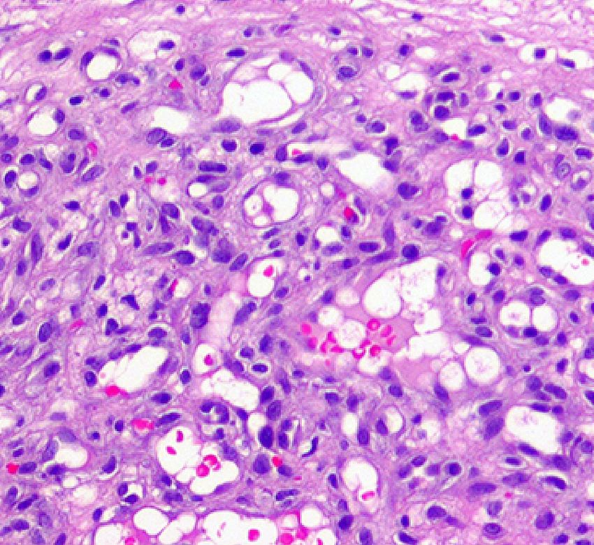
FIGURE 5: Pyogenic Granuloma Histopathology
At higher power, the lesion consists of condensed small-caliber vessels with scattered red blood cells. The stroma
is fibromyxoid with scattered inflammatory cells. Cytologic atypia and mitoses are not present.
Conclusions
Since the literature has only reported one case of PG following rhinoplasty, and the lesion was found in the
infraorbital region, this case report presents a novel case of a recurrent cutaneous pyogenic granuloma of
the nose following rhinoplasty. We found out that cutaneous PG of the nose can be a complication of this
procedure, and some factors like infections and incomplete surgical excision of these lesions can lead to
2022 Alghamdi Jr. et al. Cureus 14(2): e21849. DOI 10.7759/cureus.21849 6 of 7treatment failure or recurrence. We hope that this case draws more attention to considering PG as a
potential complication for patients undergoing rhinoplasty. In addition, we recommend conducting more
research that investigates the association between rhinoplasty and the occurrence of PG.
Additional Information
Disclosures
Human subjects: Consent was obtained or waived by all participants in this study. King Abdullah
International Research Center (KAIMRC) issued approval NRJ21/221/09. Conflicts of interest: In
compliance with the ICMJE uniform disclosure form, all authors declare the following: Payment/services
info: All authors have declared that no financial support was received from any organization for the
submitted work. Financial relationships: All authors have declared that they have no financial
relationships at present or within the previous three years with any organizations that might have an
interest in the submitted work. Other relationships: All authors have declared that there are no other
relationships or activities that could appear to have influenced the submitted work.
References
1. Sarwal P, Lapumnuaypol K: Pyogenic Granuloma. StatPearls, Treasure Island (Fl); 2021.
2. Jafarzadeh H, Sanatkhani M, Mohtasham N: Oral pyogenic granuloma: a review. J Oral Sci. 2006, 48:167-75.
10.2334/josnusd.48.167
3. Jayasree P, Kaliyadan F, Raja HM: Dermoscopy-aided diagnosis of nose ring-associated pyogenic granuloma .
Indian Dermatol Online J. 2020, 11:436-7.
4. Kumar Ghosh S, Bandyopadhyay D: Granuloma pyogenicum as a complication of decorative nose piercing:
report of eight cases from eastern India. J Cutan Med Surg. 2012, 16:197-200. 10.1177/120347541201600312
5. Kaiser B, Chijioke O, Bruder E, Takes M, Haug M, Mende K: Infraorbital subcutaneous intravascular lobular
capillary hemangioma after rhinoplasty. Ann Plast Surg. 2021, 87:239-41. 10.1097/SAP.0000000000002954
6. Buck D II: Review of Plastic Surgery . Elsevier Inc., Philadelphia, PA; 2021.
https://www.us.elsevierhealth.com/review-of-plastic-surgery-9780323775939.html.
7. Hettiaratchy S, Griffiths M, Ali F, Simmons J: Plastic Surgery. A Problem Based Approach . Springer Nature,
Switzerland; 2011. 10.1007/978-1-84882-116-3
8. Mills SE, Cooper PH, Fechner RE: Lobular capillary hemangioma: the underlying lesion of pyogenic
granuloma. A study of 73 cases from the oral and nasal mucous membranes. Am J Surg Pathol. 1980, 4:470-
9.
9. Zaballos P, Carulla M, Ozdemir F, et al.: Dermoscopy of pyogenic granuloma: a morphological study . Br J
Dermatol. 2010, 163:1229-37. 10.1111/j.1365-2133.2010.10040.x
10. Pyogenic granuloma. (2018 ). Accessed: 2/1/2022: https://ykhoa.org/d/image.htm.
11. Ardigo M, Agozzino M, Franceschini C, Lacarrubba F: Reflectance confocal microscopy algorithms for
inflammatory and hair diseases. Dermatol Clin. 2016, 34:487-96. 10.1016/j.det.2016.05.011
12. Khorsand K, Maier M, Brandling-Bennett HA: Pyogenic granuloma in a 5-month-old treated with topical
timolol. Pediatr Dermatol. 2015, 32:150-1. 10.1111/pde.12297
13. Tritton SM, Smith S, Wong LC, Zagarella S, Fischer G: Pyogenic granuloma in ten children treated with
topical imiquimod. Pediatr Dermatol. 2009, 26:269-72. 10.1111/j.1525-1470.2008.00864.x
14. Jung HJ, Eun DH, Kim JY, Lee WJ, Kim DW, Lee SJ, Jang YH: Treatment of paediatric facial pyogenic
granuloma with topical ingenol mebutate. Clin Exp Dermatol. 2017, 42:912-3. 10.1111/ced.13198
15. Mirshams M, Daneshpazhooh M, Mirshekari A, Taheri A, Mansoori P, Hekmat S: Cryotherapy in the
treatment of pyogenic granuloma. J Eur Acad Dermatol Venereol. 2006, 20:788-90. 10.1111/j.1468-
3083.2006.01615.x
16. Quitkin HM, Rosenwasser MP, Strauch RJ: The efficacy of silver nitrate cauterization for pyogenic
granuloma of the hand. J Hand Surg Am. 2003, 28:435-8. 10.1053/jhsu.2003.50092
17. Lee J, Sinno H, Tahiri Y, Gilardino MS: Treatment options for cutaneous pyogenic granulomas: a review . J
Plast Reconstr Aesthet Surg. 2011, 64:1216-20. 10.1016/j.bjps.2010.12.021
18. Folpe AL: Vascular tumors of soft tissue . Bone and Soft Tissue Pathology. Folpe AL, Inwards CY (ed):
Elsevier, Amsterdam, Netherlands; 2010. 164-92. 10.1016/B978-0-443-06688-7.00010-9
2022 Alghamdi Jr. et al. Cureus 14(2): e21849. DOI 10.7759/cureus.21849 7 of 7You can also read