Mucormycosis in Post Covid Patient - A Case Report - Open ...
←
→
Page content transcription
If your browser does not render page correctly, please read the page content below
240   Indian Journal of Forensic Medicine & Toxicology, July-September 2021, Vol. 15, No. 3

      Type of manuscript : Case Report

           Mucormycosis in Post Covid Patient - A Case Report

                            Kush Pathak1, Asha Karadwal2, Prachi Nayak3, Sushruth Nayak4
  1Associate Professor, 2Reader, 3Professor, 4Professor & Head, Department of Oral Pathology & Microbiology,
           M.M.College of Dental Science & Research, M.M(Deemed to be) University, Mullana (Ambala)

                                                                  Abstract
      Mucormycosis is considered to be a rare notorious fungal infection mainly affecting diabetics with or without
      ketoacidosis & immunocompromised patients, with high mortality rate. Occurrence of Mucormycosis as an
      implication of Covid-19 has not yet been reported. We present a case of aggressive mucormycosis in a Post
      covid-19 type-2 diabetic patient and aim to illustrate the possible Covid-19 effects on body and a dire need
      for early diagnosis.

      Keywords - Mucormycosis, Covid-19, ACE2 receptors, Type 2 Diabetes Mellitus, Iron overload

                         Introduction                                           Even though maxilla is richly vascular which
                                                                           rarely lets it getting necrosed, bacterial infections like
    Mucormycosis is a rare angioinvasive, acute
                                                                           osteomyelitis, viral and even fungal infections like
opportunistic, progressive condition caused by
                                                                           aspergillosis, mucormycosisetc can still affect/cause
saprophytic fungus. This lethal form of fungal infection,
                                                                           (necrosis) it.2,3
involves nose & paranasal sinuses. It is most commonly
seen affecting immunocompromised patients who are                               Despite of this mucormycosis on palate is considered
on steroidal or cytotoxic therapy and is life threatening                  a rare and late possibility.4
to patients with uncontrolled diabetes. Underlying
malignancy, renal failure, malnutrition can act as                              Smoking has previously been linked to
predisposing factors.                                                      mucormycosis occurring in mandible as an initiator to
                                                                           the infection.5 But relationship between COVID-19 and
    On the other hand Covid-19, which is caused                            mucormycosis has not been thoroughly explored to date.
by Severe acute Respiratory syndrome corona virus
2(SARS-COV-2) turned out to be highly contagious                                The present case report is of mucormycosis
infection with an increased mortality and economic                         presenting as a chronic ulcer on palate in a COVID-19
morbidity worldwide. It has primary symptoms of fever,                     recovered diabetic patient. The aim of this report is to
cough & fatigue leading to pneumonia and even multi                        alert the clinicians to be aware of mucormycosis as one
organ failure.1                                                            of the common side effect of COVID-19 infection.

                                                                                                Case Report

Corresponding Author:                                                           A 65-year -old male diabetic patient was presented
Dr. Kush Pathak, MDS,                                                      to the outpatient department with chief complaint of an
Associate Professor, Department of Oral Pathology                          ulcerative growth in hard palate from past 20-25 days
& Microbiology, M.M.College of Dental Science &                            with no pain. Patient also complained of headache and
Research, M.M(Deemed to be) University,                                    vomiting from last 4 days.
Mullana (Ambala)
                                                                              Past medical history revealed that the patient was a
Email: dr.kushpathak2011@gmail.com
                                                                           known case of hypertension, type II diabetes mellitus and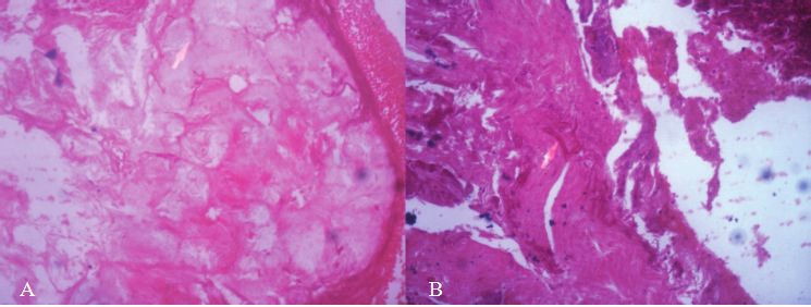
Indian Journal of Forensic Medicine & Toxicology, July-September 2021, Vol. 15, No. 3 241
was on medication. On general physical examination, of fungal infection and squamous cell carcinoma was
the patient was found to be moderately built and poorly given.
nourished with abnormal gait. His Random blood sugar
The lesion was excised. Soft tissue specimen
(RBS) was reported to be 320 mg/dl. His blood pressure
obtained was brown in color, measuring around 2 x 1.5
was 190/90 mm of Hg, pulse rate was 96 beats/min.
cm in diameter, soft to firm in consistency with irregular
Patient was also reported to be a survivor of Covid-19,
surface. After subjecting to routine processing, H & E
2 months back.
stained tissue under microscopic examination revealed
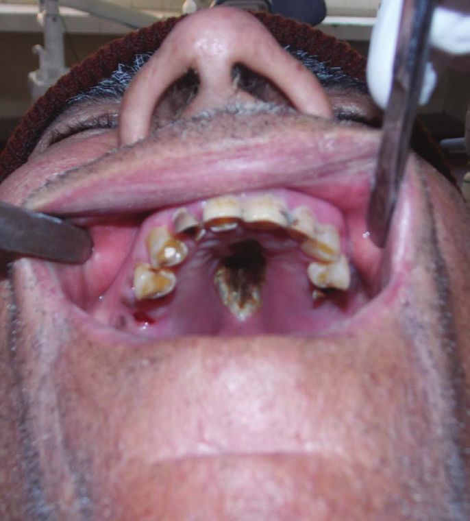
On Intraoral examination, a necrotic ulcerative the presence of numerous large, branching (at 90 degree)
lesion was noticed in the centre of the hard palate, of non septate hyphae in the background of hyalinzed
measuring approximately 3x2cm in diameter. It was connective tissue stroma. Few area showed the presence
covered in blackish gray slough with everted borders, of neural bundles. Dense chronic inflammatory cell
had a smooth surface and showed no sign of bleeding on infiltrate mainly composed of lymphocytes and plasma
inspection (Image 1). There was no pain and the ulcer cells were present at the periphery which confirmed the
was attached to the underlying bone. It was extending diagnosis of mucormycosis. (Image 2)
from palatal rougae to posterior aspect of hard palate.
After the procedure, patient was provided with an
No lymph nodes were palpable. A provisional diagnosis
obturator for the oro-nasal fistula and is being followed
of osteomyelitis was made. The differential diagnosis
up.
Image 1: Ulcerative necrosed lesion242 Indian Journal of Forensic Medicine & Toxicology, July-September 2021, Vol. 15, No. 3
Image 2 : H & E stained section showing large, non septate fungal hyphae branched at 90 degree (A)10x (B)
20x
Discussion Diabetic patients have impaired defense mechanism
Alternately coined as zygormycosis, this rare but along with increased level of iron in tissues.12,13 This can
rather notorious mucormycosis is well known for being be co-related with our present case where the patient was
fatal, especially in immunocompromised or diabetic diabetic & poorly nourished.
patients. It is an aerobic fungi of saprophytic origin Swaminathan et al in 2007 suggested that iron plays
commonly found in bread molds or decaying vegetation. a pathogenic role in diabetes and its complications such
They most commonly colonize the oral, nasal, pharyngeal as microangiopathy and atherosclerosis.14Aregbesola A.
mucosa and paranasal sinuses.6 et al in 2016 explained the excessive iron stored in body
Predisposing factors playing a role are leukemia, can play an important role in causing type 2 diabetes.
diabetes(patients with or without ketoacidosis), organ They also concluded that the strength and direction of the
transplant, AIDS & renal failure7,prolonged and severe association between body iron and glucose metabolism
neutropenia, iron overload, major trauma, prolonged is strongest among subjects in prediabetes state.15A high
use of corticosteroids, illicit intravenous drug use, 90 day mortality range of 20-58%, makes mucormycosis
neonatal prematurity and malnourishment can also lead a life threatening infection. Less traditionally it has also
to mucormycosis.8 been found to be associated with critical illness, major
surgery and pneumonia. 16
Occurrence of infections and even death due to
invasive fungal infections, is found to be greater in On the flip side Covid-19, caused by severe acute
patients who have iron overload. In fact decreased respiratory syndrome coronavirus 2 (SARS-CoV-2)
posttransplantation survival is directly related to hepatic which appeared for the first time in Wuhan, China,
iron overload.9 Researchers have found iron to be as came out to be a highly contagious newly recognized
important a nutrient to fungi as humans. Growth of infection. With human to human transmission it had a
wide range of fungi in body fluids, cells and tissues is worldwide impact. Initial symptoms of fever, cough and
stimulated by excess iron.10 fatigue that quickly progress to severe respiratory illness
& pneumonia were noted. 17
It was also found that iron free environment is
essential for proper innate and acquired immune Several patients were observed with atypical
response. Any excess of iron (iron overload) would lead manifestations such as conjunctivitis, asymptomatic
to direct damage to our natural defense system and an infections at the onset of the illness and even sudden
increase in fungal virulence.11 irreversible hearing loss post Covid-19 recovery.18,19Indian Journal of Forensic Medicine & Toxicology, July-September 2021, Vol. 15, No. 3 243
A sudden spike in reports of incidence of literature. So, we assume ours as one of the first reported
mucormycosis in Covid-19 patients throughout cases. With widespread presence of the virus, dangerous
India raised an alarm. Experts warned that Covid-19 effects of mucormycosis, and growing dental problems
patients were the most vulnerable ones facing the risk in covid-19 recovered cases, it is pertinent to investigate
of contracting the infection. Despite very low number this further. Given the mortality rate of both Covid-19
of reported cases, it is quite significant to consider the and mucormycosis we must pay heed to diagnose this
possibility of a relationship between Covid-19 and fungal infection at initial stage. It is suggested that
mucormycosis. Post Covid-19 patients are enquired about having any
dental problems and any patient with symptoms must be
In a recent study it was discovered that the SARS-
treated on urgent basis. As being noticed, Covid-19 is
CoV-2 virus damages the endothelial cells in lungs,
turning out to be a thousand piece puzzle, and now we
heart, kidneys, liver and intestine of Covid-19 patients,
as dentists need to be vigilant more than ever to identify
thus damaging the blood vessels. These endothelial
the pieces of this puzzle.
cells have an influence upon immune response. It was
concluded that may be Covid-19 is a respiratory illness Conflict of Interest - None
only to start with, but is actually a vascular illness that
Ethical Clearance - Since it was a case report, no
kills people through it’s involvement of the vasculature.20
ethical clearance was needed. But informed consent was
Teeth have an intimate connection with the rest of taken from the patient.
body. It has been suspected that the sudden spike in the
Source of Funding - Self
cases of dental deterioration in Covid-19 patients could
ultimately be related to problem with blood flow, which References
can form clots. Oral cavity gets vandalized when it’s
1. Vallamkondu J, John A, Wani WY, et al. SARS-
devoid of blood flow. Gums are extremely vascularized
CoV-2 pathophysiology and assessment of
and pulp of teeth consists of blood vessels with nerves.
coronaviruses in CNS diseases with a focus on
The reported self exfoliation of teeth in the Post Covid-19
therapeutic targets. BiochimBiophysActaMol
recovered case with no bleeding suggested that blood Basis Dis 2020;1866:165889
flow was obstructed. This can be due to vascular damage
2. Manjunatha BS, Das N, Sutariya RV, Ahmed T.
caused by Covid-19 on the body which persists even Mucormycosis of the hard palate masquerading as
after the patient has recovered.21 carcinoma. ClinPract. 2012 Feb 15;2(1):e28. doi:
10.4081/cp.2012.e28. PMID: 24765427; PMCID:
It was also found that Angiotensin converting
PMC3981330.
enzyme 2 (ACE2) receptors which are richly present
3. Auluck A. Maxillary necrosis by mucormycosis. A
in lungs are also found in abundance on the epithelial
case report and literature review. Med Oral Patol
cells of oral mucosa, not only facilitate the virus entry
Oral Cir Bucal 2007;12:E360-4.
but even affects the pathophysiological process of virus
4. Kyrmizakis DE, Doxas PG, Hajiioannou
induced acute lung injury (ALI), as well as other organ
JK, Papadakis CE. Palate ulcer due to
damage.22,23 mucormycosis. The Journal of Laryngology
and Otology. 2002 Feb;116(2):146-147. DOI:
It is thus suggested that apparently there is a
10.1258/0022215021909917.
biological pathway by which Covid-19 virus can directly
5. Paul Lee Salisbury, Ron Caloss, Julia M Cruz, Bayard
affect mouth. The presence of ACE2 receptors in oral
L Powell, Roger Cole, Robert I Kohut,Mucormycosis
cavity can provide a good habitat for Covid-19 virus to
of the mandible after dental extractions in a
encamp and replicate.
patient with acute myelogenousleukemia.Oral
Surgery, Oral Medicine, Oral Pathology, Oral
Conclusion
Radiology, andEndodontology,Volume 83, Issue
Till date, very few cases have been found in the 3,1997,340-344,https://doi.org/10.1016/S1079-244 Indian Journal of Forensic Medicine & Toxicology, July-September 2021, Vol. 15, No. 3
2104(97)90240-7.) 10.1177/0004563216646397. Epub 2016 Sep 28.
6. Bhansali A, Bhadada S, Sharma A, et al. PMID: 27166309.
Presentation and outcome of rhinoorbital-cerebral 16. Zilberberg, M. D., Shorr, A. F., Huang, H.,
mucormycosis in patients with diabetes. Postgrad Chaudhari, P., Paly, V. F., &Menzin, J. (2014).
Med J 2004;80: 670-4. Hospital days, hospitalization costs, and inpatient
7. McNulty JS. Rhinocerebralmucormycosis: mortality among patients with mucormycosis: a
predisposing factors. Laryngoscope 1982;92: retrospective analysis of US hospital discharge
1140-3. data. BMC infectious diseases, 14, 310. https://doi.
org/10.1186/1471-2334-14-310
8. Petrikkos G, Skiada A, Lortholary O, Roilides E,
Walsh T, Kontoyiannis DP. Oral epidemiology 17. J. Guan, Z.Y. Ni, Y. Hu, W.H. Liang, C.Q. Ou,
and clinical manifestations of mucormycosis. Clin J.X. He, et al.Clinical characteristics of coronavirus
Infect Dis. 2012;54:S23–34. disease 2019 in China. N Engl J Med (2020).
9. Brandhagen DJ, Alvarez W, Therneau 18. Ozturker ZK. Conjunctivitis as sole symptom of
TM, Kruckeberg KE, Thibodeau SN, Ludwig J, et COVID-19: A case report and review of literature.
al. Iron overload in cirrhosis‐HFE genotypes and Eur J Ophthalmol. 2020 Jul 24:1120672120946287)
outcome after liver transplantation. Hepatology 19. Koumpa FS, Forde CT, Manjaly JG. Sudden
2000; 31: 456–460. irreversible hearing loss post COVID-19. BMJ
10. Schaible UE, Kaufmann SH. Iron and microbial Case Rep 2020;13:e238419. doi:10.1136/bcr-
infection. Nat Rev Microbiol 2004; 2: 946–953. 2020-238419
11. Weinberg ED. Iron loading and disease 20. Varga Z. et al. Endothelial cell infection and
surveillance. Emerg Infect Dis 1999; 5: 346–352. endothelitis in Covid-19. Correspondence. 2020;
395:10234, P1417-1418.
12. Tugsel Z, Sezer B, Akalin T. Facial swelling and
palatal ulceration in a diabetic patient. Oral Surg Oral 21. Ries Julia. “Can covid 19 damage your
Med Oral Pathol Oral RadiolEndod. 2004;98:630– teeth and mouth? Here’s what you should
636. know.” Huffpost, 16 November 2020. www.
huffpost.com/entry/covid-19-damage-teeth-
13. Kontoyiannis P. Dimitrios. “Mucormycosis”.
mouth_l_5fb1d951c5b6d05e86e85b0a
National Organization of Rare diseases (NORD),
2018. https://rarediseases.org/rare-diseases/ 22. Liu M, Wang T, Zhou Y, Zhao Y, Zhang Y, Li J.
mucormycosis/ Potential Role of ACE2 in Coronavirus Disease
2019 (COVID-19) Prevention and Management.
14. Sundararaman Swaminathan, Vivian A. Fonseca,
J TranslInt Med. 2020 May 9;8(1):9-19. doi:
Muhammad G. Alam, Sudhir V. Shah. The Role
10.2478/jtim-2020-0003. PMID: 32435607;
of Iron in Diabetes and Its Complications. Diabetes
PMCID: PMC7227161.
Care Jul 2007, 30 (7) 1926-1933; DOI: 10.2337/
dc06-2625 23. Xu H, Zhong L, Deng J, et al. High expression of
ACE2 receptor of 2019‐nCoV on the epithelial
15. Aregbesola A, Voutilainen S, Virtanen JK,
cells of oral mucosa. Int J Oral Sci. 2020;12(1):8.
Mursu J, Tuomainen TP. Gender difference in
type 2 diabetes and the role of body iron stores.
Ann ClinBiochem. 2017 Jan;54(1):113-120. doi:You can also read