TRANSLATIONAL MEDICINE @ UNISA
←
→
Page content transcription
If your browser does not render page correctly, please read the page content below
Translational Medicine @ UniSa

Volume 23       Issue 4                                                                                   Article 13

October 2020

A case series of blastic plasmacytoid dendritic cell neoplasia
Valentina Giudice
Hematology and Transplant Center, University Hospital “San Giovanni di Dio e Ruggi d’Aragona”, Italy,
vgiudice@unisa.it

Follow this and additional works at: https://tmj.unisa.it/journal

    Part of the Hematology Commons

Recommended Citation
Giudice, Valentina (2020) "A case series of blastic plasmacytoid dendritic cell neoplasia," Translational
Medicine @ UniSa: Vol. 23 : Iss. 4 , Article 13.
Available at: https://doi.org/10.37825/2239-9747.1012

This Article is brought to you for free and open access by Translational Medicine @ UniSa. It has been accepted for
inclusion in Translational Medicine @ UniSa by an authorized editor of Translational Medicine @ UniSa.
A case series of blastic plasmacytoid dendritic cell neoplasia
Serio B1, Giudice V1,2,3, D’Addona M1, Guariglia R1, Gorrese M1, Bertolini A3, D’Alto F1,
Cuffa B1, Pellegrino D1, Langella M1, Selleri C1,3
1
Hematology and Transplant Center, University Hospital “San Giovanni di Dio e Ruggi D’Aragona”, Italy.
2
Clinical Pharmacology, University Hospital “San Giovanni di Dio e Ruggi D’Aragona”, Italy.
3
Department of Medicine, Surgery and Dentistry "Scuola Medica Salernitana", University of Salerno, Italy.
(email vgiudice@unisa.it)
Abstract - Blastic plasmacytoid dendritic cell also frequently reported [7-11]. To date, there are no
neoplasm (BPDCN), an extremely rare and aggressive guidelines for treatment of advanced BPDCN; however,
tumor, derives from plasmacytoid dendritic cell conventional chemotherapy used for myeloid or lymphoid
malignancies are employed with various efficacy [12-13].
precursors and is characterized by CD4 and CD56 Recently, a cytotoxin targeting CD123-expressing cells,
positivity accompanied by the expression of isolated tagraxofusp-erzs, has been approved for BPDCN with an
myeloid, B- or T-cell lineage markers. Despite the overall response rate [ORR] of 90% as frontline therapy or
recent introduction of specific targeted therapies, 67% in relapsed/refractory patients [14-15]. However,
prognosis is still poor with a median overall survival available studies show that overall survival (OS) is similar
of one year, and allogeneic bone marrow between patients receiving conventional chemotherapy and
those treated with tagraxofusp-erzs [4]. High dose
transplantation remains the only curative treatment in
cytarabine and methotrexate (CVAD) and hematopoietic
eligible patients. In this series, we described two cases stem cell transplantation (HSCT) remain the most effective
of adult BPDCN treated with high dose cytarabine and therapies for BPDCN treatment in eligible subjects [4].
methotrexate and autologous hematopoietic stem cell With the advances in diagnosis and the appliance of novel
transplantation, or fludarabine, cytarabine, and targeted regimen, the outcomes for patients with BPDCN
idarubicin achieving the first a complete lasting may be improved in the future.
remission, while the second only a transient In this series, we presented two cases of BPDCN
treated at the Hematology and Transplant Center,
improvement in skin lesions. University Hospital “San Giovanni di Dio e Ruggi
d’Aragona” of Salerno after informed consent obtained in
Keywords: blastic plasmacytoid dendritic cell neoplasm, accordance with the Declaration of Helsinki [16]. The
flow cytometry, chemotherapy, transplant. authors retrospectively reviewed all available medical
records.
I. INTRODUCTION
II. CASE 1 PRESENTATION
Blastic plasmacytoid dendritic cell neoplasm
(BPDCN), a rare and aggressive hematological disease, has Skin biopsy and immunophenotyping were
been recognized as a distinct clinical entity in 2016 revision performed in a 48-year-old female presented with
of the World Health Organization (WHO) classification of progressive erythematous lesions of lower limbs not
myeloid neoplasms and acute leukemia [1]. BPDCN mostly improving after a two-year treatment with steroids and
affects older males (mean age, 70 years) and is colchicine, and a medical history of vitiligo and pityriasis
characterized by skin lesions with or without bone marrow versicolor. She received a diagnosis of BPDCN in April
(BM) involvement and leukemic cell dissemination in 2014, and leukemic cells were positive for CD4, CD2,
secondary lymphoid organs [1-6]. In addition, 10-20% of CD38, CD56, CD123, and Ki67. No extracutaneous or BM
patients have a clinical history of other hematological involvements were found after PET-CT scanning and
malignancies, such as myelodysplastic syndrome (MDS) or marrow biopsy. She started chemotherapy according to the
chronic myeloid leukemia [1]. BPDCN cells are of hyper-CVAD scheme: cycle A with cyclophosphamide,
plasmacytoid dendritic cell origins and typically express doxorubicin, and vincristine; and four cycles B with
CD4, CD123 (interleukin-3 a receptor), HLA-DR, cTCL1 methotrexate and subcutaneous cytarabine (Ara-C). Skin
(cytoplasmic T-cell leukemia/lymphoma 1) at high levels, lesions gradually disappeared right after chemotherapy,
CD56, CD304, CD303, CD36, CD38, and CD45RA [7-8]. and she was candidate to autologous HSCT. Peripheral
Chromosomal abnormalities, such as complex karyotype blood stem cells (PBSCs) were collected at the end of the
with deletions on chromosomes 5q21 or 5q34, and somatic third cycle B, and conditioning regimen with busulfan 2
mutations in genes like CDKN1B/2A, TET2 or TP53 are mg/m2 and melphalan 130 mg/m2 was started after sevenmonths from diagnosis. Post-transplant complications were IV. DISCUSSION
persistent diarrhea, herpes zoster reactivation, severe
peripheral neuropathy, and recurrent bronchitis, all BPDCN is a rare disease that behaves like high-
successfully treated. At the time of writing, after six years risk acute leukemia with poor prognosis because no
from HSCT, the patient is alive without signs and specific therapies or consensus on frontline treatment are
symptoms of BPDCN. present, and because BPDCN frequently shows
chemoresistance. In young patients, intensive induction
III. CASE 2 PRESENTATION regimens followed by consolidation and HSCT are
considered the most effective treatment strategy, leading to
A 55-year-old man received a diagnosis of lasting responses, while treatment of elderly patients is still
refractory anemia based on 2016 World Health challenging.
Organization criteria with an International Prognostic In our experience, we presented two adult BPDCN
Scoring System (IPSS) of 0 in January 2017 [1]. Peripheral cases treated with hyper-CVAD and autologous HSCT or a
blood and BM flow cytometry immunophenotype showed FLAG-IDA protocol. The first patient who quickly
the presence of aberrant CD14+CD33+CD56+ monocytes. received HSCT achieved a long-lasting complete
The patient was treated with supportive therapy (epoetin remission; however, she had several transplant-related
alfa 40.000 IU/weekly) until April 2020 when rapid complications. The second case was treated with a reduced
progressive erythematous skin lesions appeared. Skin intensity FLAG-IDA because of low non-optimal
biopsy highlighted an infiltration of CD56+CD4+CD3+/- performance status and progression of previously
pathological cells with focal expression of MUM1 posing diagnosed MDS. This patient experienced a rapid but
for a diagnosis of BPDCN. No extracutaneous transient improvement in skin lesion; however, he died
involvements were found after PET-CT scanning, while after one month because of disease progression. Therefore,
BM biopsy confirmed the diagnosis of MDS with trisomy our results confirmed that hyper-CVAD treatment followed
8. Because of low Eastern Cooperative Oncology Group by HSCT could be the most effective therapeutic strategy
Performance Status (ECOG PS = 1), the patient started a for long-lasting remission in BPDCN patients as previously
reduced intensity FLAG-IDA protocol (fludarabine, Ara-C, reported [4]. In particular, allogeneic HSCT recipients have
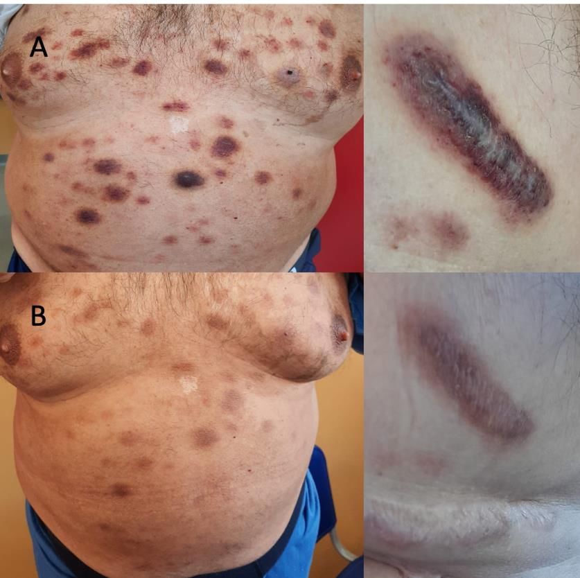
and idarubicin), and skin lesions immediately reduced in a 10-year OS of 40% with better outcomes when transplant
number and size (Figure 1). However, blood counts and is performed at the first remission [17-18]. In non-eligible
clinical conditions rapidly worsened after one month, and patients, autologous HSCT can be an effective alternative,
he died because of disease progression with neurological as shown in our case report. Tagraxofusp, bortezomib,
and hepatic involvement and severe pancytopenia. azacytidine or venetoclax could also be a valid therapeutic
option as preliminary results show significant
improvements in ORR [14,19-24]. However, further
studies are required to define the best strategy for treatment
of BPDCN and to identify candidate biomarkers of
responsiveness to therapies.
ACKNOWLEDGMENT
This research was supported by the Intramural
Program of the Department of Medicine, Surgery and
Dentistry, University of Salerno, Italy.
REFERENCES
[1] Arber DA, Orazi A, Hasserjian R, Thiele J, Borowitz
MJ, Le Beau MM, Bloomfield CD, Cazzola M, Vardiman
JW. The 2016 revision to the World Health Organization
classification of myeloid neoplasms and acute leukemia.
Blood. 2016;127(20):2391-2405.
[2] Sapienza MR, Pileri A, Derenzini E, Melle F, Motta G,
Fiori S, Calleri A, Pimpinelli N, Tabanelli V, Pileri S.
Fig. 1. Clinical presentation of Case 2. Blastic Plasmacytoid Dendritic Cell Neoplasm: State of the
(A) The patient presented with erythematous infiltrated skin lesions all Art and Prospects. Cancers (Basel). 2019;11(5):595.
over the body that were disappearing (B) just after ten days of treatment. [3] Garnache-Ottou F, Vidal C, Biichlé S, Renosi F, Poret
E, Pagadoy M, et al. How should we diagnose and treat
blastic plasmacytoid dendritic cell neoplasm patients?
Blood Adv. 2019;3(24):4238-4251.[4] Sullivan JM, Rizzieri DA. Treatment of blastic leukemic presentation: an Italian multicenter study. plasmacytoid dendritic cell neoplasm. Hematology Am Soc Haematologica. 2013;98(2):239-246. Hematol Educ Program. 2016;2016(1):16-23. [14] Pemmaraju N, Lane AA, Sweet KL, Stein AS, Vasu S, [5] Yun S, Chan O, Kerr D, Vincelette ND, Idrees A, Mo Blum W, Rizzieri DA, Wang ES, Duvic M, Sloan JM, Q, Sweet K, Lancet JE, Kharfan-Dabaja MA, Zhang L, Spence S, Shemesh S, Brooks CL, Balser J, Bergstein I, Sokol L. Survival outcomes in blastic plasmacytoid Lancet JE, Kantarjian HM, Konopleva M. Tagraxofusp in dendritic cell neoplasm by first-line treatment and stem cell Blastic Plasmacytoid Dendritic-Cell Neoplasm. N Engl J transplant. Blood Adv. 2020;4(14):3435-3442. Med. 2019;380(17):1628-1637. [6] Julia F, Petrella T, Beylot-Barry M, Bagot M, Lipsker [15] Pemmaraju N, Konopleva M. Approval of D, Machet L, Joly P, Dereure O, Wetterwald M, d'Incan M, tagraxofusp-erzs for blastic plasmacytoid dendritic cell Grange F, Cornillon J, Tertian G, Maubec E, Saiag P, neoplasm. Blood Adv. 2020;4(16):4020-4027. Barete S, Templier I, Aubin F, Dalle S. Blastic [16] World Medical Association General Assembly. World plasmacytoid dendritic cell neoplasm: clinical features in Medical Association Declaration of Helsinki: ethical 90 patients. Br J Dermatol. 2013;169(3):579-586. principles for medical research involving human subjects. [7] Riaz W, Zhang L, Horna P, Sokol L. Blastic J Int Bioethique. 2004;15(1):124-129. plasmacytoid dendritic cell neoplasm: update on molecular [17] Kharfan-Dabaja MA, Lazarus HM, Nishihori T, biology, diagnosis, and therapy. Cancer Control. Mahfouz RA, Hamadani M. Diagnostic and therapeutic 2014;21(4):279-289. advances in blastic plasmacytoid dendritic cell neoplasm: a [8] Julia F, Dalle S, Duru G, Balme B, Vergier B, Ortonne focus on hematopoietic cell transplantation. Biol Blood N, Vignon-Pennamen MD, Costes-Martineau V, Lamant L, Marrow Transplant. 2013;19(7):1006-1012. Dalac S, Delattre C, Déchelotte P, Courville P, Carlotti A, [18] Laribi K, Baugier de Materre A, Sobh M, Cerroni L, De Muret A, Fraitag S, Levy A, Mitchell A, Petrella T. Valentini CG, Aoki T, Suzuki R, Takeuchi K, Frankel AE, Blastic plasmacytoid dendritic cell neoplasms: clinico- Cota C, Ghez D, Le Calloch R, Pagano L, Petrella T. Blastic immunohistochemical correlations in a series of 91 plasmacytoid dendritic cell neoplasms: results of an patients. Am J Surg Pathol. 2014;38(5):673-680. international survey on 398 adult patients. Blood Adv. [9] Dijkman R, van Doorn R, Szuhai K, Willemze R, 2020;4(19):4838-4848. Vermeer MH, Tensen CP. Gene-expression profiling and [19] Roos-Weil D, Dietrich S, Boumendil A, Polge E, Bron array-based CGH classify CD4+CD56+ hematodermic D, Carreras E, Iriondo Atienza A, Arcese W, Beelen DW, neoplasm and cutaneous myelomonocytic leukemia as Cornelissen JJ, Kröger N, Milone G, Rossi G, Jardin F, distinct disease entities. Blood. 2007;109(4):1720-1727. Peters C, Rocha V, Sureda A, Mohty M, Dreger P; [10] Wiesner T, Obenauf AC, Cota C, Fried I, Speicher European Group for Blood and Marrow Transplantation MR, Cerroni L. Alterations of the cell-cycle inhibitors Lymphoma, Pediatric Diseases, and Acute Leukemia p27(KIP1) and p16(INK4a) are frequent in blastic Working Parties. Stem cell transplantation can provide plasmacytoid dendritic cell neoplasms. J Invest Dermatol. durable disease control in blastic plasmacytoid dendritic 2010;130(4):1152-1157. cell neoplasm: a retrospective study from the European [11] Sapienza MR, Fuligni F, Agostinelli C, Tripodo C, Group for Blood and Marrow Transplantation. Blood. Righi S, Laginestra MA, Pileri A Jr, Mancini M, Rossi M, 2013;121(3):440-6. Ricci F, Gazzola A, Melle F, Mannu C, Ulbar F, Arpinati [20] Wang S, Wang X, Liu M, Bai O. Blastic plasmacytoid M, Paulli M, Maeda T, Gibellini D, Pagano L, Pimpinelli dendritic cell neoplasm: update on therapy especially novel N, Santucci M, Cerroni L, Croce CM, Facchetti F, agents. Ann Hematol. 2018;97(4):563-572. Piccaluga PP, Pileri SA; AIRC 5xMille consortium [21] Montero J, Stephansky J, Cai T, Griffin GK, Cabal- ‘Genetics-driven targeted management of lymphoid Hierro L, Togami K, Hogdal LJ, Galinsky I, Morgan EA, malignancies and the Italian Registry on Blastic Aster JC, Davids MS, LeBoeuf NR, Stone RM, Konopleva Plasmacytoid Dendritic Cell Neoplasm. Molecular M, Pemmaraju N, Letai A, Lane AA. Blastic Plasmacytoid profiling of blastic plasmacytoid dendritic cell neoplasm Dendritic Cell Neoplasm Is Dependent on BCL2 and reveals a unique pattern and suggests selective sensitivity Sensitive to Venetoclax. Cancer Discov. 2017;7(2):156- to NF-kB pathway inhibition. Leukemia. 2014;28(8):1606- 164. 1616. [22] Yang C, Fu C, Feng Y, Zhao S, Weng H, Zhang L, Du [12] Pagano L, Valentini CG, Grammatico S, Pulsoni A. H, Zou L. Clinical efficacy of bortezomib and lenalidomide Blastic plasmacytoid dendritic cell neoplasm: diagnostic in blastic plasmacytoid dendritic cell neoplasm. Ann criteria and therapeutical approaches. Br J Haematol. Hematol. 2019;98(6):1525-1527. 2016;174(2):188-202. [23] Khwaja R, Daly A, Wong M, Mahé E, Cerquozzi S, [13] Pagano L, Valentini CG, Pulsoni A, Fisogni S, Owen C. Azacitidine in the treatment of blastic Carluccio P, Mannelli F, Lunghi M, Pica G, Onida F, plasmacytoid dendritic cell neoplasm: a report of 3 cases. Cattaneo C, Piccaluga PP, Di Bona E, Todisco E, Musto P, Leuk Lymphoma. 2016;57(11):2720-2722. Spadea A, D'Arco A, Pileri S, Leone G, Amadori S, [24] Arranto C, Tzankov A, Halter J. Blastic plasmacytoid Facchetti F; GIMEMA-ALWP (Gruppo Italiano Malattie dendritic cell neoplasm with transient response to EMatologiche dell'Adulto, Acute Leukemia Working pralatrexate. Ann Hematol. 2017;96(4):681-682. Party). Blastic plasmacytoid dendritic cell neoplasm with
You can also read