TMEM165 a new player in proteoglycan synthesis: loss of TMEM165 impairs elongation of chondroitin- and heparan-sulfate glycosaminoglycan chains ...
←
→
Page content transcription
If your browser does not render page correctly, please read the page content below
www.nature.com/cddis

                  ARTICLE                 OPEN

                  TMEM165 a new player in proteoglycan synthesis: loss of
                  TMEM165 impairs elongation of chondroitin- and heparan-
                  sulfate glycosaminoglycan chains of proteoglycans and triggers
                  early chondrocyte differentiation and hypertrophy
                                                                                                                                    1✉
                  Sajida Khan1, Malak Sbeity1, François Foulquier2, Lydia Barré1 and Mohamed Ouzzine

                  © The Author(s) 2021

                     TMEM165 deficiency leads to skeletal disorder characterized by major skeletal dysplasia and pronounced dwarfism. However, the
                     molecular mechanisms involved have not been fully understood. Here, we uncover that TMEM165 deficiency impairs the synthesis
                     of proteoglycans by producing a blockage in the elongation of chondroitin-and heparan-sulfate glycosaminoglycan chains leading
                     to the synthesis of proteoglycans with shorter glycosaminoglycan chains. We demonstrated that the blockage in elongation of
                     glycosaminoglycan chains is not due to defect in the Golgi elongating enzymes but rather to availability of the co-factor Mn2+.
1234567890();,:

                     Supplementation of cell with Mn2+ rescue the elongation process, confirming a role of TMEM165 in Mn2+ Golgi homeostasis.
                     Additionally, we showed that TMEM165 deficiency functionally impairs TGFβ and BMP signaling pathways in chondrocytes and in
                     fibroblast cells of TMEM165 deficient patients. Finally, we found that loss of TMEM165 impairs chondrogenic differentiation by
                     accelerating the timing of Ihh expression and promoting early chondrocyte maturation and hypertrophy. Collectively, our results
                     indicate that TMEM165 plays an important role in proteoglycan synthesis and underline the critical role of glycosaminoglycan
                     chains structure in the regulation of chondrogenesis. Our data also suggest that Mn2+ supplementation may be a promising
                     therapeutic strategy in the treatment of TMEM165 deficient patients.

                     Cell Death and Disease (2022)13:11 ; https://doi.org/10.1038/s41419-021-04458-1

                  INTRODUCTION                                                                            conserved motif 108ELGDKT113, the double mutations R126C/
                  Glycosylation is one of the most common and important                                   G304R and intronic splice mutation leading to skipping of the
                  posttranslational modifications of proteins [1, 2]. Genetic defects                      exon four and generation of truncated protein. The major
                  in protein glycosylation can lead to Congenital Disorders of                            clinical findings in the individuals with a homozygous splice
                  Glycosylation (CDGs) which is a group of inherited diseases                             mutation leading to total loss of TMEM165 protein are severe
                  associated with a broad variety of pathological symptoms [3].                           psychomotor retardation, major skeletal dysplasia, and pro-
                  Among these, the recently identified CDG subtype linked to                               nounced dwarfism. Interestingly, similar phenotype was
                  mutations in TMEM165 (transmembrane protein 165) [4]. Mutation                          observed in patients harboring genetic mutations in genes
                  of the yeast ortholog of TMEM165, named Gdt1 induced sensitivity                        encoding enzymes involved in the synthesis of proteoglycans
                  to high Ca2+external concentrations, suggesting its participation                       (PGs) [6–9]. Here, we generated tmem165-knockout pre-
                  to Ca2+ transport and reduction of the concentration of Ca2+ in                         chondrocyte mouse ATDC5 and human HEK293 cells and
                  the cytosol [3]. TMEM165 gene deficiency was associated with a                           showed that knockdown of TMEM165 resulted in profound
                  slight defect in sialylation and galactosylation of N-glycans in                        deficiency in polymerization of heparan-sulfate (HS) and
                  TMEM165-deficient patients. Although, the transport activity of                          chondroitin-sulfate (CS) glycosaminoglycan (GAG) chains of
                  TMEM165 in human cells have not been demonstrated yet, several                          PGs. We demonstrated that defects in GAG elongation was
                  indirect observations suggest that TMEM165 may be involved in                           rescued by supplementation of culture cell medium with the
                  maintaining Golgi Ca2+, H+, Mn2+ homeostasis [5].                                       divalent cation Mn2+, suggesting a role of TMEM165 in the
                     Different mutations were detected in TMEM165 in the                                  homeostasis of Golgi Mn2+. Importantly, we found that loss of
                  patients suffering from CDGs, including missense mutations                              TMEM165 functionally impairs TGFβ/BMP signaling pathways
                  R126H and R126C located in the putative lysosomal targeting                             and accelerates timing of Ihh expression leading to early
                  motif 124YNRL127, the mutation E108G lying in the highly                                differentiation and hypertrophy of chondrocytes.

                  1
                   UMR7365 CNRS-University of Lorraine, Biopôle, Faculty of Medicine, Vandoeuvre-lès-Nancy, Nancy, France. 2UMR8576 CNRS-University of Lille, Lille, France.
                  ✉email: mohamed.ouzzine@univ-lorraine.fr
                  Edited by Professor Anastasis Stephanou

                  Received: 23 June 2021 Revised: 19 November 2021 Accepted: 29 November 2021

                  Official journal of CDDpress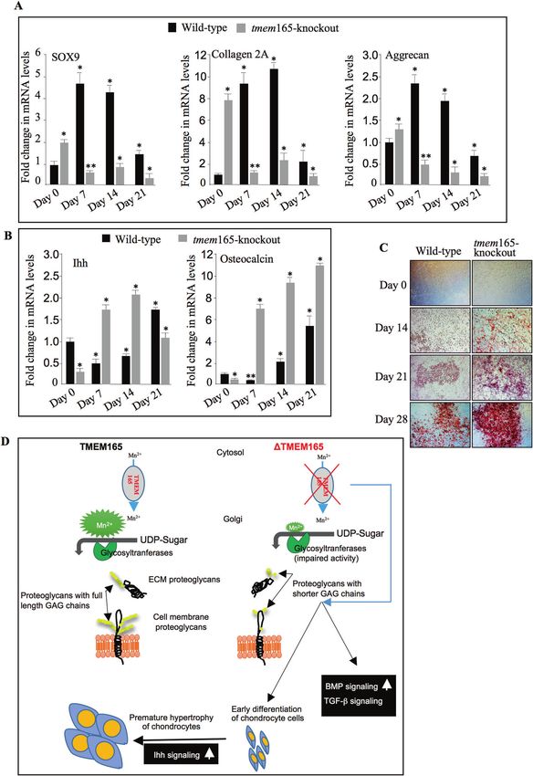
S. Khan et al.
2
Fig. 1 CRISPR/Cas9 knockdown of TMEM165. A Alignment of TMEM165 targeted sequence from wild-type and mutant mouse ATDC5 cells.
B Detection of TMEM165 in cell lysates from wild-type and tmem165-knockout mouse ATDC5 cells (mutant 1, 2, 3 and 4) using anti-TMEM165
specific antibodies. β-actin was used as loading control (n = 3). C Immunofluorescence analysis of the expression of TMEM165 in mouse
ATDC5 control and tmem165-knockout cells using antibodies against TMEM165 (green). GM130 (red) was used as a Golgi marker. The nucleus
was stained with DAPI (blue) (n = 3). Digital images were captured with an inverted microscope, Leica DMI3000. Representative images from
three independent experiments are shown. Scale bar: 50 μm.
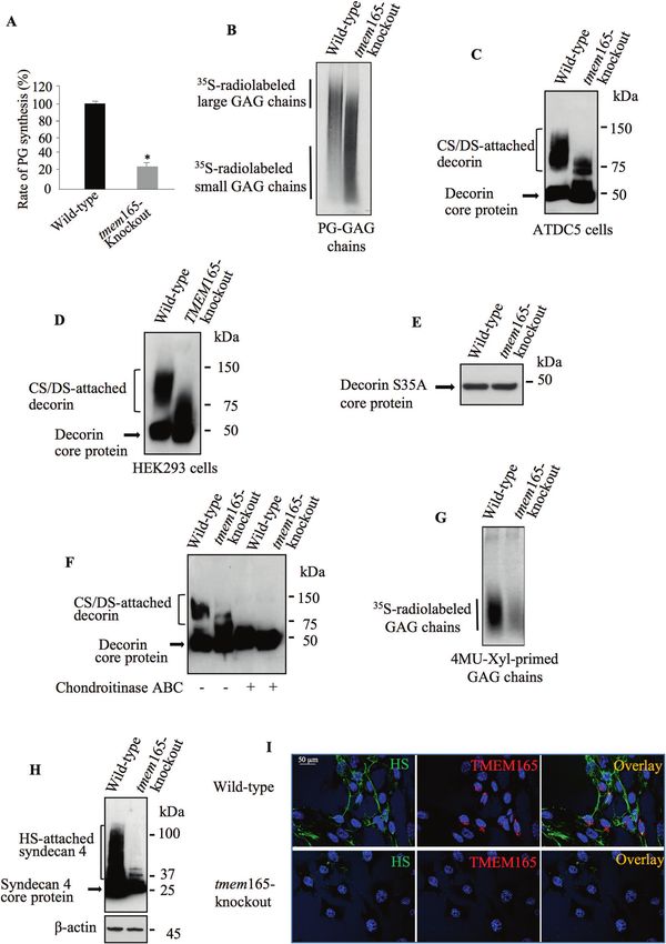
RESULTS predominance of shorter GAG chains in tmem-knockout cells,
Loss of TMEM165 impairs elongation of heparan- and compared to wild-type (Fig. 2B). To determine whether the defects
chondroitin-sulfate chains in GAG synthesis affects both CS- and HS-GAG chains, we used
Genetic mutations or knockout of glycosyltransferases involved in decorin and syndecan 4 as reporter for the synthesis of CS-PGs and
GAG synthetic pathway cause chondrodysplasia. To determine the HS-PGs, respectively. Transfection of wild-type and tmem165-
link, if any, between TMEM165 deficiency and the synthesis of PGs, knockout mouse ATDC5 cells with decorin expression vector
we generated tmem165-knockout prechondrogenic mouse ATDC5 resulted in the secretion of CS/DS-attached decorin in the culture
cell line using CRISPR-Cas9 technique. tmem165-knockout clones medium as shown by Western blot (Fig. 2C). However, there is a
harboring deletion mutations were selected for further studies loss of higher sized CS/DS-attached decorin species (80–150 kDa) in
(Fig. 1A). As expected, expression of a polypeptide of about 35 kDa tmem165-knockout mouse ATDC5 cells as evidenced by predomi-
corresponding to TMEM165 was detected in wild-type mouse nance of decorin with smaller size GAG chains (70 to 80 kDa)
ATDC5 cells, whereas no polypeptide was revealed by the anti- (Fig. 2C). Similar results were obtained using TMEM165-knockout
TMEM165 antibodies in tmem165-knockout cells (Fig. 1B). To HEK293 cells (Fig. 2D). This indicates that defects in PG synthesis
further confirm these results, immunofluorescence analysis of the produced by the loss of TMEM165 is not cell type specific. To rule
expression of TMEM165 was carried out in wild-type and in out an effect of TMEM165 on decorin core protein expression or
tmem165-knockout mouse ATDC5 cells. As shown in Fig. 1C secretion, we generated and expressed a decorin mutant lacking
TMEM165 is clearly detected in wild-type mouse ATDC5 cells GAG-attachement site by mutation of serine residue at position 34,
displaying a perinuclear Golgi distribution and colocalizes with the which is used for the attachment of the CS-GAG chain on the core
Golgi (GM130) marker. However, no staining with anti-TMEM165 protein, to alanine residue (S34A). Expression of the mutant decorin
was observed in tmem165-knockout mouse ATDC5 cells, whereas S34A in wild-type mouse ATDC5 cells led, as expected, to the
the Golgi marker GM130 was clearly detected (Fig. 1C), indicating secretion of a polypeptide of about 50 kDa corresponding to
that the expression of TMEM165 is knocked down in these cells. decorin core protein without attached GAG chain (Fig. 2E). Of note,
To uncover the link, if any, between TMEM165 and PGs, we tmem165-knockout mouse ATDC5 cells secreted mutant decorin at
evaluated the level of PG synthesis in wild-type and tmem165- similar amount to that produced by wild-type cells. These data
knockout mouse ATDC5 cells by using metabolic incorporation of indicate that TMEM165 deficiency did not affect the synthesis of
radiolabelled [35S]-sulfate into GAG chains. The results showed a decorin core protein. Altogether, these results demonstrate that the
decrease of about 70% in the rate of PG synthesis in tmem165- loss of TMEM165 affects the synthesis of GAG chain attached to
knockout cells, compared to wild-type cells (Fig. 2A). Interestingly, decorin core protein. Noteworthy, treatment of decorin, produced
SDS-PAGE analysis of radiolabelled PG-GAG chains showed in tmem165-knockout cells, with chondroitinase ABC led to change
Cell Death and Disease (2022)13:11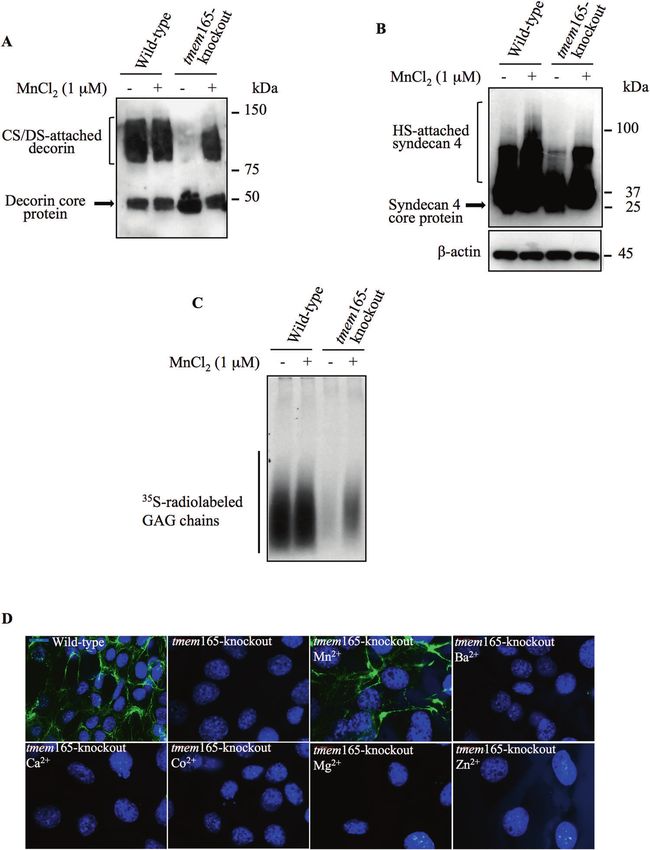
S. Khan et al.
                                                                                                                                            3

in the migration pattern in SDS-PAGE from a smear to a single band    synthesis of GAG chains in the presence of [35S]-sulfate to
corresponding to decorin core protein (Fig. 2F), indicating that      metabolically radiolabel the newly synthesized GAG chains. As
decorin from mutant cells is sensitive to chondroitinase ABC and      shown in Fig. 2G, wild-type mouse ATDC5 cells produced
therefore contains CS-GAG chains, but shorter in size. To confirm      significant amount of 4MU-Xyl primed radiolabelled GAG chains,
that GAG chain synthesis is affected, we used 4-Methylumbelliferyl-   whereas tmem165-knockout mouse ATDC5 cells produced only few
β-D-xylopyranoside (4MU-Xyl) as acceptor substrate for the            amounts, indicating that the synthesis of GAG chains is impaired in

Cell Death and Disease (2022)13:11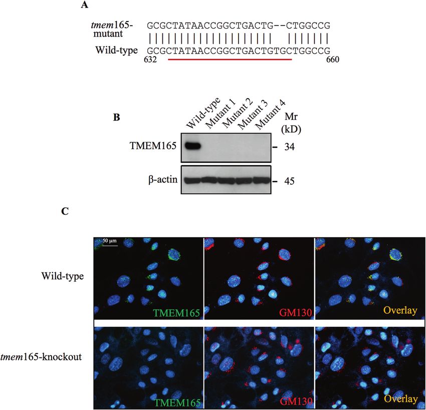
S. Khan et al.
4
    Fig. 2 GAG chain elongation is impaired in TMEM165-deficient cells. A PG anabolism evaluation in wild-type and tmem165-knockout mouse
    ATDC5 cells by measurement of the incorporation rate of [35S]-sulfate into the GAG chains (n = 3). B SDS-PAGE and autoradiography analysis
    of neosynthesized radiolabelled PG-GAG chains in wild-type and tmem165-knockout mouse ATDC5 cells (n = 3). C Detection of decorin in
    conditioned medium of wild-type and tmem165-knockout mouse ATDC5 cells and (D) in wild-type and TMEM165-knockout HEK293 cells
    (n = 3). E Detection of decorin S34A mutant lacking GAG chain in conditioned medium of wild-type and mutant mouse ATDC5 cells (n = 3).
      F Analysis of the sensitivity to degradation by chondroitinase ABC of GAG chains of decorin in conditioned medium of wild-type and
    tmem165-knockout mouse ATDC5 cells (n = 3). G SDS-PAGE and autoradiography analysis of neosynthesized radiolabelled GAG chains primed
    with 4MU-Xyl in wild-type and tmem165-knockout mouse ATDC5 cells (n = 3). H Detection of HA-syndecan 4 in cell lysates of wild-type and
    tmem165-knockout mouse ATDC5 cells transfected with HA-syndecan-4 expression vector. β-actin was used as loading control. (n = 3)
    I Immunofluorescence analysis of cell surface HS GAG chains using anti-HS specific antibodies (green) and of the expression of TMEM165 (red)
    in wild-type and tmem165-knockout mouse ATDC5 cells. The nucleus was stained with DAPI (blue). Digital images were captured with an
    inverted microscope, Leica DM13000. Representative images from three independent experiments are shown. Scale bar: 50 μm.

    tmem165-knockout cells. We next investigated whether the                 transfected with CHSY1, CHSY2 or CHSY1 and CHSY2 expression
    synthesis of HS-GAG chains is also altered in tmem165-knockout           vectors produced decorin with reduced size, compared to that
    cells. For this purpose, we expressed syndecan 4, a cell surface         produced in wild-type cells. The expression of CHSY1 and CHSY2
    HSPG, in tmem165-knockout and wild-type mouse ATDC5 cells. As            was confirmed by Western blot in all the cells transfected (Fig. 3D,
    shown by Western blot, syndecan 4 expressed in wild-type mouse           E). These data indicated that overexpression of CHSY1 and CHSY2
    ATDC5 cells exhibited a smear pattern in SDS-PAGE corresponding          individually or together did not rescue CS elongation in tmem165-
    to HS-attached syndecan 4, whereas in tmem165-knockout cells             deficient cells, suggesting that defects in CS polymerization process
    the smear was strongly reduced in size, compared to wild-type cells      did not result from reduced protein expression of the polymerizing
    indicating a loss of higher sized HS-attached syndecan 4 species in      enzymes.
    tmem165-knockout mouse ATDC5 cells (Fig. 2H). These results
    indicate that elongation of HS chains is impaired in mutant cells. To    Manganese rescues the synthesis of chondroitin-sulfate and
    further confirm this result, we carried out indirect immunofluores-        heparan-sulfate GAG chains in TMEM165-deficient cells
    cence analysis of cell surface HSPGs in wild-type and tmem165-           It has been established that Mn2+ participates in the catalytic
    knockout cells, using anti-HS monoclonal antibody 10E4. Promi-           activity of various Golgi glycosyltransferases including CS and HS
    nent staining of the cell membrane was observed in wild-type             polymerizing enzymes [10, 11]. To determine whether defects in
    cells (green), whereas very low signal could be observed in              the elongation of CS- and HS-GAG chains in tmem165-knockot
    tmem165-knockout cells (Fig. 2I). When cells were probed with anti-      cells is due to defect in Mn2+ homeostasis, cells were cultured in
    TMEM165 antibodies, efficient expression of the protein was               the absence or presence of 1μM of Mn2+. Western blot analysis of
    observed in wild-type cells (red), whereas no staining was detected      decorin produced in wild-type mouse ATDC5 cells showed similar
    in tmem165-knockout cells (Fig. 2I). Altogether, these results           pattern either in the absence or presence of Mn2+. However,
    revealed a key role of TMEM165 in the synthesis of both HS- and          decorin expressed in tmem165-knockout cells that were cultured
    CS-GAG chains of PGs.                                                    in the presence of Mn2+ was larger in size, compared to that
                                                                             produced in the absence of Mn2+ and exhibit similar pattern as
    Overexpression of polymerizing enzymes did not overcome                  decorin in wild-type (Fig. 4A). These data revealed that
    elongation defects produced by loss of TMEM165                           supplementation of the culture medium with Mn2+ rescues
    Given that CS and HS synthetic pathways involve several enzymes          elongation of decorin GAG chains in tmem165-knockout cells. As
    each of which is involved in a specific step of the synthesis process,    polymerization of HS-GAG chains are also impaired in tmem165-
    we sought to determine whether impaired elongation of CS- and            knockout cells, we investigated whether Mn2+ is able to restore
    HS-GAG chains in tmem165-knockout cells resulted from defects in         the polymerization of HS-GAG chains. Remarkably, supplementa-
    the gene expression of CS and HS polymerizing enzymes. To this           tion of Mn2+ in the culture medium of tmem165-knockout cells
    end, RT-qPCR analysis were performed using mRNA from tmem165-            resulted in the synthesis of HA-syndecan 4 with higher molecular
    knockout and wild-type mouse ATDC5 cells to evaluate gene                weight compared to that produced in the absence of Mn2+
    expression of CS polymerizing enzymes, CHSY1 and CHSY2 and of            (Fig. 4B), indicating that elongation HS-GAG chains attached to
    HS polymerizing enzymes, EXT1 and EXT2. The results showed that          HA-syndecan 4 is restored when Mn2+ is supplied in the culture
    mRNA levels of the enzymes are similar or higher in tmem165-             medium. To further confirm that Mn2+ rescues GAG chains
    knockout mouse ATDC5 cells compared to wild-type (Fig. 3A).              elongation in tmem165-knockout cells, 4MU-Xyl was used as
    Similar results were observed in TMEM165-deficient patient                exogenous substrate to monitor the synthesis of GAG chains.
    fibroblasts, compared to normal fibroblast cells (Fig. 3B), suggesting     Wild-type and tmem165-mutant mouse ADTC5 cells were cultured
    that impaired elongation of CS- and HS-GAG chains in mutant cells        in the presence or absence of Mn2+ along with 4MU-Xyl primer
    is not due to downregulation of gene expression of the                   and [35S]-sulfate to radiolabel newly synthesized GAG chains. SDS-
    polymerizing enzymes. However, we cannot rule out a decrease             PAGE analysis of radiolabelled 4MU-Xyl-primed GAG chains
    in the protein level that may result from increased protein              showed that in the absence or presence of Mn2+, high amount
    instability and/or degradation. Given that the level of protein          of GAG chains was produced in wild-type cells. In tmem165-
    expression of glycosyltransferases involved in GAG synthetic             knockout cells, a very week signal was observed in the absence of
    pathway is very low and their detection by antibodies (commer-           Mn2+ supplementation, whereas a significant amount of elon-
    cially available) is very challenging, it is, therefore, difficult to     gated GAG chains was observed when cells were supplemented
    evaluate and compare the protein levels of CS and HS polymerizing        with Mn2+ (Fig. 4C). Altogether, these data indicate that Mn2+
    enzymes in normal and mutant cells. We can however hypothesize           rescues the polymerization of both CS- and HS-GAG chains in
    that overexpression of these enzymes may overcome the instability        tmem165-knockout cells and suggest that the activity of GAG
    or degradation of these proteins, if any. We then designed               polymerizing enzymes is impaired in tmem165-knockout cells due
    expression vectors for Myc-tagged CHSY1 and HA-tagged CHSY2              to disturbed Golgi Mn2+ homeostasis.
    and used them to overexpress each of the two CS elongation                  To examine whether the addition of other ions could rescue the
    enzymes individually or together in tmem165-knockout mouse               blockage in elongation of glycosaminoglycan chains, tmem165-
    ATDC5 cells. As shown in Fig. 3C, tmem165-knockout cells                 knockout cells were cultured in presence of Mn2+, Ba2+, Ca2+,

                                                                                                 Cell Death and Disease (2022)13:11
S. Khan et al.
5
Fig. 3 Expression of CS polymerizing enzymes did not overcome GAG elongation defects in TMEM165-deficient cells. A Fold changes in
mRNA expression of CS polymerizing enzymes CHSY1 and CHSY2, and HS polymerizing enzymes EXT1 and EXT2 in tmem165-knockout mouse
ATDC5 cells normalized to wild-type mouse ATDC5 cells and B in TMEM165-deficient fibroblasts normalized to fibroblast control cells. qPCR
values were normalized for the housekeeping gene ribosomal protein S29 and are expressed as the relative expression compared with
control. Data are expressed as mean ± S.D. Statistical analysis was performed with an unpaired Student’s t test (n = 3; *p < 0.05; **p < 0.01).
C Detection of decorin in conditioned medium of wild-type and mutant ATDC5 cells transfected with empty vector, Myc-tagged CHSY1, HA-
tagged CHSY2 or Myc-tagged CHSY1 and HA-tagged CHSY2 along with decorin expression vector. D Detection of CHSY1 and (E) of CHSY2 in
wild-type and tmem165-knockout mouse ATDC5 cells. β-actin was used as loading control (n = 3). Representative images from three
independent experiments are shown.
Co2+, Mg2+ and Zn2+, respectively and endogenous cell surface cells (Fig. 4D) indicating that the knockout of tmem165 prevented
HS-GAG chains were analyzed by indirect immunofluorescence the synthesis of cell surface HS-GAG chains. Interestingly,
using anti-HS monoclonal antibody 10E4, which is commonly used supplementation of cell culture medium of tmem165-knockout
to detect HS chains of PGs. Prominent staining of the cell surface ADTC5 cells with Mn2+ rescued the synthesis of HS-GAG chains,
HS-GAGS was observed in mouse ATDC5 control cells, whereas no whereas supplementation with divalent ions Ba2+, Ca2+, Co2+,
staining could be observed in tmem165-knockout mouse ATDC5 Mg2+ or Zn2+ did not (Fig. 4D), indicating that among the divalent
Cell Death and Disease (2022)13:11S. Khan et al.
6
Fig. 4 Manganese supplementation rescue GAG elongation in TMEM165-deficient cells. A Detection of decorin in conditioned medium of
wild-type and tmem165-knockout mouse ATDC5 cells transfected with the expression vector for decorin. (B) Detection of HA-tagged syndecan
4 in cell lysates of wild-type and tmem165-knockout mouse ATDC5 cells transfected with HA-tagged syndecan 4 expression vector and grown
in the presence or absence of MnCl2 (1 µM). β-actin was used as loading control (n = 3). C SDS-PAGE and autoradiography of [35S]-sulfate
radiolabelled GAG chains primed with 4MU-Xyl in wild-type and tmem165-mutant mouse ATDC5 cells grown in the presence or absence of
MnCl2 (1 µM) (n = 3). One representative blot of three independent experiments is shown. D Immunofluorescence analysis of cell surface
HSGAG chains using anti-HS specific antibodies (green) in wild-type and tmem165-knockout mouse ATDC5 cells cultured in the absence and
presence of Mn2+, Ba2+, Ca2+, Co2+, Mg2+ and Zn2+. The nucleus was stained with DAPI (blue). Digital images were captured with an inverted
microscope, Leica DM13000. Representative images from three independent experiments are shown. Scale bar: 50 μm.
ions tested only Mn2+ is able to restore defects induced by loss of wild-type mouse ATDC5 cells revealed that the level of phospho-
TMEM165 on HS-GAG chain elongation. CaMkIIα (pCaMkIIα) was strongly increased in tmem165-knockout
cells, compared to wild-type cells (Fig. 5A) indicating that CaMkIIα
Ca2+/calmodulin-dependent protein kinase IIα pathway is was activated in TMEM165-knockout cells. Likewise, the phosphor-
activated in TMEM165-deficient cells ylation level of CaMkIIα was remarkably increased in TMEM165-
Direct implication of TMEM165 in the Golgi transport of either Ca2+ deficient patient fibroblast cells, compared to normal fibroblasts
or Mn2+ is not yet been reported. However, it is well known that (Fig. 5B). Altogether, these data indicated that the loss of TMEM165
Ca2+/calmodulin-dependent protein kinase II (CaMkII) is activated induces defects in both Ca2+ and Mn2+ homeostasis and support
when intracellular concentration in [Ca2+]i is elevated. Analysis of the notion that TMEM165 is probably involved in the regulation of
the phosphorylation status of CaMkIIα in tmem165-knockout and both Ca2+ and Mn2+ homeostasis.
Cell Death and Disease (2022)13:11S. Khan et al.
7
in Fig. 6H, a significant decrease in mRNA levels of TGFβR2 was
observed in tmem165-mutant mouse ATDC5 cells, compared with
wild-type cells. Consistent with RT-qPCR data, significant reduc-
tion in the protein level of TGFβR2 was observed in tmem165-
knockout mouse ATDC5 cells, compared to wild-type cells
(Fig. 6I). Similarly, fibroblast cells of TMEM165-deficient patient
showed strong decrease in the mRNA levels (Fig. 6J) and protein
expression of TGFβR2 (Fig. 6K), compared to normal cells.
Downregulation of TGF-β signaling may also result from up-
regulation of negative regulators such as asporin, an extracellular
protein that suppresses TGF-β signaling by direct interaction with
TGF-β thus preventing its binding to TGFβR2. Analysis of mRNA
levels of asporin (Aspn) in tmem165-knockout mouse ATDC5 cells
and in TMEM165-deficient patient fibroblasts showed a remark-
Fig. 5 Phospho-CaMKIIα is activated in TMEM165-deficient cells. able increase (4-fold) in mutant cells compared to normal cells
A Immunoblot analysis of phospho-CaMkIIα (pCaMkIIα) level in cell (Fig. 6L). Assessment of asporin protein expression showed higher
lysates of wild-type and tmem165-knockout mouse ATDC5 cells and levels of expression in tmem165-deficient mouse ATDC5 and
(B) in cell lysates of normal fibroblasts and TMEM165-deficient TMEM165-mutant fibroblast cells, compared to normal cells
fibroblasts from CDG patients, using specific antiphospho-CaMkIIα (Fig. 6M). Altogether, these data support the notion that impaired
and anti-CaMkIIα antibodies. β-actin was used as loading control TGF-β signaling in TMEM165-deficient cells is associated with
(n = 3). Representative images from three independent experiments downregulation of the expression of TGFβR2 receptor and
are shown.
upregulation TGF-β antagonist, asporin.
BMP signaling plays a key role in skeletal development and
TMEM165 deficiency impairs TGF-β and BMP signaling alterations in BMP signaling pathway are major underlying cause
pathways of human skeletal disorders [13]. To determine whether TMEM165
As a number of human genetic mutations causing a wide range of deficiency affects BMP signaling, we examined the phosphoryla-
inheritable diseases of skeletal development are related to TGF-β tion status of Smad 1,5,9 downstream mediators of BMP receptor
and BMP signaling [12] and owing to the key role of GAG chains in activation. Interestingly, immunoblot analysis showed that the
the regulation of several signaling pathways, we sought to level of phospho-Smad 1,5,9 (pSmad1,5,9) was higher in
investigate whether loss of TMEM165 affects TGF-β/BMP signaling tmem165-knockout mouse ATDC5 cells (Fig. 7A) and in
pathways. Interestingly, Western blot analysis showed that TMEM165-deficient patient fibroblast cells (Fig. 7B). Analysis of
phospho-Smad2 level was reduced in tmem165-knockout cells, the mRNA levels of Id1 gene, a BMP downstream target gene,
compared to wild-type (Fig. 6A). In addition, the mRNA levels of showed two-fold increase in tmem165-knockout cells, compared
the serpine 1 (PAI-1) gene, which is positively correlated with TGF- to wild-type cells (Fig. 7C). To further confirm the increase of BMP
β signaling activation were significantly lower in cells knocked signaling in tmem165-knockout cells, the vector pGL3-BRE-Luc
down for tmem165 (Fig. 6B), therefore indicating that the loss of containing a BMP responsive promoter element (BRE) was used to
TMEM165 perturbs TGF-β signaling in mutant cells. These data transfect wild-type and tmem165-mutant mouse ATDC5 cells. As
were further confirmed by using the p(CAGA)12-luc TGF-β shown in Fig. 7D, analysis of luciferase activity showed a
reporter plasmid which showed that the luciferase activity was significant increase (4-fold) in tmem165-knockout ATDC5 cells,
markedly (7-fold) lower in mutant cells, compared to wild-type compared to wild-type cells thus bringing evidence that BMP
cells confirming that the TGF-β/Smad axis is functionally impaired signaling is increased in tmem165-mutant cells. In an attempt to
in tmem165-knockout cells (Fig. 6C). Similar results were obtained identify the mechanism involved in the alteration of the BMP/
in TMEM165-deficient patient fibroblast cells. Indeed, phospho- Smad signaling pathway, we investigated the mRNA levels of type
Smad2 level was reduced in TMEM165-deficient fibroblasts I and type II BMP receptors ie. BMPR1A, BMR1B and BMPR2 in
compared to normal fibroblasts (Fig. 6D) and the expression of TMEM165-deficient and wild-type cells. As shown in Fig. 7E, mRNA
serpine1 gene showed significant (2.5-fold) downregulation levels of BMP receptors were increased in tmem165-knockout
(Fig. 6E). Therefore, indicating that TMEM165 deficiency impairs mouse ATDC5 cells, compared to wild-type cells. The increased
the expression of the TGF-β/Smad downstream responsive genes expression of BMPR2 at the protein level in mutant cells was
in TMEM165-deficient fibroblasts. confirmed by Western blot analysis (Fig. 7F). Similar results were
To determine whether response to TGF-β stimulation is observed, either for mRNA expression levels of BMPR1A, BMR1B
affected in mutant cells, phosphorylation level of Smad2 in and BMPR2 (Fig. 7G) or for BMPR2 protein expression (Fig. 7H) in
response to TGF-β was examined. Treatment of tmem165- TMEM165-deficient patient fibroblast cells, compared to normal
knockout mouse ATDC5 cells with TGF-β induced the phosphor- fibroblast cells. In addition, analysis of the mRNA levels of noggin,
ylation of Smad2, however the level of phospho-Smad2 is lower a BMP ligand antagonist, showed strong reduction in tmem165-
compared to that observed in wild-type cells (Fig. 6F), indicating deficient mouse ATDC5 cells (Fig. 7I) and in TMEM165-deficient
that mutant cells were less responsive to TGF-β than wild-type patient fibroblasts (Fig. 7J), compared to normal cells. These data
cells. Likewise, stimulation of TMEM165-deficient and normal suggest that upregulation of BMP receptors and downregulation
fibroblasts with TGF-β induced less phosphorylation of Smad2 in of BMP antagonist may account for the increased BMP signaling
mutant fibroblasts, compared to normal fibroblasts (Fig. 6G). observed in TMEM165-deficient cells. Overall, this study showed
Altogether, these results demonstrate that both the baseline and that TMEM165 deficiency functionally impairs TGFβ/BMP signal-
TGF-β-induced levels of phospho-Smad2 were significantly ing pathways in TMEM165-deficient cells.
reduced in mutant cells, therefore revealing for the first time We next analyzed whether supplementation with Mn2+ rescued
that TMEM165 deficiency functionally impaired the TGF-β/ TGFβ/BMP signaling. Interestingly, Western blot analysis showed
Smad2 signaling axis. that phospho-Smad2 and phospho-Smad 1,5,9 levels were
To identify the mechanism involved in the alteration of the restored to normal levels in tmem165-knockout mouse ATDC5
TGF-β/Smad signaling pathway, we investigated the mRNA cells when cultured in the presence of Mn2+ (Fig. 7K), indicating
expression levels of TGF-β receptors, TGFβR1 and TGFβR2 in that supplementation with Mn2+ restored TGFβ/BMP signaling in
tmem165-knockout and wild-type mouse ATDC5 cells. As shown tmem165-knockout cells.
Cell Death and Disease (2022)13:11S. Khan et al.
8
TMEM165 deficiency promotes early chondrocyte ATDC5 cells is affected in the absence of TMEM165, wild-type and
differentiation and hypertrophy tmem165-knockout mouse ATDC5 cells were induced by insulin
Chondrogenesis process is tightly regulated during skeletal into chondrogenic differentiation and the mRNA levels of
development and alterations in the chondrocyte maturation result chondrogenic markers including Sox9, Col2a1, AGN, Ihh, and
in defects in endochondral bone development. To investigate OCN were measured by RT-qPCR before induction and at seven,
whether chondrogenic differentiation of prechondrocyte mouse forteen and twenty-one days post induction. The results showed
Cell Death and Disease (2022)13:11S. Khan et al.
9
Fig. 6 TGF-β signaling pathway is impaired in TMEM165-deficient cells. A Detection of phosphorylated Smad2 (pSmad2) and total Smad2
(Smad2) in cell lysates from wild-type and tmem165-knockout mouse ATDC5 cells (n = 3). B Fold changes of serpine expression in tmem165-
knockout cells normalized to wild-type mouse ATDC5 cells. C Fold changes of TGF-β reporter activity in tmem165-knockout cells normalized to
wild-type mouse ATDC5. D Detection of phosphorylated Smad2 (pSmad2) and total Smad2 in cell lysates from control and TMEM165-deficient
CDG patient fibroblast cells (n = 3). E Fold changes of serpine expression in TMEM165-deficient fibroblasts normalized to normal fibroblast
cells. F Detection of p-Smad2 in cell lysates from wild-type and tmem165-knockout mouse ATDC5 cells and (G) from normal and TMEM165-
deficient CDG patient fibroblast cells treated or not with TGFβ1 (1 ng/ml) for 1 hour (n = 3). H Fold changes of TGFβR1 and TGFβR2 expression
in tmem165-knockout mouse ATDC5 cells normalized to wild-type mouse ATDC5 cells. I Detection of TGFβR2 in cell lysates from wild-type and
tmem165-mutant mouse ATDC5 (n = 3). J Fold changes of TGFβR1 and TGFβR2 expression in TMEM165-deficient fibroblasts from CDG patient
normalized to normal fibroblast cells. K Detection of TGFβR2 in cell lysates from normal fibroblasts and TMEM165-deficient CDG patient
fibroblast cells (n = 3). L Fold changes of asporin expression in tmem165-knockout cells normalized to wild-type mouse ATDC5 cells and in
TMEM165-deficient fibroblast cells normalized to normal fibroblast cells. M Detection of asporin in conditioned medium of wild-type and
tmem165-knockout mouse ATDC5 cells and of normal fibroblasts and TMEM165-deficient CDG patient fibrobast cells. β-actin was used as
loading control (n = 3). qPCR values were normalized for the housekeeping gene ribosomal protein S29 and are expressed as the relative
expression compared with control. Data are expressed as mean ± S.D. Statistical analysis was performed with an unpaired Student’s t test (n =
3; *p < 0.05; **p < 0.01). Representative images from three independent experiments are shown.
that before induction (day 0), mouse ATDC5-knockout cells express Mn2+ involves a transit across the ER compartment. Mn2+ enters
higher mRNA levels of the chondrogenic markers Sox9, Col2a1, and TMEM165-deficient HEK293 cells through plasma membrane
AGN and lower mRNA levels of pre-hypertrophic and hypertrophic transporters and reaches the ER compartment by a mechanism
markers Ihh and OCN, compared to wild-type cells (Fig. 8A, B (day involving SERCA pumps then transported from the ER to the Golgi
0)). These results indicate that loss of TMEM165 expression induces probably via SPCA pumps or other yet unknown mechanism [15].
early chondrogenic differentiation of prechondrogenic mouse Deficiency of Mn2+ results in birth defects including abnormal
ATDC5 cells. Importantly, analysis of the expression of the markers or poor bone formation and susceptibility to seizures [16].
at seven days post induction revealed that tmem165-deficient Mutations in SLC39A8 a plasma membrane protein able to
mouse ATDC5 cells undergo rapid maturation and hypertrophy as transport Mn2+ are characterized by low blood levels of Mn2+
evidenced by downregulation of chondrogenic markers Sox9, associated with delayed development, dwarfism, and profound
Col2a1, AGN and upregulation of pre-hypertrophic and hyper- psychomotor retardation [17].
trophic markers Ihh and OCN. The expression of hypertrophic Analysis of BMP and TGF-β signaling pathways revealed that
markers still increases at 14 and 21 days. In contrast, as expected, they are functionally impaired in TMEM165-deficient cells. TGF-βs
in wild-type cells the chondrogenic markers were up-regulated and BMPs play an important role in several stages of chondrogen-
at 7 and 14 days post induction, and pre-hypertrophic and esis. It has been reported that TGF-β inhibits the terminal
hypertrophic markers were downregulated. However, at 21 days differentiation of chondrocytes in high-density chondrocyte
post induction chondrogenic markers were down-regulated, and pellets or long bone cultures in vitro [18]. We found that
pre-hypertrophic and hypertrophic markers were up-regulated TMEM165 deficiency led to downregulation of TGF-β signaling
(Fig. 8A, B). associated with downregulation of TGFβRII receptor and upregu-
We also observed an early mineralization in tmem165-deficient lation of TGF-β antagonist, asporin. We showed that the TGF-β
mouse ATDC5, compared to wild-type cells. Indeed, mineralization signaling was activated when the cells were exogenously treated
was detected in tmem165-deficient mouse ATDC5 at day 14 after with TGF-β1 but at lower extent compared to wild-type cells,
initiation of differentiation, whereas it appeared only after 21 days indicating that both basal and inducible TGF-β signaling activation
of differentiation in wild-type cells (Fig. 8C). Altogether, these data is impaired in TMEM-165-deficient cells. TGF-β activates Smad2
bring evidence that TMEM165 deficiency promotes premature and Smad3 through binding to TGFβRII receptor and recruitment
chondrocyte maturation and hypertrophy, a process which affects of ALK5 receptor (TGFβRI) [19]. Noteworthy, mice lacking Tgfβr2 or
endochondral ossification and may lead to dwarfism. Alk5 in chondrocytes exhibit skeletal defects [20] and inhibition of
TGFβ signaling induces chondrocyte hypertrophy [12].
We have analyzed the BMP signaling pathway and found that
DISCUSSION phospho-Smad1,5,9 was upregulated in TMEM165-deficient cells,
Here, we generated tmem165-knockout pre-chondrocyte mouse compared to normal cells. Investigation of the mechanisms
ATDC5 and HEK293 cells and showed that loss of TMEM165 led to involved revealed that the BMP receptors BMPR2 and BMPR1B
strong defects in the synthesis of PGs. Decrease content of PGs in are upregulated and the BMP antagonist noggin is downregulated
cartilage was also reported in tmem 165-deficient zebrafish [14]. in TMEM165-deficient cells. Noteworthy, BMP regulates longitudinal
Importantly, we showed a dramatical reduction in the length of growth and excessive BMP signaling has been shown to accelerate
HS- and CS-GAG chains, revealing for the first time that elongation chondrogenesis and chondrocyte differentiation [21, 22]. Brachy-
of PG-GAG chains is impaired in TMEM165 deficient cells. We dactylies type 2 (BDB2) is a NOG mutation characterized by absence
showed that overexpression of CS elongating enzymes CHSY1 and of terminal structures of the toes and digits due to excessive BMP
CHSY2 did not rescue the elongation of CS-GAG chains; however, signaling [23]. Mutations in NOG cause inability of the antagonist to
Mn2+ supplementation restores the elongation of CS- and HS GAG bind BMP or heparin and to sequester BMPs in ECM which results in
chains of PGs in tmem165-knockout cells. Golgi glycosyltrans- excessive BMP signaling [24, 25].
ferases use UDP-sugars as a donor substrate and require Mn2+ at The increased levels of hypertrophic marker Ihh and osteo-
their catalytic site to be fully active. These findings revealed that calcin in tmem165-knockout prechondrocyte mouse ATDC5 cells
TMEM165 plays an essential role in the synthesis of PGs by during differentiation lead to early hypertrophy of chondrocytes.
regulating homeostasis of Mn2+ in the Golgi compartment. We Indeed, overexpression of Ihh induces early chondrocyte
can therefore hypothesize that GAG chains are aborted because of hypertrophy and ossification during chondrogenesis. It has been
lack of sufficient pool of the cofactor Mn2+ in the Golgi necessary suggested that TGFβ inhibits hypertrophy induced by Ihh [26].
for the synthesis of normal sized GAG chains. How Mn2+ Our study has shown that TGF-β signaling is downregulated and
supplementation rescues GAG chains elongation is not known, Ihh is upregulated in tmem165-mutant mouse ATDC5 cells,
however we previously showed that the rescue of Golgi N- suggesting accelerated chondrocyte hypertrophy. Interestingly,
glycosylation defects in TMEM165-deficient cells by extracellular we found that loss of TMEM165 induced early mineralization in
Cell Death and Disease (2022)13:11S. Khan et al.
10
ATDC5 cells. Premature transition of the chondrocyte phenotype As summarized in Fig. 8D, our findings indicate that TMEM165
from proliferating to hypertrophic chondrocytes and the early deficiency causes abnormalities in PG synthesis and aberrant
onset of mineralization may trigger a premature replacement of signaling resulting in early chondrocyte maturation and hyper-
cartilage by bone leading to defects in skeletal development and trophy. PGs and their GAG chains are key components of the ECM
hence to dwarfism. and are involved in the organization and function of the matrix.
Cell Death and Disease (2022)13:11S. Khan et al.
11
Fig. 7 BMP signaling is activated in TMEM165-deficient cells. A Detection of phosphorylated Smad1, 5, 9 (pSmad1,5,9) and total Smad in cell
lysates from wild-type and tmem165-mutant mouse ATDC5 cells and (B) from normal fibroblasts and TMEM165-deficient CDG patient
fibroblast cells (n = 3). C Fold changes of Id1 expression in tmem165-knockout cells normalized to wild-type mouse ATDC5 cells. D Fold
changes of BMP reporter activity in tmem165-knockout cells normalized to wild-type mouse ATDC5 cells. E Fold changes of BMPR1A, BMPR1B
and BMPR2 expression in tmem165-knockout cells normalized to wild-type mouse ATDC5 cells. F Detection of BMPR2 in cell lysates of wild-
type and tmem165-knockout mouse ATDC5 cells. β-actin was used as loading control (n = 3). G Fold changes of BMPR1A, BMPR1B and BMPR2
expression in TMEM165-deficient fibroblasts normalized to normal fibroblast cells. H Detection of BMPR2 in cell lysates of normal fibroblasts
and TMEM165-deficient CDG patient fibroblast cells. β-actin was used as loading control (n = 3). I Fold changes of Noggin expression in
tmem165-knockout mouse ATDC5 cells normalized to wild-type ATDC5 cells and (J) in TMEM165-deficient CDG patient fibroblasts normalized
to normal fibroblast cells. qPCR values were normalized for the housekeeping gene ribosomal protein S29 and are expressed as the relative
expression compared with control. Data are expressed as mean ± S.D. Statistical analysis was performed with an unpaired Student’s t test (n =
3; *p < 0.05; **p < 0.01). K Detection of phosphorylated Smad2 (pSmad2) and Smad1, 5, 9 (pSmad1,5,9) and of total Smad in cell lysates from
wild-type and tmem165-mutant mouse ATDC5 cells cultured in medium with or without Mn2+ supplementation. β-actin was used as loading
control (n = 3). Representative images from three independent experiments are shown.
Also, alterations in the structure of GAG chains may induce of [35S]-sulfate (Perkin Elmer, Courtabœuf, France) overnight. Then,
profound changes in the organization and function of the ECM conditioned culture medium was collected, digested with papain
and hence in cell-matrix interactions and signaling which may (1 mg/ml), and [35S]-labeled GAGs were precipitated by cetylpyridinium
lead to defects in endochondral ossification. chloride (CPC) as described by [28]. When GAG chains were primed by 4MU-
Xyl, cells were cultured in the presence of 10 µCi/ml of [35S]-sulfate and
100 μM of 4MU-Xyl overnight, and radiolabeled GAG chains were directly
precipitated from conditioned medium by CPC. The CPC precipitated
MATERIALS AND METHODS radiolabeled GAGs were separated by SDS-PAGE on a 4–20% Tris/Glycine
Cell culture and treatments gel. The gel was dried and exposed to autoradiography film.
Skin fibroblast cells were derived from skin biopsy specimens from healthy To measure the rate of sulfate incorporation into GAG chains of PGs,
controls and TMEM165-deficient patients as described by Foulquier et al, mouse ATDC5 cells were radiolabeled with 10 µCi/ml of [35S]-sulfate for 6 h
2012 and were maintained in Eagle’s minimum essential medium in then, conditioned culture medium was collected and digested with papain
humidified 37 °C incubator. Culture medium was supplemented with (1 mg/ml). [35S]-labeled GAG chains were precipitated by CPC dissolved in
10% fetal bovine serum and 1% combination of 100 U penicillin/0.1 mg/ml solvable and mixed in scintillation fluid (Perkin Elmer, MA, USA). The
streptomycin. Mouse ATDC5 cells (Riken cell RCB0565, Tsukubai, Japan) radioactivity associated with GAGs was measured by liquid scintillation
were cultured in DMEM-F12 complete medium (2 mM glutamine, 100 μg/ counting (Packard, Rungis, France).
ml streptomycin, 100 IU/ml penicillin, and 5% (v/v) foetal bovine serum)
and HEK293 cells (ATCC CRL-3216, LGC Standards, France) were cultured in
DMEM complete medium with 10% (v/v) foetal bovine serum at 37 °C in a Indirect immunofluorescence staining
humidified atmosphere supplemented with 5% CO2. Cells were seeded The mouse ATDC5 cells were grown on glass coverslips and fixed with 4%
onto six-well plates at 2 × 10⁵ cells/well and allowed to attach overnight in (w/v) paraformaldehyde in PBS for 20 min. Cells were permeabilized by
standard culture conditions. For treatment with Mn2+, mouse ATDC5 cells treatment with 0.1% (w/v) Triton X-100/PBS solution for 4 min. After
were cultured in DMEM F12 complete medium until reaching 80% extensive washing in 0.2% (w/v) fish skin gelatin in PBS, cells were then
confluency then the medium was replaced with DMEM-F12 without FBS incubated with primary antibodies anti-TMEM165 (Cat# HPA038299, 1:100,
and containing 1 μM of divalent ions, for 36 h. For treatment with growth Atlas Antibodies) or anti-HS (Cat# 370255-1, 1:100, AMSBIO) for 20 min.
factors, cells were cultured in DMEM F12 complete medium until reaching Cells were washed several times in 0.2% (w/v) fish skin gelatin in PBS and
80% confluency then the medium was replaced with DMEM-F12 without incubated with secondary antibodies coupled with Alexa Fluor 488 (Cat#
FBS and containing 1 ng/ml of TGF-β1 (R&D Systems, Minneapolis, MN A-21206 or Cat# A-11017, Molecular Probes) for 20 min. Cells were washed
USA) or vehicle (0.1% BSA in PBS) for 1 h. Cells were then washed twice with 0.2% (w/v) fish skin gelatin in PBS and incubated with primary
with PBS and stored at -80 °C prior to protein or gene expression analyses. antibodies anti-GM130 (Cat# 610822, 1:100, BD Biosciences) for 20 min.
For chondrogenic differentiation, mouse ATDC5 cells were seeded in Cover slips were then washed several times in 0.2% (w/v) fish skin gelatin
12 well/plate at 4 × 10⁴ cells/well and cultured in DMEM-F12 complete in PBS and incubated with secondary antibodies coupled with Alexa Fluor
medium containing 50 μg/ml human transferrin and 3 × 10⁻⁸ M sodium 555 (Cat# A-21428, Molecular Probes) for 20 min. Cells were washed with
selenite (Sigma, Saint Louis, MO) until confluency (day 0), then PBS and nuclei were stained with Hoechst/PBS solutions then coverslips
chondrogenesis was induced by addition of 100 μg/ml of human insulin were mounted with Moviol (National Diagnostics, U.K.) containing 1%
(Sigma, Saint Louis, MO) in the culture medium. The medium was replaced propylgallate (Sigma, Saint Louis, MO). Digital images were captured with
every second or third day. Cells were washed twice with PBS and stored at an inverted microscope Lieca DMI3000 B (Leica Microsystems, Germany).
−80 °C prior to protein or gene expression analyses. For Alizarin red
staining, cells were fixed in ethanol for 30 min then washed with PBS and CRISPR/Cas9 mutation of TMEM165 gene
1% Alizarin red solution (Sigma, pH 4.2) was added to the cell layers for Sense 5ʹCACCGCTATAACCGGCTGACTGTGC3ʹ and antisense 5ʹAAACGCA-
15 min at room temperature. Cells were washed with distilled water and CAGTCAG CCGGTTATAGC3ʹ oligonucleotides (1 μg) containing 20 bp
images were captured. sequence (underlined) targeting TMEM165 exon 2 and cohesive ends
(bold) with the vector were annealed in annealing buffer (60 mM Tris-HCl,
Gene expression analysis pH7.5; 500 mM NaCl; 60 mM MgCl2; 10 mM DTT) and ligated into BbsI sites
Total RNA from cells was extracted using TRIzol (Lifetech, Carlsbad, CA) and of pUC57-attbU6 sgRNA vector. This vector is a basic vector with U6
purified with RNeasy kit (Qiagen, Hilden, Germany) according to promoters and improved Cas9 binding sites. 1 μg of pUC57-TMEM165,
manufacturer’s instructions. The reverse transcription was performed 100 ng of SVneo vector (that confer resistance to geneticin), and 1 μg of
using 500 ng of total RNA from each sample with iScript Ready to use pSpCas-9 vector was used to transfect mouse ATDC5 cells grown at 80%
cDNA supermix (BIO-RAD, Hercules, CA). Quantitative PCR was performed confluency in six wells/plate, using Lipofectamine 2000© (Invitrogen,
with iTaq™ Universal SYBER Green Supermix kit (BIO-RAD, Hercules, CA) Carlsbad, CA) according to the instructions of the supplier. 24 h after
using StepOnePlus™ Real-Time PCR Systems (Applied Biosystems, Foster transfection, the medium was replaced by medium containing 0.2 mg/ml
city, CA) and validated primers. geneticin sulfate and cells were cultured for 48 h before trypsinized and
cloning by serial dilution in 96 wells/plate. Several clones were obtained
and amplified in six wells/plates and analyzed for the presence of
Metabolic labeling of GAG chains mutations in the targeted sequence by PCR amplification and sequencing
Metabolic labeling of GAG chains of PGs was carried out using [35S]-sulfate of the genomic DNA flanking the targeted region. TMEM165-knockout
incorporation method as described by [27]. Briefly, subconfluent mouse HEK293 cells were generated using CRISPR/cas9 technique and were
ATDC5 cells grown in six-well culture plate were radiolabeled with 10 µCi/ml previously described by Morelle et al, 2017.
Cell Death and Disease (2022)13:11S. Khan et al.
12
Plasmids and transfection seeded in 6-well culture plate until 80% confluency and transfected with 1 µg
Chsy1, Chsy2, Decorin, and HA-syndecan 4 cDNAs were generated by PCR of either pCMV-Decorin, pCMV-HA-Syndecan 4, pCMV-Decorin-S34A, pCMV-
and cloned into EcoRI and BamHI or SmaI and PstI sites of pCMV empty Myc-Chsy1, pCMV-HA-Chsy2, or pCMV-empty vector using lipofectamine
vector (Stratagene, Valencia, CA). Decorin-S34A mutant was generated 2000 transfection reagent (Invitrogen, Carlsbad, CA) according to manufac-
by site-directed mutagenesis using QuikChange XLII (Agilent, CA, USA) turer’s instructions. Expression of decorin in culture medium and of syndecan
according to the manufacturer’s instructions. For transfection, cells were 4 in cell lysate was analyzed at 48 h post transfection by Western blotting.
Cell Death and Disease (2022)13:11S. Khan et al.
13
Fig. 8 Early hypertrophic differentiation of tmem165-deficient mouse ATDC5 cells. A Fold changes of chondrogenic markers expression in
wild-type mouse ATDC5 cells and tmem165-knockout cells. RT-qPCR analysis of the mRNA levels of chondrogenic markers SOX9, Col2A, and
Aggrecan, and (B) of hypertrophic markers Ihh and OCN. qPCR values were normalized for the housekeeping gene ribosomal protein S29 and
are expressed as the relative expression compared with control. Data are expressed as mean ± S.D. Statistical analysis was performed with an
unpaired Student’s t test (n = 3; *p < 0.05; **p < 0.01). C Mineralization of wild-type and tmem165-deficient mouse ATDC5 cells analyzed by
Alizarin red staining at Days 0, 14, 21, and 28. Representative images from three independent experiments are shown. D Loss of TMEM165
function impairs Golgi Mn2+ homeostasis necessary for glycosyltransferase polymerization activities leading to blockage in the elongation of
GAG chains of proteoglycans. Blockage in the elongation of GAG chains by loss of TMEM165 may account for dysregulation of TGF/BMP and
Ihh signaling, and therefore in defects in chondrocyte differentiation and maturation. However, other mechanisms can’t be ruled-out.
TGF-β and BMP luciferase reporter activity assays 5. Morelle W, Potelle S, Witters P, Wong S, Climer L, Lupashin V, et al. Galactose
p(CAGA)12-luc and pGL3-BRE-Luc Wild-type and tmem165-knockout supplementation in patients with TMEM165-CDG rescues the glycosylation
mouse ATDC5 cells were plated onto twenty-four-well plates and grown defects. J Clin Endocrinol Metab. 2017;102:1375–86.
to 80% confluency. Cells were transfected with 500 ng of p(CAGA)12-luc 6. Domowicz MS, Cortes M, Henry JG, Schwartz NB. Aggrecan modulation of growth
and pGL3-BRE-Luc promoter constructs, respectively along with 25 ng of plate morphogenesis. Dev Biol. 2009;329:242–57.
pRL-TK vector (Promega, Madison, WI) using. Twenty-four hours after 7. Hilton MJ, Gutierrez L, Martinez DA, Wells DE. EXT1 regulates chondrocyte pro-
transfection, Firefly and Renilla luciferase activities in cells of each well liferation and differentiation during endochondral bone development. Bone
were measured with the Dual-Luciferase Assay System (Promega) using a 2005;36:379–86.
Berthold (Bad Wildbad, Germany) luminometer. Luciferase activities were 8. Watanabe Y, Takeuchi K, Higa Onaga S, Sato M, Tsujita M, Abe M, et al. Chon-
normalized to pRL-TK vector activity. droitin sulfate N-acetylgalactosaminyltransferase-1 is required for normal carti-
lage development. Biochem J. 2010;432:47–55.
9. Wilson DG, Phamluong K, Lin WY, Barck K, Carano RA, Diehl L, et al. Chondroitin
Western blotting sulfate synthase 1 (Chsy1) is required for bone development and digit patterning.
Total protein from cells was extracted using RIPA buffer (150 mM NaCl, Dev Biol. 2012;363:413–25.
50 mM Tris-HCl, pH 7.5, 1% deoxycholate, 0.1% SDS, 1% Triton X-100)
10. Yada T, Gotoh M, Sato T, Shionyu M, Go M, Kaseyama H, et al. Chondroitin sulfate
supplemented with protease and phosphatase inhibitors (Roche Diagnos-
synthase-2. Molecular cloning and characterization of a novel human glycosyl-
tics, Indianapolis, IN, USA). Cell lysates were sonicated on ice and protein transferase homologous to chondroitin sulfate glucuronyltransferase, which has
concentration of the samples was determined by the Bradford method. dual enzymatic activities. J Biol Chem. 2003;278:30235–47.
Proteins (50 μg/lane) were separated on 10% SDS-PAGE gels, transferred to 11. Yada T, Sato T, Kaseyama H, Gotoh M, Iwasaki H, Kikuchi N, et al. Chondroitin
a PVDF membrane (Millipore, Eschborn, Germany), and subsequently sulfate synthase-3. Molecular cloning and characterization. J Biol Chem.
blocked in PBS-Tween 20 containing 5% nonfat milk or 5% BSA.
2003;278:39711–25.
Membranes were then incubated overnight with primary antibodies
12. Wu M, Chen G, Li YP. TGF-beta and BMP signaling in osteoblast, skeletal devel-
directed against TMEM165 (Cat# HPA038299, 1:1000, Atlas Antibodies), opment, and bone formation, homeostasis and disease. Bone Res. 2016;4:16009.
decorin (Cat# MAB143, 1:1000, R&D Systems), HA (Cat# 901501, 1:10000, 13. Salazar VS, Gamer LW, Rosen V. BMP signalling in skeletal development, disease
BioLegend), Myc (Cat# 2276, 1:1000, CST), Smad2 (Cat# 5339, 1:1000, CST), and repair. Nat Rev Endocrinol. 2016;12:203–21.
pSmad2 (Cat# 3104, 1:1000, CST), Smad1 (Cat# 6944, 1:1000, CST), 14. Bammens R, Mehta N, Race V, Foulquier F, Jaeken J, Tiemeyer M, et al. Abnormal
pSmad1,5,9, (Cat# 13820, 1:1000, CST) CamKIIα (Cat# 50049, 1ː1000, CST), cartilage development and altered N-glycosylation in Tmem165-deficient zeb-
pCamKII α (Cat# sc-12886-R, 1ː1000, Santa Cruz Biotechnology) p44/42
rafish mirrors the phenotypes associated with TMEM165-CDG. Glycobiology
MAPK (Cat# 4695, 1:1000, CST) phospho-p44/42 (Cat# 4370, 1:1000, CST),
2015;25:669–82.
β-actin, (Cat# 3700, 1:1000, CST), asporin (Cat# ab58741, 1:1000, Abcam), 15. Houdou M, Lebredonchel E, Garat A, Duvet S, Legrand D, Decool V, et al. Invol-
TGFβR2 (Cat# GTX37527, 1:1000, GeneTex) or BMPR2 (Cat# GTX60415, vement of thapsigargin- and cyclopiazonic acid-sensitive pumps in the rescue of
1:1000, GeneTex) followed by incubation with horseradish peroxidase- TMEM165-associated glycosylation defects by Mn(2). FASEB J. 2019;33:2669–79.
conjugated secondary antibodies (Cat# 7074, 1:2000, CST or Cat# 7076, 16. Boycott KM, Beaulieu CL, Kernohan KD, Gebril OH, Mhanni A, Chudley AE, et al.
1:2000 CST). Antibodies were diluted in 5% BSA/0.01% tween 20 in PBS.
Autosomal-recessive intellectual disability with cerebellar atrophy syndrome
The blots were then developed using Clarity Western ECL substrate (BIO-
caused by mutation of the manganese and zinc transporter gene SLC39A8. Am J
RAD, Hercules, CA) according to the instructions of the manufacturer. Hum Genet. 2015;97:886–93.
17. Park C-Y, Min KN, Son J-Y, Park S-Y, Nam J-S, Kim D-K, et al. An novel inhibitor of
Data analysis and statistical procedures TGF-β type I receptor, IN-1130, blocks breast cancer lung metastasis through
Each experiment was repeated at least three times independently. inhibition of epithelial–mesenchymal transition. Cancer Lett. 2014;351:72–80.
Quantitative data were expressed as mean ± S.D. Statistical analysis was 18. Ballock RT, Heydemann A, Wakefield LM, Flanders KC, Roberts AB, Sporn MB.
performed with an unpaired two-tailed Student’s t-test, and effects were TGF-beta 1 prevents hypertrophy of epiphyseal chondrocytes: regulation of
considered statistically significant at *P < 0.05. One representative immu- gene expression for cartilage matrix proteins and metalloproteases. Dev Biol.
noblot of three independent experiments was shown in results. 1993;158:414–29.
19. Derynck R, Akhurst RJ, Balmain A. TGF-beta signaling in tumor suppression and
cancer progression. Nat Genet. 2001;29:117–29.
20. Baffi MO, Slattery E, Sohn P, Moses HL, Chytil A, Serra R. Conditional deletion of
DATA AVAILABILITY
the TGF-beta type II receptor in Col2a expressing cells results in defects in the
All data needed to evaluate the conclusions in the paper are present in the paper.
axial skeleton without alterations in chondrocyte differentiation or embryonic
Additional data related to this paper may be requested from the corresponding
development of long bones. Dev Biol. 2004;276:124–42.
author.
21. Freire-Maia N, Maia NA, Pacheco CN. Mohr-Wriedt (A2) brachydactyly: analysis of
a large Brazilian kindred. Hum Hered. 1980;30:225–31.
22. Zhang C, Richon V, Ni X, Talpur R, Duvic M. Selective induction of apoptosis by
REFERENCES histone deacetylase inhibitor SAHA in cutaneous T-cell lymphoma cells:
1. Varki A. Biological roles of oligosaccharides: all of the theories are correct. Gly- relevance to mechanism of therapeutic action. J investigative Dermatol.
cobiology 1993;3:97–130. 2005;125:1045–52.
2. Sharon N, Lis H. Lectins as cell recognition molecules. Science 1989;246:227–34. 23. Lehmann K, Seemann P, Silan F, Goecke TO, Irgang S, Kjaer KW, et al. A new
3. Demaegd D, Foulquier F, Colinet AS, Gremillon L, Legrand D, Mariot P, et al. subtype of brachydactyly type B caused by point mutations in the bone mor-
Newly characterized Golgi-localized family of proteins is involved in calcium and phogenetic protein antagonist NOGGIN. Am J Hum Genet. 2007;81:388–96.
pH homeostasis in yeast and human cells. Proc Natl Acad Sci USA. 24. Pang X, Wang Z, Chai Y, Chen H, Li L, Sun L, et al. A Novel Missense Mutation of
2013;110:6859–64. NOG Interferes With the Dimerization of NOG and Causes Proximal Symphalan-
4. Foulquier F, Amyere M, Jaeken J, Zeevaert R, Schollen E, Race V, et al. TMEM165 gism Syndrome in a Chinese Family. Ann Otol Rhinol Laryngol. 2015;124:745–51.
deficiency causes a congenital disorder of glycosylation. Am J Hum Genet. 25. Masuda S, Namba K, Mutai H, Usui S, Miyanaga Y, Kaneko H, et al. A mutation in
2012;91:15–26. the heparin-binding site of noggin as a novel mechanism of proximal
Cell Death and Disease (2022)13:11You can also read


