The pro-inflammatory effect of Staphylokinase contributes to community-associated Staphylococcus aureus pneumonia - Nature
←
→
Page content transcription
If your browser does not render page correctly, please read the page content below
ARTICLE
                  https://doi.org/10.1038/s42003-022-03571-x                OPEN

                  The pro-inflammatory effect of Staphylokinase
                  contributes to community-associated
                  Staphylococcus aureus pneumonia
                  Yanan Wang            1,3,   Na Zhao1,3, Ying Jian1, Yao Liu1, Lin Zhao1, Lei He                    1,   Qian Liu    1✉   & Min Li       1,2 ✉

                  Pneumonia caused by community-associated Staphylococcus aureus (CA-SA) has high mor-
                  bidity and mortality, but its pathogenic mechanism remains to be further investigated. Herein,
                  we identify that staphylokinase (SAK) is significantly induced in CA-SA and inhibits biofilm
1234567890():,;

                  formation in a plasminogen-dependent manner. Importantly, SAK can enhance CA-SA-
                  mediated pneumonia in both wild-type and cathelicidins-related antimicrobial peptide
                  knockout (CRAMP−/−) mice, suggesting that SAK exacerbates pneumonia in a CRAMP-
                  independent manner. Mechanistically, SAK induces pro-inflammatory effects, especially in
                  the priming step of NLRP3 inflammasome activation. Moreover, we demonstrate that SAK
                  can increase K+ efflux, production of reactive oxygen species production, and activation of
                  NF-κB signaling. Furthermore, the NLRP3 inflammasome inhibitor can counteract the effec-
                  tive of SAK induced CA-SA lung infection in mice. Taken together, we speculate that SAK
                  exacerbates CA-SA-induced pneumonia by promoting NLRP3 inflammasome activation,
                  providing new insights into the pathogenesis of highly virulent CA-SA and emphasizes the
                  importance of controlling inflammation in acute pneumonia.

                  1 Department of Laboratory Medicine, Ren Ji Hospital, Shanghai Jiao Tong University School of Medicine, Shanghai, China. 2 Faculty of Medical Laboratory

                  Science, College of Health Science and Technology, Shanghai Jiao Tong University School of Medicine, Shanghai, China. 3These authors contributed equally:
                  Yanan Wang, Na Zhao. ✉email: qq2005011@163.com; ruth_limin@126.com

                  COMMUNICATIONS BIOLOGY | (2022)5:618 | https://doi.org/10.1038/s42003-022-03571-x | www.nature.com/commsbio                                            1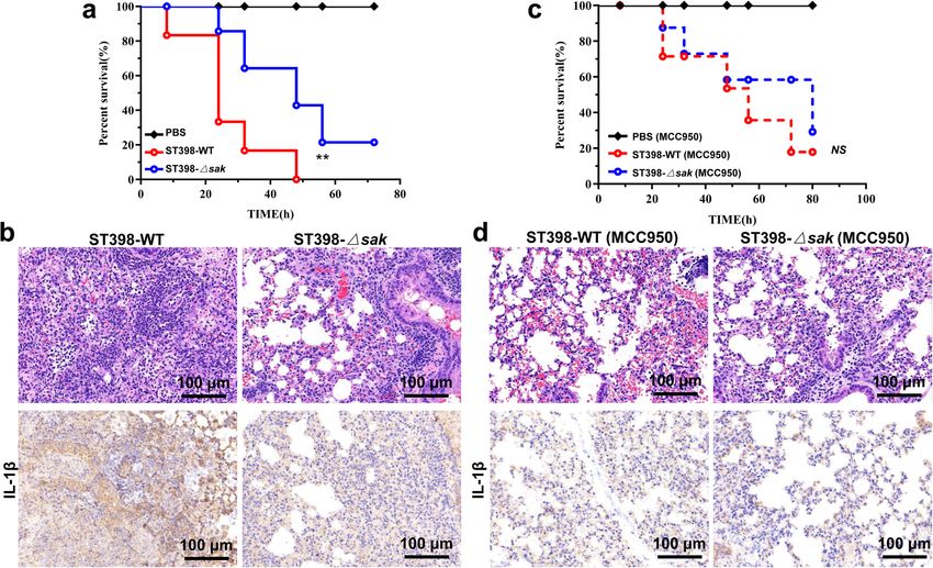
ARTICLE                                                             COMMUNICATIONS BIOLOGY | https://doi.org/10.1038/s42003-022-03571-x

S
        taphylococcus aureus (S. aureus) is one of the most                   Although the sak gene is conserved in HO-SA, we found that
        important pathogens and can cause a wide variety of                the expression level of sak in CA-SA was significantly higher than
        infections1,2. During the past decade, S. aureus has become        that in HA-SA (Fig. 1b). It has been shown that the increased
an important cause of community acquired pneumonia3–5. In                  expression of pathogenicity-related genes is responsible for the
contrast with hospital-associated S. aureus (HA-SA), which                 high virulence of CA-SA21,22. We speculate that the high
usually infects patients with impaired immunity, community-                expression of SAK may also play an important role in promoting
associated S. aureus (CA-SA) infections can occur in other                 the pathogenesis of CA-SA. In addition, we used a chromogenic
healthy individuals, suggesting that these bacterial strains have          assay to measure the level of SAK secreted by bacteria in cultures
greater virulence6.                                                        with different incubation times. The results showed that the high
   High expression of virulence genes that promote invasive                SAK secretion level of CA-SA reached its peak in the logarithmic
infections plays an important role in the pulmonary pathology              growth phase and remained stable (Fig. 1c). Clinical isolates of S.
associated with CA-SA related pneumonia5,7,8. However, excessive           aureus exhibit similar growth states under in vitro culture
local inflammation and tissue damage can also impede bacterial              conditions (Supplementary Fig. 1a), and we also quantified SAK
clearance9. Mouse pneumonia models lacking innate immune                   levels in stationary phase (12 h) supernatants by using standard
signal components showed significantly improved outcomes when               dilutions of recombinant SAK and found that CA-SA secreted
infected by S. aureus10,11. The NLRP3 inflammasome is essential             SAK concentrations of approximately 3 μg/ml, at least 5-fold
for the host’s immune defense against bacterial, fungal and viral          higher than HA-SA (Supplementary Fig. 1b).
infections12. But, when NLRP3 is dysregulated or overactivated, the
activated Caspase-1 promotes the maturation of IL-1β and IL-18,
                                                                           SAK negatively regulates the biofilm formation in
facilitates the recruitment of neutrophils and participates in the
                                                                           plasminogen-dependent manner. SAK can promote the activa-
inflammatory process of acute lung injury damage9,13–15.
                                                                           tion of human plasminogen and exert its fibrinolytic function.
   Recent evidence suggests that the epidemic HA-SA clones in
                                                                           However, SAK can only activate plasminogen in some specific
China mainly belong to ST5 and ST239, while the main prevalent
                                                                           hosts, including humans, rabbits, and sheep, but not mice26.
clones of CA-SA include ST59, ST398, ST188 and ST116–18. The
                                                                           Studies in mice expressing human plasminogen showed that SAK
ST398 clone was previously considered to be livestock-associated,
                                                                           can inhibit the formation of biofilms26. Biofilms are involved in
but more and more epidemiological studies have shown that
                                                                           antibiotic resistance of bacteria and host immune defenses31. But
ST398 isolates spread in community populations are usually not
                                                                           the highly virulent S. aureus often causes invasive infections21
related to animal infections19. The isolation rate of ST398 con-
                                                                           through high expression of virulence-related genes, and their
tinues to increase, and it has become one of the most prevalent
                                                                           biofilm formation ability is usually weaker than HA-SA (Fig. 1d).
CA-SA clones in Shanghai, China16,19,20. We have reported the
                                                                              By detecting the activation effect of SAK on plasminogen, we
effects of high expression levels of virulence genes such as the
                                                                           confirmed the successful construction of sak gene knockout strain
ESAT-6 secretion system on the pathogenicity of ST39821,22. In
                                                                           and the sak complementary strain (Supplementary Fig. 1c). We
addition, prophage also plays an important role in bacterial
                                                                           found that sak deletion did not affect the growth of ST398
pathogenicity and evolution23,24. The gene encoding staphyloki-
                                                                           (Supplementary Fig. 1d) and the formation of biofilm in TSB
nase (SAK) is located on prophage 325 and it is conserved in
                                                                           medium (Fig. 1e). Furthermore, the addition of SAK protein did
human adapted S. aureus (HO-SA)19. SAK was originally found
                                                                           not inhibit the formation of biofilms, but after adding human
to promote the activation of plasminogen and exert its fibrinolytic
                                                                           plasma, significant differences were observed between the wild-
function in specific hosts26,27. Moreover, studies have shown that
                                                                           type and the sak knockout strain (Fig. 1e). This indicates that the
SAK can bind to α-defensins28, human cathelicidin LL-37 and
                                                                           ability of SAK to inhibit biofilm formation is achieved primarily
mouse cathelicidins-related antimicrobial peptide (CRAMP)29 to
                                                                           by promoting fibrinolysis in specific hosts, and these results are
regulate fibrinolysis and evade innate immunity defenses. While
                                                                           consistent with the research of Kwiecinski et al26. Although CA-
the research on SAK has often focused on its fibrinolytic function,
                                                                           SA with high SAK expression showed weak biofilm formation
the role of SAK in the pathogenesis of S. aureus remains insuf-
                                                                           ability (Fig. 1b–d), we speculated that this is more likely due to
ficient and controversial.
                                                                           the high expression level of the important virulence regulator
   In the present study, our results showed increased expression of
                                                                           system, such as accessory gene regulator (Agr) system (Supple-
SAK in ST398 and ST59 isolates, pointing to a potential role of SAK
                                                                           mentary Fig. 2a). High expression of the agr locus can
in the virulence of CA-SA. Moreover, we demonstrated a sig-
                                                                           significantly inhibit the formation of biofilms32, which is
nificant role of SAK in ST398 infection-induced acute pneumonia
                                                                           consistent with our results (Supplementary Fig. 2b). The Agr
model, which is mainly achieved by activating innate immune
                                                                           system positively regulates a variety of virulence-related genes,
signals, especially NLRP3 inflammasome-related pathways.
                                                                           and the expression of sak is also regulated by it, but the knockout
                                                                           of sak gene does not affect the expression of agr (Supplementary
                                                                           Fig. 2c, d). Therefore, we speculated that the inhibitory effect of
Results
                                                                           SAK on biofilm formation mainly depends on fibrinolysis.
sak is conserved in HO-SA and highly expressed in CA-SA.
Prophage 3 is considered as a potential molecular marker to
distinguish livestock-adapted S. aureus (LA-SA) from HO-SA30.              SAK of ST398 enhances the severity of acute lung infection in
In the present study, we detected the carrying rate of sak in the          mice. It is reported that SAK can directly bind to mouse CRAMP,
predominant HA-SA (ST5, ST239), CA-SA (ST59, ST188, ST398)                 helping to promote fibrinolysis and evasion of the host’s innate
and LA-SA (ST188, ST398, ST97, ST520) isolates of China. The               immunity29, so we used both wild-type and CRAMP−/− mice for
results showed that 96-100% of HO-SA carried the sak gene, and             study. The lung tissues of ST398-infected wild-type mice showed
there was no significant difference between HA-SA and CA-SA,                obvious congestion and edema (Fig. 2a). HE staining of the lung
while the detection rate of sak in LA-SA was only 0–28% (Fig. 1a).         tissues showed that the alveolar structure of mice was destroyed
Among these LA-SA, ST398 and ST188, which carry a higher                   after infection with the ST398 strain, with infiltration of a large
proportion of sak than ST97 and ST520, can be isolated from                number of inflammatory cells (Fig. 2a). Immunohistochemistry
both humans and animals. These results further confirmed that               analysis of lung tissues also proved that ST398-infected mice had
the carrier rate of sak is related to human infection.                     more macrophage infiltration, as shown by increased staining of

2                        COMMUNICATIONS BIOLOGY | (2022)5:618 | https://doi.org/10.1038/s42003-022-03571-x | www.nature.com/commsbio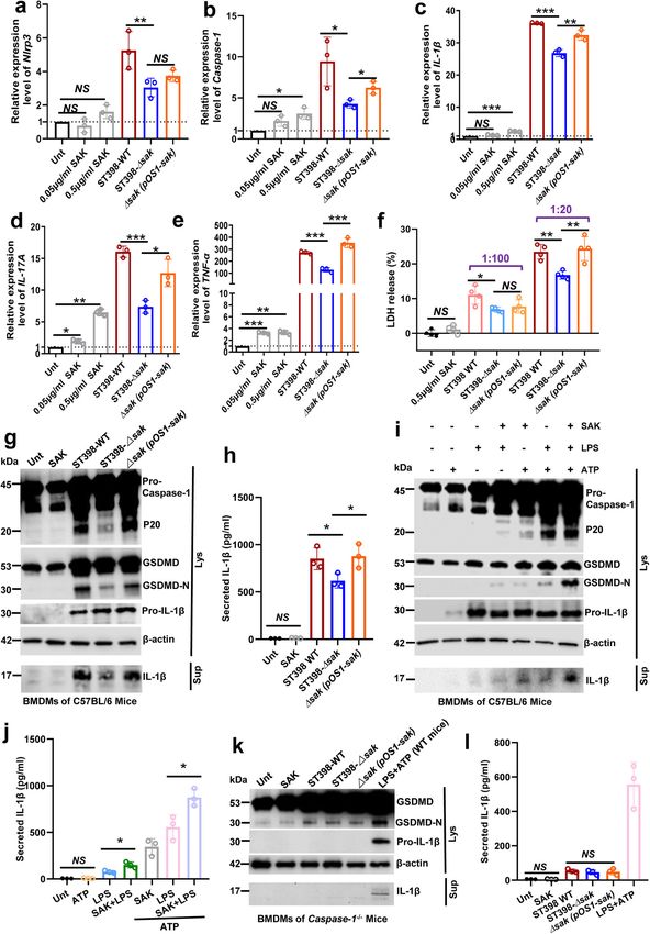
COMMUNICATIONS BIOLOGY | https://doi.org/10.1038/s42003-022-03571-x ARTICLE
Fig. 1 SAK is high expressed in CA-SA and can reduce the biofilm formation in plasminogen-dependent manner. a The prevalence of sak gene in
S. aureus (SA) epidemic isolates. Deep blue: humans-adapted (HO) isolates (including CA-SA and HA-SA); Light blue: livestock-adapted (LA) isolates.
Unpaired t test was used for statistical analyses between HO-SA and LA-SA after Shapiro–Wilk normality test. b qRT-PCR analysis of sak gene expression
in randomly selected clinical CA-SA (ST398, ST59) and HA-SA (ST239, ST5) isolates, at 4 h of in vitro growth. Relative mRNA levels were calculated using
gyrb as control and expressed as 2^(−ΔΔCt). Unpaired t test was used for statistical analyses between CA-SA and HA-SA after Shapiro–Wilk normality
test. c Measurement of SAK secretion at different incubation times. Two-way ANOVA with Bonferroni’s multiple comparison post-test was used for
statistical analyses (at the 12th h). d Biofilm formation ability of CA-SA (ST398, ST59) and HA-SA (ST239, ST5). Unpaired t test was used for statistical
analyses between CA-SA and HA-SA after Shapiro–Wilk normality test. e The effect of adding SAK protein, human plasma and mice plasma on the biofilm
formation of ST398-WT, sak deletion mutant isolate and the complemented isolate. Two-way ANOVA with Bonferroni’s multiple comparison post-test was
used for statistical analyses. All data in Fig. 1 are presented as mean ± SD and *p < 0.05, ** p < 0.01, *** p < 0.001.
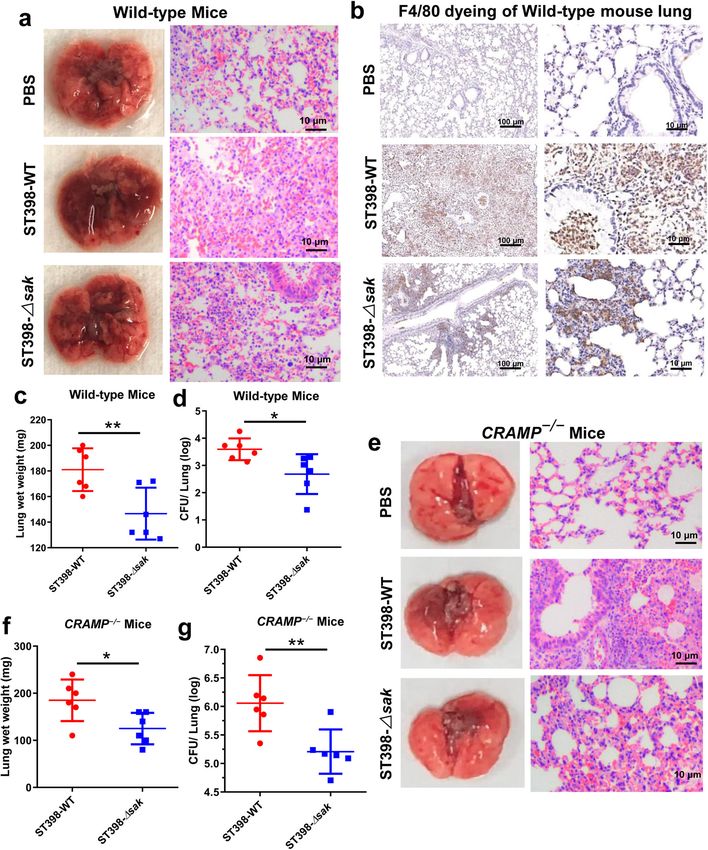
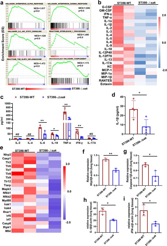
F4/80, a well characterized macrophage marker (Fig. 2b). Fur- such as interferon-γ (IFN-γ), interferon-α (IFN-α), interleukin-6
thermore, the wet weight of the lungs and the colony-forming (IL-6), interleukin-1 (IL-1), complement-related genes, and
unit (CFU) counts in lung tissues of ST398-infected mice NLRP3 inflammasome-related genes were enriched in the lung
increased significantly (Fig. 2c, d). Interestingly, similar results tissues of wild-type infected mice (Fig. 3a). Furthermore, we
can be seen in CRAMP−/− mice (Fig. 2e–g). After infection with detected 23 cytokines in mouse serum and 8 cytokines in mouse
the sak knockout strain, both wild-type mice and CRAMP−/− bronchoalveolar lavage fluid (BALF), and the results showed that
mice had milder lung pathological changes. Therefore, we mice infected with wild-type ST398 had higher levels of most
speculated that SAK can increase the severity of acute lung cytokines in both serum (Fig. 3b) and BALF (Fig. 3c, d). In the
infection in mice, and the effect of combining with CRAMP is not host’s confrontation with pathogens, excessive inflammation can
the main mechanism of SAK’s pathogenicity. cause tissue damage and poor prognosis. The activation of
NLRP3 has been shown to promote cytokine production and cell
pyrolysis. We further compared the expression of toll-like
SAK promotes NLRP3 inflammasome-related gene transcrip- receptors and other NLRP3 inflammasome activation-related
tion and cytokine release in CA-SA pneumonia. In order to genes in the two groups. Consistent with the higher bacterial load,
explore the mechanism of SAK in the process of S. aureus nearly all of these molecules are highly expressed in the lung
infection, we analyzed the RNA sequencing data of lung tissues tissues of mice infected with wild-type ST398 (Fig. 3e). In addi-
from wild-type and sak gene knockout ST398 infected mice tion, we detected the expression of IL-1β, IFN-α, Nlrp3 and
(C57BL/6 wild-type). A total of 580 genes were differentially Caspase-1 in lung tissues by qRT-PCR, and the results were
expressed between the two groups (See Supplementary data 1 for consistent with RNA sequencing (Fig. 3f–i). Combined with
the top 50 significantly different genes). In addition, the results of the bacterial load and pathological changes in mouse lung tissue,
GSEA analysis identified that genes related to cytokine response, we speculate that the lung tissue of mice infected with wild-type
COMMUNICATIONS BIOLOGY | (2022)5:618 | https://doi.org/10.1038/s42003-022-03571-x | www.nature.com/commsbio 3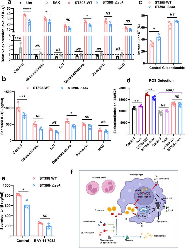
ARTICLE                                                                  COMMUNICATIONS BIOLOGY | https://doi.org/10.1038/s42003-022-03571-x

Fig. 2 SAK enhances CA-SA-mediated lung infection in both wild-type and CRAMP−/− mice. a–g 2 × 10⁸ CFU of S. aureus was pipetted into the nares of
anesthetized C57BL/6 wild-type mice a–d and CRAMP−/− mice e–g (n = 6), and mice were euthanized 48 h after inoculation with S. aureus.
a, e Photographs of lungs and corresponding H&E stained sections of mice 48 h after infection. b Immunohistochemical staining (F4/80) was performed
on the lung tissue of infected mice to observe the infiltration of macrophages. c, f Lung wet weight of infected mice. d, g The left lung was homogenized and
plated on TSB agar for CFU determination. Unpaired t test was used for statistical analyses between ST398-WT and sak deletion mutant isolate infected
mice after Shapiro–Wilk normality test. Data are presented as mean ± SD and *p < 0.05, ** p < 0.01.

4                            COMMUNICATIONS BIOLOGY | (2022)5:618 | https://doi.org/10.1038/s42003-022-03571-x | www.nature.com/commsbio
COMMUNICATIONS BIOLOGY | https://doi.org/10.1038/s42003-022-03571-x                                                           ARTICLE

ST398 is in a more active state of inflammatory response than                   bacterial culture supernatant or SAK protein and extracted the
that of sak gene knockout ST398 infected mice.                                 mRNA of the cells. Gene expression was detected by qRT-PCR
                                                                               after reverse transcription, and the results showed that SAK
                                                                               can effectively promote the expression of innate immune
SAK mainly play a role in the priming step of NLRP3                            inflammation-related factors, such as cytokines (IL-17A, TNF-α,
inflammasome activation and promotes pyroptosis. We incu-                       IL-1β) and NLRP3 inflammasome-related genes (Fig. 4a–e). In
bated bone marrow-derived macrophages (BMDMs) of mice with                     addition, we assessed cell death by measuring LDH released by

COMMUNICATIONS BIOLOGY | (2022)5:618 | https://doi.org/10.1038/s42003-022-03571-x | www.nature.com/commsbio                                5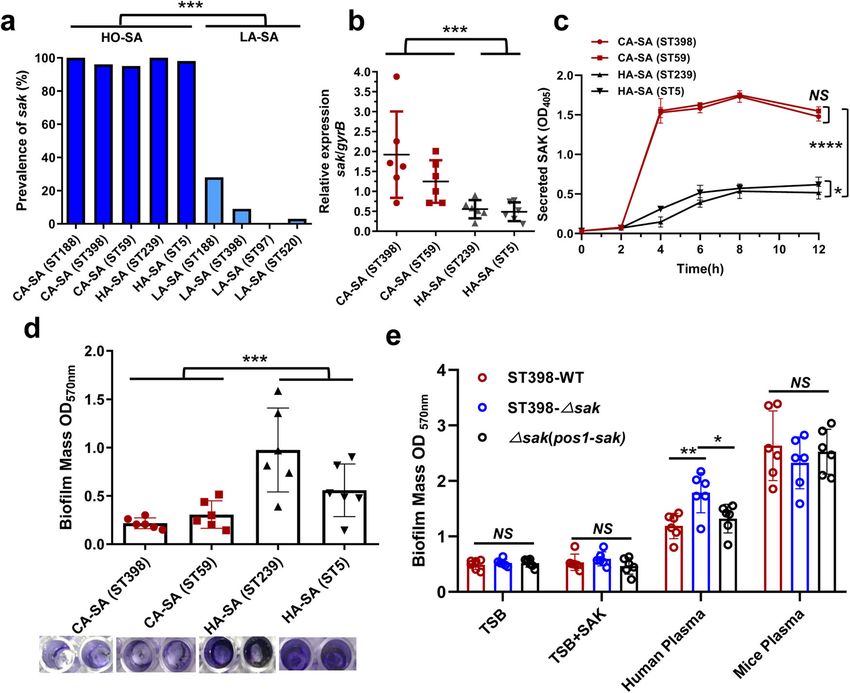
ARTICLE                                                                  COMMUNICATIONS BIOLOGY | https://doi.org/10.1038/s42003-022-03571-x

Fig. 3 SAK promotes NLRP3 inflammasome-related gene transcription and cytokine release in CA-SA pneumonia. a–i 2 × 10⁸ CFU of S. aureus was
pipetted into the nares of anesthetized C57BL/6 wild-type mice. Bronchoalveolar lavage fluid (BALF) was taken 48 h after infection of mice, and total RNA
was extracted from lung tissue at the same time. a GSEA analysis identified cytokine related genes (IFN-α, IFN-γ, IL-6, IL-1), complement related genes and
NLRP3 inflammasome pathway enrichment in the ST398-WT infected mice. b The level of mouse plasma cytokine was detected by Bio-Plex Pro Mouse
Cytokine 23-plex Assay. The data were normalized by the Z score according to the standard deviation from the mean. c Cytokine levels in mouse BALF
were detected by BD Cytometric Bead Array (CBA) Mouse Th1/Th2/Th17 Cytokine Kit (n = 4). Unpaired t test was used for statistical analyses after
Shapiro–Wilk normality test and data are presented as mean ± SD. d Detection of IL-1β in mouse BALF by ELISA (n = 4). Unpaired t test was used for
statistical analyses after Shapiro–Wilk normality test and data are presented as mean ± SD. e Comparison of the expression levels of NLRP3 inflammasome
activation-related molecules between the two groups. The data were normalized by the Z score according to the standard deviation from the mean.
f–i qRT-PCR analysis of Nlrp3, Caspase-1, IL-1β, and IFN-α in the lung tissues of wild-type or sak deletion mutants infected mice. Relative mRNA levels were
calculated using β-actin as control and expressed as 2^(-ΔΔCt). Unpaired t test was used for statistical analyses after Shapiro–Wilk normality test and data
are presented as mean ± SD. See Supplementary methods for the abbreviated list of cytokines or genes in Fig. 3b, e. *p < 0.05, ** p < 0.01.

BMDMs after co-incubation with SAK or S. aureus supernatants,                  sections of mice infected with wild-type ST398 showed a high
and the results showed that sak gene knockout significantly                     degree of inflammatory cell infiltration, and immunohistochem-
reduced macrophage death (Fig. 4f).                                            istry analysis showed high levels of IL-1β (Fig. 5b). In addition,
   We further detected the ability of SAK to active NLRP3                      the NLRP3 inflammasome can be inhibited by intraperitoneal
inflammasome by western blot. It should be noted that the sak                   injection of MCC950 to mice. When mice were given NLRP3
gene knockout attenuated the activation of NLRP3 inflamma-                      inhibitors, their survival was prolonged, and there was no sig-
some by bacterial supernatants, which was reflected by reduced                  nificant difference in the survival rate of mice infected with wild-
levels of Caspase-1 maturation, gasdermin D (GSDMD) cleavage                   type ST398 and sak knockout strains (Fig. 5c). This indicates that
(Fig. 4g) and decreased IL-1β secretion (Fig. 4h). But we did not              NLRP3 inflammasome activation plays an important role in
observe a significant increase in the activation of NLRP3                       SAK’s promotion of the pathogenicity of ST398. Furthermore, the
inflammasome after SAK protein treatment (Fig. 4g, h,                           inhibitory effect of MCC950 on the NLRP3 inflammasome was
Supplementary Fig. 3a). The classic NLRP3 inflammasome                          confirmed by immunohistochemical staining of IL-1β, and HE
activation is considered to be a two-step process involving                    staining of lung tissue sections showed that inhibiting NLRP3
priming and activation33. We first examined the role of SAK in                  inflammasomes can reduce the recruitment of inflammatory cells
NLRP3 priming and the results showed increased expression of                   and weaken the inflammatory damage caused by infection
pro-Caspase-1, GSDMD and pro-IL-1β in SAK-treated BMDMs                        (Fig. 5d).
upon addition of ATP. Furthermore, we could observe the
maturation of Caspase-1 and GSDMD, as well as the release of
IL-1β, which also indicated that the NLRP3 inflammasome was                     SAK promotes NLRP3 inflammasome activation mainly by
successfully activated, although the effect was not as strong as that          increasing K+ efflux, reactive oxygen species (ROS) produc-
of lipopolysaccharide (LPS) (Fig. 4i, j, Supplementary Fig. 3b).               tion, and activating NF-κB signaling. The NLRP3 inflamma-
Interestingly, we also found that the addition of SAK could                    some can be activated by a variety of stimuli, such as ion flux,
further promote the activation of the NLRP3 inflammasome by                     production of ROS, and lysosomal damage12. Most NLRP3
LPS (Fig. 4i, j, Supplementary Fig. 3b), implying that SAK may                 inflammasome activators lead to K+ efflux, and in some studies,
have additive effects with other NLRP3 inflammasome activators,                 K+ efflux has even been considered a necessary upstream event
helping to further amplify inflammasome signaling. In addition,                 for NLRP3 activation34. We pretreated cells with the K+ efflux
we primed the BMDMs for 3 h with LPS and then treated the                      inhibitor glibenclamide and high concentrations of extracellular
LPS-primed cells with SAK for determining whether purified SAK                  K+, and then tested whether SAK could effectively promote the
can activate NLRP3 inflammasome. Our results suggest a limited                  expression and release of IL-1β in BMDMs. The results showed
role for SAK in the activation step of NLPR3 inflammasome                       that although glibenclamide failed to eliminate the difference in
(Supplementary Fig. 3c–e). Although the level of IL-1β release in              IL-1β expression in macrophages caused by the supernatant of
the SAK-treated group showed an upward trend, the difference                   ST398 wild-type and sak gene knockout strain (Fig. 6a), glib-
was not statistically significant, as was the result of western blot            enclamide could inhibit SAK from promoting the secretion of
densitometry (Supplementary Fig. 3d, e). Moreover, when the                    IL-1β from cells (Fig. 6b). Moreover, under the effect of high
BMDMs of Caspase-1−/− mice was incubated with purified SAK                      concentrations of extracellular K+, SAK no longer exhibited
or bacterial supernatants, SAK failed to promote GSDMD                         additional effects on promoting IL-1β expression and release
cleavage and IL-1β maturation (Fig. 4k, l, Supplementary Fig. 3f).             (Fig. 6a, b). In addition, we also examined intracellular K+ levels
Furthermore, by using the NLRP3 inhibitor MCC950, BMDMs of                     and found that the presence of SAK promoted K+ efflux, which
wild-type mice yielded similar results to Caspase-1−/− mice                    could also be inhibited by glibenclamide (Fig. 6c).
(Supplementary Fig. 3g–i). These results suggest that SAK may                     Lysosomal disruption results in the release of proteases and
enhance the host’s inflammatory response after S. aureus                        phagocytic granules from the lysosome, which promotes the
infection and that SAK may play a role in the priming step of                  activation of the NLRP3 inflammasome35. We pretreated
NLRP3 inflammasome activation to promote pyroptosis and IL-                     BMDMs with dexamethasone as a stabilizer for lysosomal
1β release.                                                                    membranes and found that SAK may not promote IL-1β
                                                                               expression and release by disrupting lysosome (Fig. 6a, b).
                                                                                  NADPH oxidase is one of the main sources of ROS in
NLRP3 inflammasome is required for the effective of SAK                         macrophages, and to explore the role of SAK in ROS production,
induced CA-SA lung tissue damage in mice. We constructed a                     we used the NADPH oxidase inhibitor apocynin and the ROS
lethal mouse model of pneumonia to visually show the effect of                 scavenger N-acetyl-L-cysteine (NAC) to pretreat BMDMs. The
SAK on the pathogenicity of ST398. The results showed that sak                 results showed that both apocynin and NAC could effectively
gene knockout can improve the survival rate of ST398-infected                  inhibit the promoting effect of SAK on the expression and release
mice (Fig. 5a). Correspondingly, HE staining of lung tissue                    of IL-1β (Fig. 6a, b). In addition, purified SAK protein could

6                            COMMUNICATIONS BIOLOGY | (2022)5:618 | https://doi.org/10.1038/s42003-022-03571-x | www.nature.com/commsbio
COMMUNICATIONS BIOLOGY | https://doi.org/10.1038/s42003-022-03571-x ARTICLE directly promote the production of ROS in BMDMs, and its effect findings. Furthermore, we measured the cytokines released by was also inhibited by NAC (Fig. 6d). human THP-1 cells (human monocyte derived cell line) after Notably, all the above inhibitors significantly inhibited the stimulation with bacterial culture supernatant. Similar to the promoting effect of purified SAK on IL-1β expression, we results in mouse models, SAK can also stimulate the release of speculate that this result is due to the stronger effect of the inflammatory factors (IL-1β, TNF-α, IL-17A) in THP-1 cells inhibitor than SAK and also indicates that purified SAK is not a (Supplementary Fig. 4a–c). Moreover, SAK can also promote the potent NLRP3 activator, which is consistent with our previous production of ROS in THP-1 cells, and this effect can be inhibited COMMUNICATIONS BIOLOGY | (2022)5:618 | https://doi.org/10.1038/s42003-022-03571-x | www.nature.com/commsbio 7
ARTICLE                                                                 COMMUNICATIONS BIOLOGY | https://doi.org/10.1038/s42003-022-03571-x

Fig. 4 SAK mainly play a role in the priming step of NLRP3 inflammasome activation and promotes pyroptosis. a–e BMDMs of mice were incubated
with bacterial culture supernatant (stationary phase, 1:20 dilution) or SAK (0.05/0.5 μg/ml) for 3 h. The mRNA of the cells was extracted and gene
expression relative to the untreated (Unt) group was detected by qRT-PCR after reverse transcription. Relative mRNA levels were calculated using β-actin
as internal control and expressed as 2^(-ΔΔCt). Unpaired t-test was used to compare the differences between the variables after Shapiro–Wilk normality
test. f Determination of macrophage viability by LDH assay. BMDMs were incubated with bacterial culture supernatant (stationary phase, 1:100 or 1:20
dilution) or SAK protein (0.5 μg/ml) for 3 h. Unpaired Student’s t test was used for statistical analyses of differences between groups after Shapiro–Wilk
normality test. g, h BMDMs of mice were treated with SAK (0.5 μg/ml) or bacterial supernatants (stationary phase, 1:20 dilution) for 3 h. Immunoblot
analysis g of cell lysates or culture supernatants. Secreted IL-1β from BMDMs was detected by ELISA h. i, j BMDMs of mice were treated with SAK
(0.5 μg/ml, 3 h) or LPS (0.2 μg/ml, 3 h), followed by the treatment with ATP (2.5 mM, 30 min). Immunoblots i of cell lysates or culture supernatants from
mouse BMDMs. Secreted IL-1β from BMDMs was detected by ELISA j. k, l BMDMs from Caspase-1−/− mice were treated with SAK (0.5 μg/ml, 3 h) or
bacterial supernatants (stationary phase, 1:20 dilution, 3 h). Immunoblot analysis k of cell lysates or culture supernatants. Secreted IL-1β from BMDMs of
Caspase-1−/− mice was detected by ELISA l. Cell lysates or culture supernatants of wild-type mouse BMDMs treated with LPS (0.2 μg/ml, 3 h) and ATP
(2.5 mM, 30 min) were used as positive control. Unpaired t test was used to compare the differences between the variables after Shapiro–Wilk normality
test. All data are presented as mean ± SD and *p < 0.05, ** p < 0.01, *** p < 0.001.

Fig. 5 Inhibition of the NLRP3 inflammasome can reduce lung infection in mice. a–d 109 CFU of S. aureus was pipetted into the nares of anesthetized
C57BL/6 mice (n = 6), and 50 mg/kg MCC950 sodium (NLRP3 inflammasome inhibitor) or PBS was injected intraperitoneally to mice 2 h before infection.
a, c Survival curves were compared using a log-rank (Mantel-Cox) test. b, d Tissue sections (48 h after infection) for HE staining and immunohistochemical
staining (IL-1β). **p < 0.01.

by NAC (Supplementary Fig. 4d). This indicates that the                       inflammation-related factors by activating NF-κB, which may
activation of the host inflammatory response by SAK is also                    also be one of the mechanisms by which SAK acts on the priming
applicable to human S. aureus infections.                                     step of the NLRP3 inflammasome.
   Activation of NF-κB can promote the expression of various
inflammatory factors and has been shown to play an important
role in the activation of NLRP3, especially in the priming step36.            Discussion
In the present study, we found that purified SAK could promote                 SAK has been demonstrated to play an important role in the
the phosphorylation of NF-κB by western blot, and sak gene                    activation of plasminogen in specific hosts26. Previous studies
knockout significantly reduced NF-κB activation in ST398                       showed that SAK can directly bind to human α-defensins28 and
supernatant-stimulated BMDMs (Supplementary Fig. 5a). In                      murine CRAMP, and play a role in evading the host’s innate
addition, the effect of SAK was attenuated after the application of           immunity29. Our results showed that SAK is highly expressed in
the NF-κB pathway inhibitor BAY 11-7082 (Supplementary                        the highly virulent CA-SA, however, the role of SAK in the
Fig. 5b), and there was no statistically significant difference in the         pathogenesis of S. aureus remains largely unknown. In this study,
release of IL-1β from BMDMs after treatment with the super-                   our data suggest that SAK negatively regulates biofilm formation
natant of ST398 wild strain and sak knockout strain (Fig. 6e).                in a plasminogen-dependent manner. Furthermore, we found
These results suggest that SAK may promote the transcription of               that SAK could promote CA-SA-mediated lung infection in the

8                           COMMUNICATIONS BIOLOGY | (2022)5:618 | https://doi.org/10.1038/s42003-022-03571-x | www.nature.com/commsbio
COMMUNICATIONS BIOLOGY | https://doi.org/10.1038/s42003-022-03571-x                                                               ARTICLE

CRAMP-independent manner. In our present study, our results                    more likely due to the high expression of the Agr system. In
showed that SAK appears to play an important role in acute                     addition, the expression of SAK is also positively regulated by the
necrotizing pneumonia by exerting pro-inflammatory effects,                     Agr system. Furthermore, it has been proven that SAK can
particularly by promoting the activation of NLRP3 inflamma-                     directly bind to human α-defensins28 and murine CRAMP, and
some. Mechanistical investigation reveal that SAK contributes the              play a role in evading the host’s innate immunity29. In this study,
priming step of NLRP3 inflammasome activation through                           our data indicate that high expression of SAK may promote lung
increasing K+ efflux and ROS production, and activating NF-κB                   infection of ST398 isolates in both wild-type and CRAMP−/−
signaling (Fig. 6f). Taken together, our findings suggest that SAK              mice. Due to the limited effect of SAK on plasminogen in mice,
exacerbates CA-SA-induced lung infection by promoting NLRP3                    we speculate that promoting fibrinolysis and evading host innate
inflammasome activation.                                                        immunity may not be the main mechanism of SAK aggravating
   The effect of SAK on biofilm formation is human plasmino-                    pneumonia in ST398-infected mice.
gen-dependent, which is similar with other reports26,27. It is                    Antimicrobial peptides act primarily in the early stages of
worth noting that although the SAK highly expressed CA-SA                      infection and are thought to be essential innate immune effectors
biofilm formation ability is weak, there is no evidence that SAK                against bacterial infections37. In addition, inflammasomes also
can directly inhibit the formation of S. aureus biofilm. It has been            play important roles in the detection and clearance of pathogens
found that the increased expression of virulence factors controlled            during infection38. S. aureus has a variety of factors that promote
by Agr is responsible for the high pathogenicity of ST398, and we              infection, and different infection sites trigger different immune
speculate that the weak biofilm formation ability of CA-SA is                   responses in the host. Research shows that NLRP3 signaling plays

COMMUNICATIONS BIOLOGY | (2022)5:618 | https://doi.org/10.1038/s42003-022-03571-x | www.nature.com/commsbio                                     9
ARTICLE                                                                  COMMUNICATIONS BIOLOGY | https://doi.org/10.1038/s42003-022-03571-x

Fig. 6 SAK promotes the activation of NLRP3 inflammasome mainly through increasing K+ efflux and ROS production, and activating NF-κB signaling.
a–d BMDMs were pretreated with glibenclamide (10 μM), high concentrations of extracellular K+ (25 mM), apocynin (200 μM), dexamethasone
(200 nM), and NAC (1 mM) for 30 min, and then incubated with SAK (0.5 μg/ml) or bacterial secretion supernatant for 3 h (stationary phase, 1:20
dilution). Two-way ANOVA with Bonferroni’s multiple comparison post-test was used to compare the differences between the variables. a Relative mRNA
levels of IL-1β were calculated using β-actin as internal control and expressed as 2^(-ΔΔCt). The Y-axis represents the fold change of the IL-1β gene
expression level in the experimental group relative to the untreated group. b The secretion of IL-1β in BMDMs treated with bacterial culture filtrate was
detected by ELISA. Two-way ANOVA with Bonferroni’s multiple comparison post-test was used to compare the differences between the variables.
c Detection of intracellular K+ of BMDMs. Two-way ANOVA with Bonferroni’s multiple comparison post-test was used to compare the differences
between the variables. d Detection of ROS production by BMDMs. BMDMs were pretreated with DMSO or NAC (1 mM) for 30 min. Two-way ANOVA
with Bonferroni’s multiple comparison post-test was used to compare the differences between the variables. e Mouse BMDMs were pretreated with BAY
11-7082 (10 μM, 30 min) or DMSO, then cells were treated with SAK (0.5 μg/ml) or bacterial secretory supernatant for 3 h (stationary phase, 1:20
dilution). The secretion of IL-1β in BMDMs was detected by ELISA. Two-way ANOVA with Bonferroni’s multiple comparison post-test was used to
compare the differences between the variables. All data in Fig. 6 are presented as mean ± SD and *p < 0.05, ** p < 0.01, *** p < 0.001. f The role of SAK in
Staphylococcus aureus infection. ① SAK can neutralize AMPs and regulate fibrinolysis. ②SAK promotes transcription of inflammation-related genes and
activation of NLRP3 inflammasome (this study). Research has shown that SAK can promote the activation of plasminogen in specific hosts (human, rabbits,
and sheep) and exert its fibrinolytic function (blue arrows). The epithelium expresses antimicrobial peptides to kill extracellular bacteria. Studies have
shown that SAK can bind to α-defensins, human cathelicidin LL-37 and mouse CRAMP to regulate fibrinolysis and evade innate immunity defenses (green
arrows). In this study, we speculate through preliminary studies that SAK can promote the transcription of inflammatory response-related molecules and
the release of cytokines, help recruit polymorphonuclear leukocytes (PMNs) and contribute to a more active state of inflammatory response in the host.
Our results suggest that SAK may contribute to the activation of NF-κB and promote K+ efflux and ROS production, thereby playing a role in the activation
of the NLRP3 inflammasome (red arrows) and promoting acute lung infection. This figure was drawn by using BioRender (Agreement number:
JV23RYUB4V. https://app.biorender.com/) and Photoshop.

an important role in the body’s defense against skin and soft                  knockout strain. Due to the presence of many virulence factors in
tissue infections38–40. However, in acute pneumonia caused by                  bacterial supernatants, SAK may have additive effects with other
highly virulent CA-SA, inflammasome signaling is not necessary                  NLRP3 activators, ultimately promoting further activation of the
for clearance of S. aureus from mouse lung tissue38, and even                  NLRP3 inflammasome. In addition, SAK can also promote
leads to inflammation-related cell death41. In acute lung infec-                cytokine release and ROS production in human THP-1 cells,
tions caused by high virulence CA-SA, uncontrolled excessive and               suggesting that the activation of host inflammatory response by
long-term inflammation induced by virulence factors can lead to                 SAK is also applicable to human infection. The mechanism
cell death and tissue damage9,41,42, thereby hindering bacterial               by which SAK helps S. aureus evade host innate immunity by
clearance11,43. In particular, alveolar macrophage loss and                    inhibiting antimicrobial peptides is different from SAK’s pro-
immunomodulatory dysfunction caused by Caspase-1-dependent                     motion of inflammasome activation in this study, but both con-
pyroptosis amplify the pathological consequences of                            tribute to the promotion of S. aureus infection. Notably, the
infection13,41. It is worth noting that RNA sequencing analysis of             supernatant of the sak knockout strain could still activate the
mouse lung tissue showed that SAK can promote the activation of                NLRP3 inflammasome, leading to pyroptosis and IL-1β release,
the innate immune inflammatory response including the NLRP3                     which may be driven by other important virulence factors
inflammasome. In addition, SAK can also increase the content of                 secreted by S. aureus. In fact, multiple studies have shown that
cytokines in serum and BALF of mice after S. aureus infection.                 α-toxin45, Panton-Valentine leukocidin46 and other S. aureus
These features of inflammatory hyperactivation are consistent                   exocrine toxins can effectively promote the activation of the
with stronger inflammatory cell infiltration and pulmonary                       NLRP3 inflammasome. Although classical NLRP3 inflammasome
edema in the lung tissue of wild-type ST398-infected mice.                     activation is thought to be achieved through a two-step process of
   The canonical NLRP3 inflammasome activation process is                       priming and activation, studies have shown that LPS alone can
thought to require a priming step prior to activation. Various                 also directly activate NLRP3 through an unknown mechanism47.
stimuli including bacterial toxins can activate the NLRP3                      Furthermore, the cellular events of NLRP3 activation are com-
inflammasome, but no direct interaction between NLRP3 and                       plicated by the fact that NLRP3 activators are able to trigger
these stimuli has been observed44. It seems that they may activate             multiple cellular signals and that there are also interactions
NLRP3 by inducing various cell signal transduction events,                     between these signals33.
including K+ efflux, ROS production and lysosomal damage12.                        The inhibition of NLRP3 may diminish pathology in S. aureus
Our results suggest that although SAK cannot directly activate the             pulmonary infection. This may be due to the fact that lung
NLRP3 inflammasome, SAK may play a role in the priming step                     infection is prone to severe pathological changes, and tissue
of NLRP3 inflammasome activation. This may be due to the fact                   destruction caused by excessive inflammation can lead to extre-
that SAK can promote the activation of NF-κB, thereby indirectly               mely impaired lung function48. Therefore, a balance of anti-
up-regulating the expression levels of inflammasome-related                     microbial immunity and overall inflammation is required to
molecules. Our further studies showed that SAK can promote                     reduce inflammatory damage to lung tissue and improve patient
ROS production and K+ efflux, which are important upstream                      outcomes45. We emphasize that in the pneumonia caused by
events of NLRP3 inflammasome activation, and may also be the                    CA-SA, the control of inflammation is as important as the anti-
mechanism by which SAK promotes NLRP3 inflammasome                              infection strategy, and that inhibition of the NLRP3 inflamma-
activation. Interestingly, although purified SAK played a modest                some may serve as a mechanism for severe acute S. aureus
role in priming the NLRP3 inflammasome, in the presence of an                   pneumonia adjuvant therapy.
effective NLRP3 inflammasome activator (such as LPS), SAK may                      In this study, we investigate the pathogenic mechanism by
further promote Caspase-1 maturation and IL-1β release. This                   which the pro-inflammatory effect of SAK promotes CA-SA
was also indicated by the ST398 secretion supernatant stimulated               infection in a mouse model of severe pneumonia. Previous studies
BMDMs to release higher levels of IL-1β compared to the sak                    pay more attention to the promoting effect of SAK on fibrinolysis.

10                           COMMUNICATIONS BIOLOGY | (2022)5:618 | https://doi.org/10.1038/s42003-022-03571-x | www.nature.com/commsbio
COMMUNICATIONS BIOLOGY | https://doi.org/10.1038/s42003-022-03571-x                                                                                       ARTICLE

Our study shows that SAK is also an effective pro-inflammatory                            (Qiagen). The 7500 Sequence Detector (Applied Biosystems) was used to perform
factor for acute infections caused by CA-SA. Our results suggest                         reactions in MicroAmp Optical 96-well reaction plates. Oligonucleotides used in
                                                                                         this study are presented in Supplementary Table 2.
that SAK may contribute to the activation of NF-κB and promote
K+ efflux and ROS production, thereby playing a role in the
activation of the NLRP3 inflammasome. This enriches our                                   RNA Sequencing. Mice were euthanized 48 h after infection and their lungs were
                                                                                         dissected. Total RNA was extracted from lung tissues (Qiagen). Nanodrop2000 was
understanding of the function of SAK, and also provides new                              used to detect the concentration and purity of the extracted RNA, agarose gel
ideas for the pathogenic mechanism of high virulence CA-SA.                              electrophoresis was used to detect RNA integrity, and the Agilent 2100 was used to
                                                                                         determine the RIN value. The library was constructed using the Illumina Tru-
                                                                                         seqTM RNA sample prep Kit method, and finally sequenced on the Illumina
Methods                                                                                  Novaseq 6000 platform (Shanghai Majorbio Bio-pharm Technology Co., Ltd).
Bacterial isolates. S. aureus strains were isolated from adult patients of Renji
Hospital affiliated with Shanghai Jiaotong University. The concentration of anti-
biotics used when constructing gene knockout strains is as follows: ampicillin,          Western blot. Overnight cultures (12–15 h) were diluted 1:100 into fresh TSB and
100 μg/ml; chloramphenicol, 10 μg/ml. Please see the Supplementary methods for           incubated at 37 °C with shaking until stationary phase (OD600nm = 5.0). The
bacterial growth conditions and molecular typing. In addition, bacterial strains and     purified SAK (0.5 μg/ml) or LPS (0.2 μg/ml) or S. aureus culture filtrates (1/20-fold
plasmids used in this study can be found in Supplementary Table 1.                       dilution) were added into mouse BMDMs cultured in a 6-well plate and incubate
                                                                                         for 3 h. Some groups need to add 2.5 mM ATP to incubate for another 30 min. In
                                                                                         some experiments, BMDMs were primed with LPS for 3 h, and then LPS-primed
Measurement of SAK secretion by S. aureus. Overnight cultures (12–15 h) were
                                                                                         cells were stimulated with SAK (0.5 μg/ml, 30 min) or ATP (2.5 mM, 30 min, as a
diluted 1:100 into fresh TSB and incubated at 37 °C with shaking at 200 rpm.
                                                                                         positive control) to determine whether purified SAK could activate the inflam-
Bacterial suspension at different time points was centrifuged (4000 rpm for 10 min
                                                                                         masome. Cells were collected after being washed by sterile PBS, and boiled in
at 4 °C) and the supernatant was collected after filtration. Human plasminogen
                                                                                         loading buffer. Then the samples were subjected to western blot experiment with
(Roche) was mixed with supernatant and incubated at 37 °C for 30 min, then a
                                                                                         Caspase-1 (Abcam, ab207802, 1:1000), GSDMD (Abcam, ab209845, 1:1000)
plasmin-specific chromogenic substrate S-2251 (3 mmol/L, Chromogenix) was
                                                                                         interleukin-1β (IL-1β, Abcam, ab234437, 1:1000), β-actin (Abcam, ab8227, 1:3000),
added and incubation was continued at 37 °C for 60 min. The amount of SAK
                                                                                         NF-κB p65(Cell Signaling Technology, 8242, 1:1000), Phospho-NF-κB p65 (Cell
secreted by the strains was read with a micro enzyme-linked immunosorbent assay
                                                                                         Signaling Technology, 3033, 1:1000) antibodies. The densitometry of each band
(ELISA) autoreader (BioRad) at 405 nm.
                                                                                         was quantified by Image J.

Semiquantitative biofilm assay. Overnight cultures (12–15 h) were diluted into
fresh TSB (with 0.5% glucose) to a final optical density of 0.1 (600 nm). The diluted     Cytokine detection. Anesthetize the mice with 2,2,2-tribromoethanol (3.75–5 mg/
S. aureus cultures were dispensed into 96-well flat-bottom tissue culture plates          25 g), then slowly pipet the inoculum with 2 × 10⁸ CFU into the nostrils of the
(200 μL/well), and incubated at 37 °C for 24 h without shaking. In some experi-          mice. The orbital venous plexus blood of mice was collected 48 h after infection,
ments, 10% plasma or purified SAK (2 µg/ml) were added into TSB medium (with              and the BALF was collected after euthanizing the mice. The level of mouse plasma
0.5% glucose). Heparin anticoagulated human plasma was provided voluntarily by           cytokine was detected by Bio-Plex Pro Mouse Cytokine 23-plex Assay in accor-
healthy subjects. Mouse plasma came from the experimental C57BL/6 mice. Both             dance with the manufacturer’s instructions (Bio-Rad, Luminex 200™ System).
human and mouse plasma were incubated at 56 °C for 30 min before use to                  Cytokine levels in mouse BALF were detected by BD Cytometric Bead Array (CBA)
inactivate complement. After incubation, the culture supernatant was gently              Mouse Th1/Th2/Th17 Cytokine Kit. BD FACSCantoII was used for data collection
removed and the wells were washed twice with sterile phosphate buffered saline           and FCAP Array V3.0 was used for data analysis. IL-1β in the BALF and mouse
(PBS). Bouin’s fixative was added to the bottom of the well, and after 1 hour of          BMDMs supernatants was measured by using ELISA kit (R&D systems). IL-1β and
fixation, the well was washed three times with PBS, and then the biofilm was               tumor necrosis factor α (TNF-α) released by THP-1 cells were detected by Human
stained with crystal violet. The floating stain was washed off with running water         IL-1β or TNF-α ELISA Kit (Sangon Biotech), and interleukin-17A (IL-17A)
and the ability of biofilm formation was reflected by the absorbance at 570 nm             released by THP-1 was measured by Human IL-17 Quantikine HS ELISA Kit
(Micro ELISA autoreader, BioRad).                                                        (R&D systems).

Lung infection model in mice. C57BL/6 wild-type (Shanghai JSJ Laboratory                 Cytotoxicity detection (LDH release). BMDMs were incubated for 3 h with
Animal Co, Ltd.) or CRAMP−/− (Jackson Laboratory) female mice (7–8 weeks old)            purified SAK or a 1/20 or 1/100 dilution of each S. aureus supernatant (stationary
were used for the lung infection model. S. aureus strains were grown to mid-             phase). BMDMs lysis was measured using a lactate dehydrogenase (LDH) cyto-
logarithmic phase, washed once and then resuspended with sterile PBS at 5 × 10⁶          toxicity assay kit according to the manufacturer’s protocol (Roche). The released
CFU/μl. Mice were anesthetized with 2,2,2-tribromoethanol (3.75–5 mg /25 g), and         LDH was normalized to the total LDH content measured in BMDMs samples
then 40 μl inoculum was pipetted into the nares of the anesthetized mice slowly.         infiltrated with 2% Triton X-100.
The mice were euthanized 48 h after infection, and their lungs were dissected out.
The left lung was homogenized in 0.5 ml of TSB, the homogenized tissue was               Measurement of intracellular K+. Cells were quickly washed and resuspended
diluted and plated on TSB agar for CFU determination. The right lung was fixed in         (by scraping the cells) in nuclease-free water for three freeze-thaw cycles. Lysates
4% formalin and tissue sections were prepared for HE staining and immunohis-             were centrifuged at 16,000 g for 10 min at 4 °C53. Supernatants were taken and K+
tochemical staining. The remaining right lungs were used for RNA sequencing. For         concentrations were quantified by indirect potentiometric methods using a Cobas
the fatal pneumonia model, 50 mg/kg MCC950 sodium (NLRP3 inflammasome                     8000 with ISE module (Automatic biochemistry analyzer, Roche).
inhibitor, Selleck) or sterile PBS was injected intraperitoneally into mice 2 h before
infection by 109 CFU (2 × 10⁷ CFU/μl, 50 μl) S. aureus. Lung tissues were taken out
immediately after the mouse died.                                                        Detection of ROS. SAK (0.5 μg/ml) was added into the medium of BMDMs or
                                                                                         THP-1 cells, and NAC (1 mM, Sigma) or PBS was added at the same time in 12-
                                                                                         well plates of cultured cells at 37 °C, and incubated for 5 h, after which the
Cell culture. BMDMs were isolated from bone marrow of C57BL/6 wild-type or               fluorescent dye 2,7-Dichlorodi-hydrofluorescein diacetate (DCFH-DA, Sigma) was
Caspase-1−/− (Jackson Laboratory) female mice (7–8 weeks old) and differentiated         added to the cell culture medium and incubated for 30 min at 37 °C in the dark.
for 7 days in RPMI 1640 medium supplemented with 10% fetal bovine serum                  After washing off the excess dye, the fluorescence signal was collected at an exci-
(Gibco), penicillin (100 U/ml) and streptomycin (0.1 mg/ml) and 20 ng/ml human           tation wavelength of 480 nm and emission wavelength of 525 nm, and the cells
M-CSF (R&D Systems). THP-1 (Cell Bank of Shanghai Institutes of Biological               placed under a fluorescence microscope for observation.
Sciences, Chinese Academy of Sciences) were cultured in RPMI 1640 medium with
FBS and penicillin and streptomycin. Phorbol 12-myristate 13-acetate (100 ng/ml,
24 h) was used in differentiation of THP-1 into macrophages. The additives or            Statistics and reproducibility. Statistical analyses were performed with Graph-
inhibitors used in in vitro experiments are as follows: purified SAK (0.5 μg/ml),         Pad Prism, version 8.0. The comparison of survival curves was carried out by log-
lipopolysaccharide (LPS, 0.2 μg/ml), ATP (5 mM), MCC950 (1 μM, Selleck),                 rank (Mantel-Cox) test. Unpaired t tests and two-way ANOVA were used to
potassium channel blocker glibenclamide49 (10 μM, Sigma), KCl (25 mM),                   compare the differences between the variables. All error bars in the graphs show
NADPH oxidase inhibitor apocynin50 (200 μM, Selleck), lysosome membrane                  standard deviation (±SD). P < 0.05 was regarded as statistically significant. Sample
stabilizer dexamethasone51 (200 nM, Sangon Biotech), ROS scavenger NAC52                 sizes and replicates are described in the corresponding legends.
(1 mM, Sigma), and NF-κB inhibitor BAY 11-7082 (10 μM, Sigma).
                                                                                         Ethics statement. All animal experiments were performed in accordance with the
Quantitative reverse-transcription (qRT) PCR. Total RNA of S. aureus or cells            laboratory animal care and use guidelines of the Chinese Association for Labora-
was extracted, and complementary DNA was synthesized from total RNA by using             tory Animal Sciences (CALAS). Heparinized venous blood was donated voluntarily
the QuantiTect reverse transcription system (Qiagen). Amplification of the                by healthy subjects and S. aureus strains were isolated from patients. All partici-
resulting complementary DNA sample utilized the QuantiTect SYBR green PCR kit            pants or their legal guardians have provided written informed consent to take part

COMMUNICATIONS BIOLOGY | (2022)5:618 | https://doi.org/10.1038/s42003-022-03571-x | www.nature.com/commsbio                                                               11
ARTICLE COMMUNICATIONS BIOLOGY | https://doi.org/10.1038/s42003-022-03571-x
in the study. This study was approved by the ethics committee of Renji Hospital, 22. Dai, Y. et al. A novel ESAT-6 secretion system-secreted protein EsxX of
School of Medicine, Shanghai Jiao Tong University, Shanghai (RA-2020-229). community-associated Staphylococcus aureus lineage ST398 contributes to
immune evasion and virulence. Front Microbiol. 8, 819 (2017).
23. Xia, G. & Wolz, C. Phages of Staphylococcus aureus and their impact on host
Data availability evolution. Infect. Genet. Evol. 21, 593–601 (2014).
RNA-sequence data of this study have been submitted to NCBI (Accession: PRJNA767166, 24. Coombs, G. W., Baines, S. L., Howden, B. P., Swenson, K. M. & O’Brien, F. G.
Sample: SRR16107127, SRR16107128, SRR16107129, SRR16107130). Details of methods are Diversity of bacteriophages encoding Panton-Valentine leukocidin in
described in Supplementary Methods. Top 50 significantly different genes in RNA-seq temporally and geographically related Staphylococcus aureus. PLoS ONE 15,
analysis of mouse lung tissue are showed in Supplementary data 1. The source data e0228676 (2020).
underlying the figures presented in this manuscript are provided in Supplementary data 2. 25. van Wamel, W. J., Rooijakkers, S. H., Ruyken, M., van Kessel, K. P. & van Strijp,
Extra data are available from the corresponding author upon request. J. A. The innate immune modulators staphylococcal complement inhibitor and
chemotaxis inhibitory protein of Staphylococcus aureus are located on beta-
hemolysin-converting bacteriophages. J. Bacteriol. 188, 1310–1315 (2006).
Received: 16 November 2021; Accepted: 9 June 2022; 26. Kwiecinski, J. et al. Staphylokinase control of Staphylococcus aureus biofilm
formation and detachment through host plasminogen activation. J. Infect. Dis.
213, 139–148 (2016).
27. Bokarewa, M. I., Jin, T. & Tarkowski, A. Staphylococcus aureus:
staphylokinase. Int J. Biochem Cell Biol. 38, 504–509 (2006).
References 28. Jin, T. et al. Staphylococcus aureus resists human defensins by production of
1. McGuire, E., Boyd, A. & Woods, K. Staphylococcus aureus Bacteremia. Clin. staphylokinase, a novel bacterial evasion mechanism. J. Immunol. 172,
Infect. Dis. 71, 2765–2766 (2020). 1169–1176 (2004).
2. Galar, A., Weil, A. A., Dudzinski, D. M., Munoz, P. & Siedner, M. J. 29. Braff, M. H., Jones, A. L., Skerrett, S. J. & Rubens, C. E. Staphylococcus aureus
Methicillin-Resistant Staphylococcus aureus Prosthetic Valve Endocarditis: exploits cathelicidin antimicrobial peptides produced during early pneumonia
Pathophysiology, Epidemiology, Clinical Presentation, Diagnosis, and to promote staphylokinase-dependent fibrinolysis. J. Infect. Dis. 195,
Management. Clinical microbiology reviews 32, https://doi.org/10.1128/CMR. 1365–1372 (2007).
00041-18 (2019). 30. Verkaik, N. J. et al. Immune evasion cluster-positive bacteriophages are highly
3. Bacterial and viral co-infections complicating severe influenza: Incidence and prevalent among human Staphylococcus aureus strains, but they are not
impact among 507 U.S. patients, 2013–14. Journal of Clinical Virology the essential in the first stages of nasal colonization. Clin. Microbiol Infect. 17,
Official Publication of the Pan American Society for Clinical Virology 80, 12–19 343–348 (2011).
(2016). 31. Van, A. H., Van, D. P. & Coenye, T. Molecular mechanisms of antimicrobial
4. Allou, N., Larsen, K., Dubernet, A., Traversier, N. & Coolen-Allou, N. Co- tolerance and resistance in bacterial and fungal biofilms. Trends Microbiol. 22,
infection in patients with hypoxemic pneumonia due to COVID-19 in 326–333 (2014).
Reunion Island. Med. (Baltim.) 100, e24524 (2021). 32. Boles, B. R., Horswill, A. R. & Cossart, P. agr-Mediated Dispersal of
5. Grousd, J. A., Rich, H. E. & Alcorn, J. F. Host-pathogen interactions in gram- Staphylococcus aureus biofilms. PLoS Pathog. 4, e1000052 (2008).
positive bacterial Pneumonia. Clin. Microbiol. Rev. 32, https://doi.org/10.1128/ 33. Swanson, K. V., Deng, M. & Ting, J. P. The NLRP3 inflammasome: molecular
cmr.00107-18 (2019). activation and regulation to therapeutics. Nat. Rev. Immunol. 19, 477–489
6. Otto, M. Basis of virulence in community-associated methicillin-resistant (2019).
Staphylococcus aureus. Annu Rev. Microbiol 64, 143–162 (2010). 34. Muñoz-Planillo, R. et al. K+ efflux is the common trigger of NLRP3
7. DuMont, A. L. et al. Staphylococcus aureus LukAB cytotoxin kills human inflammasome activation by bacterial toxins and particulate matter. Immunity
neutrophils by targeting the CD11b subunit of the integrin Mac-1. Proc. Natl 38, 1142–1153 (2013).
Acad. Sci. USA 110, 10794–10799 (2013). 35. Hornung, V. et al. Silica crystals and aluminum salts activate the NALP3
8. Inoshima, I. et al. A Staphylococcus aureus pore-forming toxin subverts the inflammasome through phagosomal destabilization. Nat. Immunol. 9,
activity of ADAM10 to cause lethal infection in mice. Nat. Med. 17, 847–856 (2008).
1310–1314 (2011). 36. Bauernfeind, F. G. et al. Cutting edge: NF-kappaB activating pattern
9. Kitur, K. et al. Toxin-induced necroptosis is a major mechanism of recognition and cytokine receptors license NLRP3 inflammasome activation
Staphylococcus aureus lung damage. PLoS Pathog. 11, e1004820 (2015). by regulating NLRP3 expression. J. Immunol. 183, 787–791 (2009).
10. Gomez, M. I. et al. Staphylococcus aureus protein A induces airway epithelial 37. Ventura, C. L., Higdon, R., Kolker, E., Skerrett, S. J. & Rubens, C. E. Host
inflammatory responses by activating TNFR1. Nat. Med. 10, 842–848 (2004). airway proteins interact with Staphylococcus aureus during early pneumonia.
11. Cohen, T. S. & Prince, A. S. Bacterial pathogens activate a common Infect. Immun. 76, 888–898 (2008).
inflammatory pathway through IFNlambda regulation of PDCD4. PLoS 38. Melehani, J. H. & Duncan, J. A. Inflammasome activation can mediate tissue-
Pathog. 9, e1003682 (2013). specific pathogenesis or protection in Staphylococcus aureus infection. Curr.
12. Kelley, N., Jeltema, D., Duan, Y. & He, Y. The NLRP3 inflammasome: an Top. Microbiol. Immunol. 397, 257–282 (2016).
overview of mechanisms of activation and regulation. Int. J. Mol. Sci. 20, 39. Miller, L. S. et al. Inflammasome-mediated production of IL-1beta is required
https://doi.org/10.3390/ijms20133328 (2019). for neutrophil recruitment against Staphylococcus aureus in vivo. J. Immunol.
13. Lamkanfi, M. & Dixit, V. M. Mechanisms and functions of inflammasomes. 179, 6933–6942 (2007).
Cell 157, 1013–1022 (2014). 40. Maher, B. M. et al. Nlrp-3-driven interleukin 17 production by γδT cells
14. Chen, J. et al. RIP3 dependent NLRP3 inflammasome activation is implicated controls infection outcomes during Staphylococcus aureus surgical site
in acute lung injury in mice. J. Transl. Med. 16, 233 (2018). infection. Infect. Immun. 81, 4478–4489 (2013).
15. Saleh, M. The machinery of Nod-like receptors: refining the paths to 41. Pinkerton, J. W. et al. Inflammasomes in the lung. Mol. Immunol. 86, 44–55
immunity and cell death. Immunol. Rev. 243, 235–246 (2011). (2017).
16. Dai, Y. et al. Decreasing methicillin-resistant Staphylococcus aureus (MRSA) 42. Pasparakis, M. & Vandenabeele, P. Necroptosis and its role in inflammation.
infections is attributable to the disappearance of predominant MRSA ST239 Nature 517, 311–320 (2015).
clones, Shanghai, 2008-2017. Emerg. Microbes Infect. 8, 471–478 (2019). 43. Kapetanovic, R. et al. Contribution of NOD2 to lung inflammation during
17. Xiao, M. et al. National surveillance of methicillin-resistant Staphylococcus Staphylococcus aureus-induced pneumonia. Microbes Infect. 12, 759–767 (2010).
aureus in China highlights a still-evolving epidemiology with 15 novel 44. He, Y., Hara, H. & Núñez, G. Mechanism and regulation of NLRP3
emerging multilocus sequence types. J. Clin. Microbiol. 51, 3638–3644 (2013). inflammasome activation. Trends Biochem. Sci. 41, 1012–1021 (2016).
18. Li, T., Song, Y., Zhu, Y., Du, X. & Li, M. Current status of Staphylococcus 45. Kebaier, C. et al. Staphylococcus aureus α-hemolysin mediates virulence in a
aureus infection in a central teaching hospital in Shanghai, China. BMC murine model of severe pneumonia through activation of the NLRP3
Microbiol. 13, 153 (2013). inflammasome. J. Infect. Dis. 205, 807–817 (2012).
19. He, L. et al. Detection and analysis of methicillin-resistant human-adapted 46. Holzinger, D. et al. Staphylococcus aureus Panton-Valentine leukocidin
sequence type 398 allows insight into community-associated methicillin- induces an inflammatory response in human phagocytes via the NLRP3
resistant Staphylococcus aureus evolution. Genome Med 10, 5 (2018). inflammasome. J. Leukoc. Biol. 92, 1069–1081 (2012).
20. Chuang, Y.-Y. & Huang, Y.-C. Molecular epidemiology of community- 47. He, Y., Franchi, L. & Núñez, G. TLR agonists stimulate Nlrp3-dependent IL-
associated meticillin-resistant Staphylococcus aureus in Asia. Lancet Infect. 1β production independently of the purinergic P2X7 receptor in dendritic cells
Dis. 13, 698–708 (2013). and in vivo. J. Immunol. 190, 334–339 (2013).
21. Wang, Y. et al. Role of the ESAT-6 secretion system in virulence of the 48. Kumar, V. Pulmonary innate immune response determines the outcome of
emerging community-associated Staphylococcus aureus lineage ST398. Sci. inflammation during pneumonia and sepsis-associated acute lung injury.
Rep. 6, 25163 (2016). Front. Immunol. 11, 1722 (2020).
12 COMMUNICATIONS BIOLOGY | (2022)5:618 | https://doi.org/10.1038/s42003-022-03571-x | www.nature.com/commsbioYou can also read


