The Role of Exercise, Diet, and Cytokines in Preventing Obesity and Improving Adipose Tissue
←
→
Page content transcription
If your browser does not render page correctly, please read the page content below
nutrients
Review
The Role of Exercise, Diet, and Cytokines in Preventing Obesity
and Improving Adipose Tissue
Muhammed Mustafa Atakan 1 , Şükran Nazan Koşar 1 , Yasemin Güzel 1 , Hiu Tung Tin 2 and Xu Yan 2,3, *
1 Division of Exercise Nutrition and Metabolism, Faculty of Sport Sciences, Hacettepe University,
06800 Ankara, Turkey; muhammed.atakan@hacettepe.edu.tr (M.M.A.); nazank@hacettepe.edu.tr (Ş.N.K.);
yasmin@hacettepe.edu.tr (Y.G.)
2 Institute for Health and Sport (iHeS), Victoria University, P.O. Box 14428, Melbourne 8001, Australia;
hiu.tin@live.vu.edu.au
3 Sarcopenia Research Program, Australia Institute for Musculoskeletal Sciences (AIMSS), Melbourne 3021,
Australia
* Correspondence: xu.yan@vu.edu.au; Tel.: +61-3-9919-4024; Fax: +61-3-9919-5615
Abstract: The prevalence of obesity continues to rise worldwide despite evidence-based public health
recommendations. The promise to adopt a healthy lifestyle is increasingly important for tackling
this global epidemic. Calorie restriction or regular exercise or a combination of the two is accepted
as an effective strategy in preventing or treating obesity. Furthermore, the benefits conferred by
regular exercise to overcome obesity are attributed not only to reduced adiposity or reduced levels
of circulating lipids but also to the proteins, peptides, enzymes, and metabolites that are released
from contracting skeletal muscle or other organs. The secretion of these molecules called cytokines in
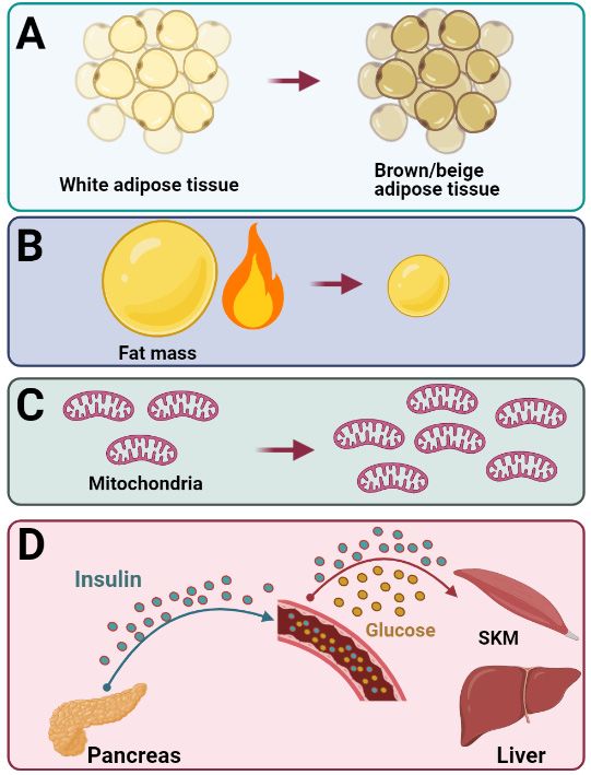
response to exercise induces browning of white adipose tissue by increasing the expression of brown
adipocyte-specific genes within the white adipose tissue, suggesting that exercise-induced cytokines
may play a significant role in preventing obesity. In this review, we present research-based evidence
Citation: Atakan, M.M.; Koşar, Ş.N.;
supporting the effects of exercise and various diet interventions on preventing obesity and adipose
Güzel, Y.; Tin, H.T.; Yan, X. The Role
tissue health. We also discuss the interplay between adipose tissue and the cytokines secreted from
of Exercise, Diet, and Cytokines in
Preventing Obesity and Improving
skeletal muscle and other organs that are known to affect adipose tissue and metabolism.
Adipose Tissue. Nutrients 2021, 13,
1459. https://doi.org/10.3390/ Keywords: obesity; adipose tissue; exercise; diet; cytokines
nu13051459
Academic Editor: Dyan Sellayah
1. Introduction
Received: 15 March 2021 According to the World Health Organization (WHO), worldwide obesity has nearly
Accepted: 22 April 2021
tripled since 1975 and reached a global epidemic [1,2]. In 2016, more than 1.9 billion (about
Published: 25 April 2021
39%) adults worldwide were overweight and, among them, about 650 million (about 13%)
were obese [2]. The increase in the prevalence of being overweight and obese has been
Publisher’s Note: MDPI stays neutral
attributed to an imbalance between energy intake and expenditure due to an increasingly
with regard to jurisdictional claims in
sedentary lifestyle, and a nutritional transition to processed foods and high-calorie diets
published maps and institutional affil-
over the last 30 years [3]. Obesity is considered a multisystem chronic relapsing progressive
iations.
disease process [4,5] adversely affecting almost all physiological functions of the body and
leading to increased morbidity and mortality [5–7]. Furthermore, obesity is associated with
many metabolic dysfunctions and comorbidities [8–15] that interfere with the quality of
life and work productivity, and increases healthcare costs [16–18].
Copyright: © 2021 by the authors.
A positive association has been found between body mass index (BMI) over 24.9 kg/m2
Licensee MDPI, Basel, Switzerland.
and overall mortality [6,7]. The associations are stronger at younger ages compared to older
This article is an open access article
ages and the hazard ratio is greater in men than women [6,7]. Additionally, a population-
distributed under the terms and
based cohort study of 3.6 million adults in the UK revealed that life expectancy from age
conditions of the Creative Commons
40 years was 4.2 and 3.5 years shorter in men and women with obesity (BMI ≥ 30.0 kg/m2 ),
Attribution (CC BY) license (https://
respectively, than individuals with healthy weight (BMI 18.5–24.9 kg/m2 ) [7]. A recent
creativecommons.org/licenses/by/
4.0/).
study by Dai et al. [19] revealed that, in 2017, high BMI caused 2.4 million deaths and 70.7
Nutrients 2021, 13, 1459. https://doi.org/10.3390/nu13051459 https://www.mdpi.com/journal/nutrientsNutrients 2021, 13, 1459 2 of 56
disability-adjusted life years (DALYs) in females, and 2.3 million deaths and 77.0 million
DALYs in males globally, based on the data from 195 countries and territories. The study
showed that, although the age-standardized rate of high-BMI-related DALYs increased
by only 12.7% for females and 26.8% for males, the global number of high-BMI-related
DALYs has more than doubled for both sexes between 1990 and 2017 [19]. Cardiovascular
disease was the leading cause of high-BMI-related DALYs, followed by diabetes and kidney
diseases, and neoplasms, together accounting for 89.3% of all high-BMI-related DALYs [19].
In addition to morbidity and life expectancy, obesity is a major burden on the healthcare
system due to both direct and indirect costs [16].
Adipose tissue is a highly metabolically active organ that performs many functions
such as lipid storage, mechanical protection, thermal insulation, immune responses, en-
docrine functions, and non-shivering thermogenesis [20,21]. It has a substantial capacity to
control its size and function in response to several internal and external stimuli including
nutritional status and temperature, accordingly. It plays an important role in the regulation
of systemic nutrient and energy homeostasis [20]. Although WHO defines overweight
and obesity as the abnormal or excessive fat accumulation that may impair health (WHO
fact sheet 2021), BMI is preferentially used to define overweight factors (BMI ≥ 25) and
obesity (BMI ≥ 30) in epidemiological studies. However, BMI is not sensitive enough to
differentiate the level or the distribution of adipose tissue mass. Furthermore, the percent
of body fat (BF) for any given BMI value varies greatly among individuals based on age,
sex, and ethnicity. In addition, for any given amount of BF, greater cardiometabolic risk
has been associated with the localization of excess fat in the visceral adipose tissue (VAT)
and ectopic depots (such as muscle, liver, and pancreas) [1,22]. Furthermore, the balance
of hypertrophic expansion of existing adipocytes and adipogenesis within an individual
profoundly affects metabolic health. Partly due to hypoxia and mechanical stress, large
adipocytes are associated with impaired metabolic health while small adipocytes are associ-
ated with a reduced risk of metabolic decline [23]. Compared to small adipocytes, increased
lipolysis and inflammatory cytokine secretion, and reduced secretion of anti-inflammatory
adipokines have been observed in hypertrophic adipocytes [23]. Moreover, a subgroup of
individuals with obesity, named metabolically healthy obese (MHO), is protected against
cardiometabolic disturbances as compared to individuals with metabolically unhealthy
obesity (MUO) [24,25], suggesting that adipose tissue distribution and dysfunction, rather
than the amount of fat mass, are the crucial factors in the pathophysiology of obesity-
related metabolic and cardiovascular diseases [24–26]. Furthermore, lower subcutaneous
fat mass, adipocyte hypertrophy, and an impaired fat storage capacity of adipose tissue are
the common features of MUO individuals [24,25], which may lead to ectopic fat deposition
and inflammation in VAT [24,25]. On the other hand, MHO, which is more common among
young, physically active individuals with a better nutritional status, is characterized by
a lower degree of systemic inflammation and a favourable immune and liver function
profile [24,25].
Given the underlying reasons for the dramatic increase in the prevalence of being
overweight and obese during the last 40 years, population-based preventive strategies
that improve social and physical environmental contexts for healthy eating and physical
activity (PA) have been suggested. These preventive strategies require a multisectoral
joint effort, including policymakers, educators, health professionals, food producers, city
planners, etc. [27]. As a complex chronic disease, the management of obesity requires a
holistic approach. Although pharmacotherapy and bariatric surgery are indicated with
severe obesity, diet, exercise, and cognitive behavioural therapy are the primary strategies
for the lifelong management of obesity [5,28,29]. Kheniser et al. [29] stated that two years
of lifestyle interventions can facilitate a 5% weight loss and that, although a weight regain
occurs, both diet and exercise interventions have substantial effects on obesity-associated
comorbidities [5,29] and adipose tissue remodelling [30]. Moreover, both regular aerobic
exercise and the consumption of a hypocaloric diet are associated with a substantial
reduction in VAT and liver fat independent of age, biological sex, or ethnicity [31,32].Nutrients 2021, 13, 1459 3 of 56
Although diet is more effective in reducing total body weight (BW) loss, exercise is superior
at reducing VAT [32]. Furthermore, it has been reported that moderate-to-vigorous intensity
of regular exercise for 4 to 6 months combined with a balanced, healthful diet resulted in a
substantial decrease in VAT (15–20%) and that 5% to 10% of weight loss can be achieved
with reasonable reductions in caloric intake with or without exercise [31]. Additionally,
several organs secrete biochemicals in response to low caloric intake and exercise as well as
several other factors, which contributes to the browning of white adipose tissue (WAT), and
is, therefore, considered a potential therapeutic approach against obesity and associated
metabolic dysfunctions [33]. Therefore, diet and exercise are the key components of weight
loss and maintenance program.
Over the last 20 years, molecules secreted from skeletal muscle and other organs have
been the focus of much research in terms of their therapeutic role as circulatory factors
with effects on metabolically active tissue and organs. Some of these molecules released in
response to muscle contraction have been reported to mediate some of the beneficial effects
of exercise in other organs, such as the liver and the adipose tissue [34], such as browning
of WAT and increasing thermogenesis and energy expenditure (EE), which make cytokines
appealing therapeutic targets for metabolic diseases.
In this review, we provide an overview of the research-based evidence supporting
the effects of exercise and various diet interventions on preventing obesity and adipose
tissue health. The interplay between adipose tissue and the cytokines secreted from skeletal
muscle and other organs that are known to affect adipose tissue was also discussed.
2. Adipose Tissue Biology: Why Our Body Is a Fat-Storing Machine?
Adipose tissue is a connective tissue mainly composed of lipid-rich cells named
adipocytes [35]. It has long been believed that adipose tissue’s main function is to store
energy as triglycerides while energy excess, which can then be broken down into free fatty
acid and glycerol during starvation or fasting [35,36]. Since the body has a limited capacity
to store glycogen, long-term imbalances between energy intake, and EE lead to a substantial
increase in the amount of triacylglycerol stored in adipocytes, causing obesity [37]. Recent
research has unveiled that adipose tissue also functions as an endocrine organ [38,39], which
expresses and secretes factors called adipocytokines or adipokines [37,38]. Adipose tissue
is a complex and essential tissue as demonstrated by the adverse metabolic consequences
resulting from either excessive or deficient adipose tissue [38]. An excess of adipose
tissue leads to the development of obesity and metabolic syndrome, while adipose tissue
deficiency (lipodystrophy) can also cause a metabolic syndrome [40]. There are two main
types of adipose tissue: WAT and brown adipose tissue (BAT). These are briefly outlined in
the following section.
2.1. White Adipose Tissue
WAT generally stores excess energy in the form of triglycerides and makes up the
majority of the human BF percentage (BF%) [41,42]. Additionally, the main functions of
WAT are to protect organs against mechanical damage and release adipokines regulating
various biological processes, including inflammatory reactions [43]. Adipose tissue accu-
mulation around the abdominal cavity and mediastinum is referred to as VAT, whereas it
can also be found in the hypodermis layer as subcutaneous adipose tissue (SAT) [44]. At a
molecular level, WAT takes the form of single lipid droplets and has a limited number of
mitochondria. WAT is not a static form of connective tissue, as it regularly remodels and
changes its number of adipocytes depending on nutritional availability as well as hormonal
signals [44]. Additionally, WAT is an endocrine organ capable of actively secreting free
fatty acids and adipocytokines, which have autocrine, paracrine, and endocrine effects on
other organs, such as skeletal muscles, the brain, and the liver [42]. Concretely, WAT is
essential for energy homeostasis and metabolic regulation [35].Nutrients 2021, 13, 1459 4 of 56
2.2. Brown and Beige Adipose Tissue
BAT, on the other hand, is mainly utilized for insulation against a cold climate. It
achieves this by generating heat as a result of dissipating energy [41]. BAT is characterized
by a high abundance of uncoupling protein 1 (UCP1), which is the protein responsible for
non-shivering thermogenesis, along with many other genes including cell death-inducing
DNA fragmentation factor-like effector A (CIDEA), PR domain containing 16 (Prdm16),
and Type 2 Deiodinase (DIO2), which are all important in mitochondrial biogenesis [45].
BAT is also capable of mediating adaptive thermogenesis and, thus, contributes to the
maintenance of body temperature. The role of BAT in adults has yet to be fully realized,
with some studies suggesting that it may play a key role in energy homeostasis. Generally,
as the BW increases with age, the amount of total BAT decreases, showing an inverse
relationship between BAT and BW [41]. BAT can be found in small amounts in the neck,
supraclavicular, axillar, paravertebral, perirenal/adrenal, and para-ventral regions, as well
as the major vessels surrounding the heart [42]. Some studies have found that BAT can also
reside in skeletal muscle tissues and even WAT [42]. At a molecular level, BAT takes the
form of multiple small vacuoles and has large amounts of mitochondria [41].
More recently, another unique type of adipose tissue, beige adipose tissue, has been
identified in rodents and humans [46]. Beige adipocytes are found within the WAT depots,
but with similar morphology to brown adipocytes and large amounts of mitochondria [47].
In rodents, beige adipocytes can be induced by cold-exposure [48], exercise [49,50], and
hypothalamic brain-derived neurotrophic factor (BDNF) [51]. To a smaller extent, beige
adipocytes have been observed in humans [46], which is known to be induced by chronic
exposure to peroxisome proliferator-activated receptor gamma (PPARγ) agonists [52].
3. Exercise Strategies to Prevent Obesity and Improve Adipose Tissue Health
Exercise plays an important role in human health as a non-pharmacological elixir that
helps prevent obesity by increasing EE for weight loss, lower metabolic risk factors, and
enhance adipose tissue health [53,54]. Exercise can be sub-categorised into two types: acute
and chronic/training [55]. Acute exercise refers to one session of PA, while chronic/training
includes repeated exercise sessions weekly or monthly [55]. Acute exercise and chronic
training studies can demonstrate the short-term and long-term effects of exercise on the
human body, respectively. Moreover, acute exercise interventions have been used to study
the mechanistic adaptations to exercise. For instance, an acute bout of exercise increases
blood flow through adipose tissue and fat mobilization, leading to the delivery of fatty
acids to skeletal muscles, which is mainly based on the exercise intensity and metabolic
requirements [37]. Furthermore, following an acute exercise, dietary fat stored in adipose
tissue decreases as a result of the mobilization of fatty acids stimulated by β-adrenergic
activation from adipose tissue to other tissues, such as skeletal muscle [37]. In addition,
regular exercise/training is known to alter adipose tissue physiology, which results in
enhanced fat mobilization during acute exercise [37]. It is, however, not fully elucidated
whether the structural changes in adipose tissue are induced by exercise training or negative
energy balance [37,45,56] and remain an important area of investigation. The relationship
between PA and adiposity has been comprehensively investigated in longitudinal cohort
studies that have documented a strong inverse association between measures of PA and
measures of fat mass and distribution [57,58]. Thus, incorporating well-designed exercise
training routines into a weight loss program are efficient strategies. In the following section,
the effects of different types of exercise models on obesity and adipose tissue and the
underlying molecular mechanisms are reviewed. The exercise studies included in the
following sections are described in greater detail in Table 1.
3.1. Continuous Exercise and Adipose Tissue
Over the last 20 years, the effects of regular endurance training on adipose tissue have
gained momentum and have been comprehensively investigated in numerous studies. In
light of the findings of these studies, it is accepted that exercise seems to reduce fat mass,Nutrients 2021, 13, 1459 5 of 56
which significantly depends on the net energy deficit induced by exercise interventions.
It is also noteworthy that the effect of PA without a calorie restriction diet on fat loss
might be relatively small or modest [59,60]. Furthermore, a substantial energy deficit
created by increased PA results in a loss of fat mass from significant depots, such as
SAT and VAT. For example, an increase of daily steps from 7013 to 8840 decreased SAT
and VAT as well as BF% in men with obesity [61]. Similarly, one year of training at 58%
of maximal oxygen consumption (VO2max ) (6 sessions/week) reduced total fat mass as
well as abdominal visceral and SAT in nonobese women and men [60]. An experiment
consisting of moderate to high-intensity aerobic training (3 sessions/week, 40 min/session,
total distance 12 miles/week at 75% VO2max ) for 8 to 9 months in individuals who are
overweight and obese have reported a significant reduction in thigh SAT in both men and
women who are overweight, but VAT decreased only in men [62].
Furthermore, it is well known that training studies with shorter durations exert
profound effects on adipose tissue as well. For example, 24 weeks of moderate-intensity
training at a low amount (LAMI, 5 days/week, 31 min/session, 50% VO2max ) resulted in a
decrease of waist circumference, similar to the moderate-intensity high amount (HAMI,
58 min/session) and high-intensity high amount (HAHI, 40 min/session, 75% VO2max ) [63].
A follow-up study has confirmed similar reductions in total BF, SAT, and VAT among the
three training groups [64]. However, there was an individual response to training in total
and abdominal fat with a greater proportion of subjects “very likely” to decrease fat in the
HAMI (total BF) and HAHI groups (total BF and SAT) [64]. Wilmore et al. determined
the extent of changes in SAT and VAT in subjects who are overweight after 20 weeks of
chronic training (3 sessions/week, at 50–75% VO2max for 30–50 min) [65]. The findings
implied that males had a greater loss in abdominal SAT and VAT than females. A higher
rate of decline was also seen in abdominal SAT compared to VAT [65]. A study investigated
16 weeks moderate-intensity (≤lactate threshold) and high-intensity (>lactate threshold)
training in women with metabolic syndrome, but no significant changes of SAT and VAT
were observed in the moderate-intensity group [66]. The high-intensity group resulted in
reduced total abdominal fat, subcutaneous abdominal fat, and abdominal visceral fat [66],
showing that high-intensity exercise is more effective than moderate-intensity exercise
training in reducing fat storage in women with obesity and metabolic syndrome.
Furthermore, research has investigated the differences between 12 weeks of moderate-
intensity (4 to 5 sessions/week, EE of 1000 kcal/week, 50% VO2max ) and high-intensity (4 to
5 sessions/week, EE of 1000 kcal/week, 75% VO2max ) exercise on regional fat distribution
in elderly adults who are overweight [67]. The findings showed a remarkable reduction in
VAT in the high-intensity group exhibited, while no change was observed in the moderate
group [67]. A recent study reported that vigorous-intensity physical activities are associated
with high BAT density in humans, suggesting that long-term high-intensity physical
activities might positively influence BAT content [68]. Collectively, the intensity of exercise
training seems to play an essential role in changing adipose tissue. Another study that
aimed at revealing the effect of training on adiposity in children with obesity reported a
significant decrease in BF%, total BF, and SAT mass [69], following 4 months of moderate
intensity training (5 sessions/week, 40 min/day at 70–75% maximal heart rate (HRmax ),
equivalent to 58–66% VO2max ). Similarly, with the diet controlled, 12 weeks of daily
exercise (brisk walking or light jogging no more than 70% VO2max ) resulted in a decrease
in both SAT and VAT in men with obesity. The reduction in total fat was greater in the
training group when compared with the diet-induced weight loss group (22% decrease in
energy intake) [59]. Even in the exercise without a weight loss group (with 23% increase
in energy intake), there was a decrease in abdominal adipose tissue and VAT [59]. Even
without changes in total BW, 13 weeks of moderate-intensity training (5 sessions/week,
60 min/session, −60% peak oxygen uptake (VO2peak )) led to significant reductions in total,
abdominal subcutaneous factors, and visceral fat in men with obesity with and without
type 2 diabetes (T2D), as well as in the lean control group [70]. The reduction in VAT was
greater in the groups with obesity and T2D when compared with the lean group [70]. OnlyNutrients 2021, 13, 1459 6 of 56
8 weeks of training at moderate intensities (70% VO2peak ) reduced liver fat and VAT [71].
Interestingly, 45 min of training at 50% VO2max (three sessions/week) seems to be enough
for these reductions, as an increase of volume (to 60 min/session, 4 sessions/week) or
intensity (to 70% VO2max ) did not result in further reductions [71]. Moreover, Christiansen
and colleagues compared the independent and combined effects of 12 weeks of regular
exercise and diet-induced weight loss on BF distribution in subjects with obesity [72]. They
reported that there was a significant decrease in BW (3.5 kg) and VAT (18%) [72]. More
importantly, a hypocaloric-diet and exercise resulted in a markedly higher reduction in VAT
(30–37%) and BW (12.3 kg) [72], compared to the exercise group, showing a hypocaloric
diet to be more effective in reducing the VAT depot, compared to exercise only. Walhin et al.
reported that 3 weeks (5 sessions/week) of moderate-intensity (50% VO2max ) and vigorous-
intensity exercise training (70% VO2max ) combined with caloric restriction (5000 kcal/week)
led to similar reductions in total fat and abdominal fat mass [73]. In addition, both exercise
interventions with simultaneous restricted energy intake similarly affected the expression
of the lipogenic enzymes [73].
In summary, regular exercise, especially moderate to high-intensity exercise for eight
weeks to one year, decreases total BF, SAT, and VAT. Furthermore, exercise training com-
bined with a hypocaloric-diet is more effective compared to exercise intervention alone in
preventing and reducing BF.
Molecular Mechanisms Underlying the Reduction in Total BF, SAT, and VAT with Exercise
In vivo studies and adipose tissue biopsies following acute and chronic exercise trials
have provided mechanistic insight into the molecular mechanisms that are responsible for
the reduction in total BF, SAT, and VAT in response to exercise training. For example, a
single session of 30 min of continuous running at 65% VO2max was reported to increase
whole-body fat oxidation during the post-exercise recovery period in young men [74].
Acute moderate-intensity continuous exercise at 45–70% VO2max increased the oxidation of
total lipid and plasma fatty acid (~60%) [75] and the amount of the adipose tissue lipopro-
tein lipase (56%) in men [76] but not women, and increased the number of the adipose
tissue progenitor cell phenotype in adults with obesity [77]. Similarly, one hour of acute
exercise at 55% VO2max has been shown to modify adipose tissue mRNA and interstitial
cytokine concentration in males who are overweight [78]. In addition, an increased con-
centration of interstitial adiponectin and interleukin (IL)-6 was detected [78], while the
response at the mRNA level was different, with IL-6 mRNA increasing but adiponectin
mRNA decreasing [78]. Another similar study reported increased SAT mRNA expression
of vascular endothelial growth factor A (VEGFA), which is an important regulator of
angiogenesis and capillary growth, in adults who are overweight/obese following acute
moderate-intensity exercise at 65% VO2max [79]. Furthermore, a decrease of preadipocyte
content was shown in the stromal vascular cells fraction of SAT twelve hours after sixty
minutes of moderate-intensity endurance exercise in adults with obesity [77]. It was also
reported that a single session of 15 min exercise at 80% VO2max has induced more than
3800 genes in adipose tissue from individuals who are or are not overweight, among them
are the genes responsible for monocyte infiltration [80].
There are limited long-term training studies that have investigated the effects of exer-
cise training on molecular mechanisms involved in exercise-induced changes in adipose
tissue biology. One of these studies aimed to reveal gene expression changes in adipose
tissue following 6 months of diet-induced and/or exercise-induced weight loss in post-
menopausal women who are overweight/obese [81]. The authors showed that the mRNA
expression of candidate genes in the SAT did not change in the intervention groups [81]. On
the other hand, those participants with greater weight loss showed decreased expression
of the leptin gene [81]. Finally, microarray analyses revealed the association of weight
loss with adipose tissue gene expression involved in the synthesis of sex hormones in
adipose tissue, whereas there was no impact of weight reduction with diet or diet plus
exercise on genes related to inflammation in SAT in obese people [81], indicating thatNutrients 2021, 13, 1459 7 of 56
changes in energy balance following diet and/or exercise factors can have a limited impact
on adipose tissue inflammation [82]. This field remains a fertile area of research in the
near future. Furthermore, 12 weeks of endurance training (2 sessions/week supervised,
3 times/week home-based exercise at 50% VO2max ) did not change genes involved in
the control of SAT lipolysis [83] or gene expression of adipocytokines in women with
obesity [84], while a decrease of plasma leptin was detected [84]. Eight weeks of exercise
training (3 sessions/week, 30 min/session at 70% VO2max ) reduced adipose tissue IL-18
mRNA content by 20% in obese individuals [85]. In addition, the mRNA expression of
adipose tissue adiponectin and adiponectin receptors increased significantly after 12 weeks
of training (3 sessions/week, 60 to 75 min/session at 70% of heart rate reserve) in obese
men and women [86].
Findings on the browning of WAT in response to exercise come from both rodent and
human studies. As reviewed comprehensively by Stanford and Goodyear [87] in rodents, it
is well documented that exercise training can induce browning of WAT and the recruitment
of brown-like adipocytes within WAT via exercise-induced cytokines such as irisin and
IL-6, which triggered the interest in investigating WAT browning in humans. Current
evidence has shown that eleven days of voluntary running resulted in increased expression
of many beige adipocyte marker genes in rodent SAT [49]. Moreover, 30 days of swimming
(90 min daily) increased expression of UCP1 and Prdm16 in mice SAT, suggesting browning
of SAT by training in rodents [88]. While evidence from rodents seems promising, the
reports of human studies are not very conclusive. First, it seems that the existence of
brown/beige adipose tissue in adult humans is not very common and decreases with
age [89]. Second, it was shown that endurance-trained athletes had a lower metabolic
activity of BAT compared to lean sedentary individuals [90]. Furthermore, gene expression
of classical brown and beige adipocyte markers in subcutaneous WAT, plasma irisin, and
Il-6 levels during mild cold exposure were similar in trained and sedentary individuals [90].
Conversely, 12 weeks of cycling (3 sessions/week, 60 min/session at 43% to 70% VO2max )
induced the mRNA expression of beige/BAT markers of UCP1, T-box transcription factor
1 (TBX1), and carnitine palmitoyltransferase-1B (CPT1B) in SAT of sedentary subjects,
suggesting browning of SAT by training [91].
In summary, acute exercise interventions have shown that low-moderate-intensity
exercise can increase whole-body fat oxidation, possibly by regulating adipose tissue lipol-
ysis, gene expression of adipocytokines, or changing the cell composition of adipose tissue.
However, exercise intervention may not be associated with brown and beige adipocyte
recruitment in humans. Rather, endurance training can lead to the lower metabolic activity
of BAT in humans. More work is needed to reveal whether particular groups or populations
experience beneficial changes in adipose tissue from exercise training.
3.2. High-Intensity Interval Training and Adipose Tissue
For the management of obesity, it is recommended to be physically active such as 150
to 250 min/week or up to 60 min/day [92]. However, current epidemiological data indicate
that the majority of the adult population does not meet the recommended PA guidelines
mainly due to lack of time [93], and there is, therefore, a need to establish the efficacy of
time-efficient doses of exercise that overcome the health risks associated with obesity with
less time commitment. High-intensity interval training (HIIT) is characterized as a short
period that must be performed over the lactate threshold, near VO2max , and interspersed
with light exercise or rest so that extra high-intensity bouts can be performed [53,55]. HIIT is
based on the Wingate test, which consists of “supra-maximal power output” [94]. A typical
HIIT protocol is considered as sprint interval training (SIT), in which individuals will have
to complete “all-out” several times (≥100% maximal workload capacity) performance
with recovery time in between the intensive exercise sessions on a cycle ergometer [55].
Therefore, a customized low-volume HIIT protocol (near the maximal corresponding to
≥75% toNutrients 2021, 13, 1459 8 of 56
studies [95,96] and is prone to be more workable for individuals than the Wingate-based
HIIT model [55].
There is robust evidence that HIIT can reduce adiposity and abdominal visceral fat
despite the discrepancies available in the previous studies that are attributed to training
protocols, exercise protocol, obesity status, and gender. For example, a study compared the
impacts of 12 weeks (3 to 4 sessions/week) of prolonged moderate-intensity continuous
training (MICT 60% VO2max ) with HIIT (90% VO2max , repeated 4 min bout with 3 min
recovery) on abdominal adipose tissue reduction in young women with obesity [97]. The
findings showed a similar reduction in abdominal SAT and VAT in both groups [97]. Ten
weeks of endurance exercise training (a combination of continuous and HIIT) improved
adipose tissue insulin sensitivity. However, changes in adipose tissue composition was not
reported [98]. Six weeks of HIIT (3 sessions/week, 7 × 1 min at 95–100% VO2max , with
1 min recovery), which resulted in increased skeletal muscle mitochondrial respiratory
capacity, did not change BF% and reduced the mitochondrial respiratory capacity in SAT
in overweight subjects [99]. Another study by Leggate et al. examined two weeks of
HIIT (3 sessions/week, 10 × 4 min at 85% VO2max , 2 min rest) in sedentary males with
overweight/obesity, and they found a decrease in waist circumference, as well as reductions
in IL6 and fatty acid synthase content in SAT biopsies [100]. A recent experiment by Islam
et al. investigated the impacts of acute high-intensity interval exercise (HIIE 10 × 4 min
at 90% of HRmax , separated by 2 min recovery) on SAT and whole-body fat oxidation in
women who are overweight [101]. They showed that, despite a significant increase in
whole-body fat oxidation, β-adrenergic and insulin signalling in subcutaneous adnominal
adipose tissue remained unchanged following acute HIIE [101], suggesting that HIIE
does not alter intracellular signalling pathways controlling fat mobilization or storage in
subcutaneous abdominal adipose tissue. Another study comparing the effects of 12 weeks
(3 sessions/week, 6 to 10 × 60 s intervals) moderate intensity interval training (60–80%
maximal workload, with 60 s of active recovery at 40 W) with HIIT (80–90% maximal
workload, with 75 s active recovery at 40 W) reported an increased fat oxidation rate in
sedentary women with normal weight, overweight, and obesity [102]. However, none
of the training intensity affected BW, BF%, or circumferences of waist and hip [102]. A
recent study by Taylor et al. compared the impacts of HIIT with MICT on VAT and liver fat
reduction in patients with coronary artery disease for 4 weeks, followed by three home-
based sessions/week for 11 months [103]. The authors documented that both exercise
interventions reduced VAT over 3 and 12 months, while HIIT resulted in a slightly greater
reduction in liver fat when compared to MICT [103]. A meta-analysis by Keating and
colleagues that reviewed 28 trials with 873 participants reported that HIIT and MICT
present similar benefits for eliciting small reductions in total BF [104]. In addition, it was
reported that 6 weeks of SIT (3 sessions/week, 5 × 60 s at ~128% of peak power, 90 s
recovery) did not alter BF% or adipose tissue mitochondrial function [105], while it resulted
in a greater loss in total BF and android fat than MICT (3 sessions/week, 45–55% HRmax , for
20–30 min) cycling in young women who are overweight [94]. Two weeks of Wingate-based
SIT (3 sessions/week) significantly reduced waist and hip circumference, and increased the
resting fat oxidation rate in sedentary men who are overweight/obese [106]. Another study
reported two weeks of Wingate-based SIT (3 sessions/week) resulted in a similar reduction
in BF%, abdominal SAT, and VAT compared to MICT (40–60 min at 60% VO2max ) in healthy
subjects with insulin resistance [107]. Furthermore, both training interventions decreased
CD26 and ANGPTL4 gene expression in SAT [107]. Finally, Cooper et al. reported no
significant change in FM or abdominal VAT following 12 weeks (3 sessions/week) of SIT
interventions consisting of 4 to 10 × 30 s sprint efforts in men who are overweight [108],
raising further questions regarding if exercise training without caloric restriction could
facilitate favourable changes in body composition and abdominal VAT.
In summary, it is apparent that interval training models improve adipose tissue despite
the inconsistent and controversial findings that existed. Moreover, HIIT seems to be an
alternative to MICT for reducing visceral and liver fat. More work that combines HIIT withNutrients 2021, 13, 1459 9 of 56
hypocaloric diets is needed. The findings of further studies can open up new time-efficient
therapeutic potential in developing new strategies for the prevention and management
of obesity.
3.3. Resistance Exercise Training and Adipose Tissue
Resistance exercise training (RT) consists of various types of physical exercise that
causes the skeletal muscles to contract against an external resistance [109,110] that develops
the strength and size of muscles, and increases bone mass [111–113]. The metabolic effects
of reduced muscle mass has been reported to result in a high prevalence of obesity, insulin
resistance, and T2D [114,115]. Therefore, RT and subsequent increases in muscle mass are
likely to reduce metabolic disease risk factors [114,116]. Although, the aerobic exercise
has traditionally been recommended for preventing and managing obesity and associated
metabolic risk factors [116,117], recently, RT has also been suggested to be a feasible and
efficacious alternative to aerobic exercise for weight control due to its multiple therapeutic
effects [114,116]. For example, the age-related decline in resting EE is closely associated
with the loss of skeletal muscle mass [118], which can be reversed by RT that leads to
increased muscle mass based on the training duration and intensity. However, despite no
clinically important change in resting EE following RT, maintenance of muscle mass with
RT helps prevent age-associated fat mass gains by promoting an active lifestyle [119].
Several studies have reported that RT can reduce FM and VAT in men [120] and
women [109,121] independent of dietary caloric restriction [122]. A study that assessed
body composition in older women reported significant decreases in visceral fat after
16 weeks of RT [123]. Similarly, another study investigated the effects of 16 weeks of RT
combined with diet interventions on FM and VAT in middle-aged men with obesity. The
findings showed that there was a 40% reduction in visceral fat in the RT combined diet
group [122]. Hunter et al. showed that 25 weeks of chronic RT resulted in an improvement
in fat-free mass and a reduction in BF in older males and females [124]. There was also
a substantial loss of intra-abdominal adipose tissue and abdominal SAT in women but
not in men who are overweight [124]. Ku and colleagues documented that 12 weeks
of RT (5 sessions/week elastic band exercise) decreased SAT, which was comparable to
12 weeks of aerobic training (5 sessions/week, walking for 60 min at moderate-intensity
[3.6–5.2 metabolic equivalents]) in individuals with T2D [110]. However, only RT decreased
subfascial adipose tissue at the mid-thigh level [110]. Rosety et al. highlighted 12 weeks
of resistance circuit training (3 sessions/week), which resulted in a reduced thickness
of epicardial adipose tissue in obese women [125]. Ross et al. reported a substantial
similar decrease in the volume ratio of VAT to SAT after 16 weeks of RT and aerobic
training (3 sessions/week) combined with caloric restriction (reduced by 1000 kcal) in
obese women [126]. Moreover, within the VAT depot, a remarkable reduction was observed
for both intraperitoneal and extraperitoneal adipose tissue [126]. Slentz et al. compared
the effects of high-intensity aerobic training (12 miles/week at 75% VO2max ) and RT
(3 times/week, 3 sets of 8–12 repetitions/set) in adults who are overweight [127]. They
reported high-intensity training provided a greater reduction in VAT and total abdominal
fat than RT [127], indicating high-intensity aerobic exercise to be a more effective exercise
mode to reduce visceral fat.
The effects of acute resistance exercise (RE) on adipose tissue have also been investi-
gated by a limited number of studies that documented a transient increase in adipose tissue
lipolysis. For example, one study with trained men reported that acute RE (3 sets of 10
repetitions with a load at 85–100% of the individual’s 1 maximum repetition (1RM), 90 sec
rest periods between all sets and exercises, for a total of 40–45 min) increased SAT lipolysis
during RE, while SAT lipolysis and whole-body fat oxidation were higher immediately
post RE [128]. Another acute RE (one set of 10 repetitions at 40% 1RM and three sets of
10 repetitions at 65% 1RM) study in trained women reported an increase in post-exercise
whole-body fat oxidation and SAT lipolysis [129]. Chatzinikolaou et al. investigated the
effect of performing 30 min of acute circuit RE (3 cycles on 10 machines selected to stressNutrients 2021, 13, 1459 10 of 56
the major muscle groups, 10–12 repetitions/set at 70–75% of 1RM with 30 s rests between
sets, and 2 min rests between cycles) on adipose tissue lipolysis in lean men and men
with obesity [130]. The authors documented that adipose tissue triacylglycerol lipase
activity was elevated by 18-fold after 5 min of exercise in lean subjects, whereas a 16-fold
increase was observed 10 min after exercise in males with obesity [130]. In summary, the
overall available body of literature indicates that RT with or without diet modification is
an effective way to reduce BF and control obesity.
3.4. Concurrent Training and Adipose Tissue
Concurrent training (CT) is a designed exercise model involving aerobic and anaer-
obic metabolic pathways so that it can enhance the effects of both aerobic and RT mod-
els [131–133]. Although CT has been used among athletes for multiple decades to enhance
performance in a variety of sports, it has recently grown in popularity [134,135]. As a
combined form of endurance and strength exercise modes, CT induces changes in the car-
diovascular and the neuromuscular systems, providing widespread disturbances occurring
in local and systemic homeostasis that, in turn, results in remarkable adaptation in human
physiology. In addition to providing traditional physiological adaptations known to be
induced by traditional endurance exercise, CT can also improve body composition and
health-related outcomes [132].
The effect of CT on FM and adiposity has been addressed in a variety of studies that
have yielded contrasting results. These discrepancies may be partially due to potential
factors known to alter one’s energy balance, such as caloric intake or EE, which were
not usually considered in previous studies. Furthermore, some studies have reported a
similar improvement in adiposity following CT or aerobic exercise [127,136,137], whereas
other studies documented that CT elicited greater improvement [138]. For example, a
one-year intervention (3 sessions/week) of aerobic (30 min of aerobic exercise at 50–70%
VO2max ) plus RT (30 min of RT) induced higher changes in body composition, waist cir-
cumference, and BF in adolescents with obesity rather than aerobic exercise by itself [139].
Similarly, Dâmaso and colleagues compared the effect of aerobic exercise alone or aerobic
plus RT on visceral fat and its role on pro-inflammatory/anti-inflammatory adipokines
in obese adolescents [138]. They reported that aerobic plus RT provided a greater re-
duction in visceral fat and pro-inflammatory adipokines than an aerobic training alone
intervention [138], showing CT to be a more effective strategy to control central obesity
in adolescents. Slentz et al. reported similar significant reductions in VAT, SAT, and
total abdominal fat following aerobic plus RT (3 sessions/week, 12 miles/week at 75%
VO2max plus 3 × 8–12 repetitions/set, 3 sessions/week) and aerobic training alone (3 ses-
sions/week, 12 miles/week at 75% VO2max ) in overweight adults [127]. Similarly, Monteiro
et al. reported a significant reduction in waist circumferences and BF% after 20 weeks of
CT (3 times/week, 60 min at 50% of 1RM, followed by 30 min at between 65% and 85%
VO2max aerobic training) and aerobic training (3 times/week, 50 min of continuous exercise
between 65% and 85% VO2max ) [136]. Another study reported a significant reduction of
waist circumference (~3%), VAT (~10%), and SAT (~10%) in obese adolescents following
16 weeks of CT (twice/week, 30–45 min/session 70–85% HRmax plus 30–45 min, 12–14
repetitions, low-heavy weights) [140]. Conversely, Norheim et al. investigated the effect
of 12 weeks of CT on human abdominal subcutaneous fat in adults with normal weight
and overweight [141]. The CT program consisted of two aerobic exercise sessions plus
two RT exercise sessions per week. The obtained findings following the training program
showed that there was no significant change in the mRNA level of PPARγ coactivator-1α
(PGC-1α) of SAT, the brown-fat-selective gene Prdm16, or other known browning genes
TBX1, transmembrane protein 26 (TMEM26), or tumor necrosis factor receptor superfamily
member 9 (CD137) [141]. Stinkens et al. reported similar findings showing that 12 weeks
of the CT program did not change abdominal subcutaneous adipocyte size, β2-adrenergic
sensitivity of lipolysis, and adipose tissue gene expression of markers involved in brown-
ing and lipolysis in obese subjects [142]. Collectively, 12 weeks of CT does not seem toNutrients 2021, 13, 1459 11 of 56
provide enough stimulus to induce adipocyte morphology and adipose tissue gene/protein
expression in humans [142].
Taken together, it is evident that CT is a preventative and therapeutic exercise model
capable of inducing similar or even superior improvement in adipose tissue and obesity
to traditional endurance exercise. Given that long-term CT increases fat-free mass that
results in a reduction of BF% independent of changes in fat stores, CT can be regarded as an
alternative exercise mode able to decrease BF%. Health authorities should be encouraged to
recommend the incorporation of CT into exercise routines. Furthermore, the effect of CT on
adipose tissue morphology remains equivocal and awaits determination in further studies.Nutrients 2021, 13, 1459 12 of 56
Table 1. Description of exercise studies that are presented in the exercise section.
Continuous Exercise and Adipose Tissue
Participants
. Duration,
Author Year (VO2max ) n (M/F) Protocols Main Findings Ref
Frequency, Mode
(mL/kg/min)
Group 1: Diet Reduction in total fat was
(reducing total daily energy intake to 700 greater in group 2
kcal/day) compared with group 1.
Group 2: Exercise Group 2: Substantial
Obese males 12 weeks, daily, brisk walking (80% of HRmax until 700 kcal is expended) decreased in both SAT and
1 Ross et al. 2000 (52/0) [59]
(NR) or light jogging Group 3: Exercise without weight loss VAT
(enough calories given to compensate for the Group 3: Attenuation in
energy expended during the daily exercise abdominal fat and
sessions) prevented further weight
Group 4: Control group gain.
Significantly decreased in
Miyatake Obese males 1 year follow up study, daily,
2 2002 (31/0) An increase of daily steps from 7013 to 8840 SAT, VAT, and body [61]
et al. (NR) walking
composition.
1 year,
Healthy, 6 days/wk, Group 1: 20% calorically-restricted diet Significant reduction in fat
Racette
3 2006 non-obese adults (18/30) running/cycling/rowing Group 2: Training at 58% of VO2max mass, SAT, and VAT for [60]
et al.
(NR) ergometers/elliptical Group 3: Healthy lifestyle control group both group 1 and 2.
machines/stairclimbersNutrients 2021, 13, 1459 13 of 56
Table 1. Cont.
Continuous Exercise and Adipose Tissue
Participants
. Duration,
Author Year (VO2max ) n (M/F) Protocols Main Findings Ref
Frequency, Mode
(mL/kg/min)
Sedentary, Group 1: ∼20 miles/wk of jogging
dyslipidemic, (65–80% VO2max ) Significantly reduced in
8–9 months,
Durheim overweight males Group 2: 12 miles/wk of jogging thigh SAT for all three
4 2008 (40/33) 3 days/wk, [62]
et al. (~32.8 VO2peak) (65%-80% VO2max ) groups, but VAT decreased
aerobic training
females Group 3: 12 miles/wk of brisk walking substantially in men only.
(~23.9 VO2peak) (40–55% VO2max )
Group 1: Training at a low-amount,
moderate-intensity exercise at 50% VO2max
(31 min/session)
24 weeks, Group 2: Training at a high-amount, Similar reductions were
Abdominally
5 days/wk, moderate-intensity exercise at 50% VO2max resulted in total BF, SAT,
5 Ross et al. 2015 obese adults (104/196) [63]
walking/jogging/treadmill (58 min/session) and VAT in all training
(NR)
training Group 3: Training at a high-amount, groups.
high-intensity exercise at 75% VO2max
(40-min/session)
Group 4: Control group
Males had a greater loss in
abdominal SAT and VAT
20 weeks,
Wilmore Overweight adults Training at 55% VO2max to at 75% VO2max than females. A higher rate
6 1999 (258/299) 3 days/wk, [65]
et al. (NR) for 30 min to 50 min. of decline was also seen in
cycling
abdominal SAT compared
to VAT.
Group 1: Moderate-intensity training (5 No significant changes of
days per week at an intensity ≤ LT SAT and VAT were
Middle-aged 16 weeks,
Group 2: High-intensity training (3 days per observed in group 1,
7 Irving et al. 2008 obese women (0/27) 5 days/wk, [66]
week at an intensity > LT and 2 days per whereas group 2 resulted in
(~21 VO2peak) aerobic training
week ≤ LT) reduced total abdominal fat,
Group 3: No-exercise training SAT, and VAT.Nutrients 2021, 13, 1459 14 of 56
Table 1. Cont.
Continuous Exercise and Adipose Tissue
Participants
. Duration,
Author Year (VO2max ) n (M/F) Protocols Main Findings Ref
Frequency, Mode
(mL/kg/min)
A remarkable reduction in
Overweight 12 weeks, VAT in the high-intensity
Group 1: Moderate-intensity (50% VO2peak)
8 Coker et al. 2009 elderly adults (9/9) 4–5 days/wk, group exhibited, while no [67]
Group 2: High-intensity (75% VO2peak)
(NR) aerobic training change was observed in the
moderate group.
VPA activities resulted in
high BAT density,
4 months, Group 1: WM particularly in men.
Tanaka Healthy adults
9 2020 (87/145) NR, Group 2: WM + vigorous-intensity physical BAT-density is related to [68]
et al. (NR)
walking/aerobic training (VPA) activities (VWM) visceral fat area and VWM
in men, and related to body
fat percentage in women.
4 months,
Significant decrease in BF%,
Owens Obese children 5 days/wk, Group 1: 40 min/day at 70–75% HRmax
10 1999 (25/49) total BF, and SAT for group [69]
et al. (NR) exercising on machines and Group 2: Control group
1.
sports activities
Significant reductions in
total abdominal SAT and
VAT in all groups (lean and
Lean and obese obese males with and
13 weeks:
male with and All participants trained for 60 min/day at a without T2D).
11 Lee et al. 2005 (24/0) 5 days/wk, [70]
without T2D moderate intensity (∼60% VO2peak) Reduction in VAT was
aerobic training
(~61.2% VO2peak) greater in the obese and
T2D groups when
compared with the lean
group.Nutrients 2021, 13, 1459 15 of 56
Table 1. Cont.
Continuous Exercise and Adipose Tissue
Participants
. Duration,
Author Year (VO2max ) n (M/F) Protocols Main Findings Ref
Frequency, Mode
(mL/kg/min)
Group 1: Cycling and brisk walk at 50%
VO2peak for 3 days and 1 day/wk,
respectively. (From 45 min in week one to
60 min by the 3rd week, totaling
180–240 min/wk)
Group 2: Cycling and brisk walk at 50%
Inactive and VO2peak for 2 days and 1 day/wk,
8 weeks, Reduction in liver fat and
Keating overweight/obese respectively. (From 30 min in week one to
12 2015 (17/31) 3–4 days/wk, VAT for all three groups. [71]
et al. adults 45 min by the 3rd week, totaling
brisk walking/cycling
(~22.4 VO2peak) 90–135 min/wk)
Group 3: Cycling and brisk walk at 60–70%
VO2peak for 2 days and 1 day/wk,
respectively. (From 30 min in week one to
45 min at 70% VO2peak by the third week,
totaling 90–135 min/wk)
Group 4: Control group
Group 1: exercise Reduction in BW 3.5 kg and
12 weeks, (60–75 min at 70% VO2max per training VAT 18% in group 1. Higher
Christiansen Obese adults
13 2009 79 3 days/wk, session) reduction in BW (12.3 kg) [72]
et al. (NR)
aerobic training Group 2: hypocaloric diet (600 kcal/day) and VAT (30–37%) in group
Group 3: hypocaloric diet and exercise 2 and 3 than group 1.
Sedentary Group 1: Moderate intensity training (50% Both groups resulted
overweight men VO2max ) with caloric restriction similar reductions in total
3 weeks,
Walhin and (5000 kcal/wk) fat and abdominal fat mass,
14 2016 (24/14) 5 days/wk, [73]
et al. postmenopausal Group 2: Vigorous-intensity training (70% as well as similarly affected
treadmill
women VO2max ) with caloric restriction the expression of the
(31.5 VO2max ) (5000 kcal/wk) lipogenic enzymes.Nutrients 2021, 13, 1459 16 of 56
Table 1. Cont.
Continuous Exercise and Adipose Tissue
Participants
. Duration,
Author Year (VO2max ) n (M/F) Protocols Main Findings Ref
Frequency, Mode
(mL/kg/min)
Group 1: 30 min continuous running at 65% Increased whole-body fat
VO2max oxidation during the
Active young 1 day,
Group 2: 30 min of running at 85% VO2max post-exercise recovery
15 Islam et al. 2018 males (8/0) acute session, [74]
Group 3: 4 × 30 s “all-out” sprints with 4 period in all exercise groups
(NR) running
min of rest (SIT) and it was greatest in group
Group 4: No exercise 3.
Substantial increased for
the oxidation of total lipid
and plasma fatty acid in
Healthy males both groups.
1 day,
Henderson (56.6% VO2peak) Group 1: 90 min of exercise at 45% VO2peak Women was more
16 2007 (10/8) acute session, [75]
et al. and females Group 2: 60 min of exercise at 65% VO2peak dependent on lipid during
aerobic exercise
(48.9% VO2peak) exercise, whereas during
recovery, lipid metabolism
is accentuated to a greater
extent in men.
Healthy lean Significantly increased the
males (59.4 1 day, amount of the adipose
Perreault
17 2004 VO2max ) and (10/10) acute session, Exercised at 85% LT for 90 min tissue lipoprotein lipase [76]
et al.
females (60 aerobic exercise (56%) in men but not
VO2max ) women.
Increased the number of the
adipose tissue progenitor
cell phenotype in exercise
1 day, Group 1: 60 min acute session at 80% group, as well as decreased
Ludzki Obese adults
18 2020 (3/7) acute session, HRpeak of preadipocyte content was [77]
et al. (NR)
aerobic exercise Group 2: No acute exercise session shown in the stromal
vascular cells fraction of
SAT twelve hours after
exercise.Nutrients 2021, 13, 1459 17 of 56
Table 1. Cont.
Continuous Exercise and Adipose Tissue
Participants
. Duration,
Author Year (VO2max ) n (M/F) Protocols Main Findings Ref
Frequency, Mode
(mL/kg/min)
Modification of adipose
tissue mRNA and
interstitial cytokine
concentration in overweight
Overweight males.
1 day,
Hojbjerre (54.6 VO2max ) and An increased concentration
19 2007 (16/0) acute session, Exercised for 1 h at 55% of VO2max [78]
et al. lean males of interstitial adiponectin
aerobic exercise
(57.1 VO2max ) and IL-6, while the response
at the mRNA level was
different, with IL-6 mRNA
increasing but adiponectin
mRNA decreasing.
Overweight and
obese adults that
1 day,
active 60 min of acute moderate-intensity exercise Increased SAT mRNA
20 Van et al. 2017 (8/12) acute session, [79]
(51 VO2peak) and at 65% VO2max expression of VEGFA.
aerobic exercise
sedentary
(42 VO2peak)
Induction of more than 3800
genes in adipose tissue
Healthy young 1 day,
A single session of 15 min exercise at 80% from lean and overweight
21 Fabre et al. 2018 males (46.88 (15/0) acute session, [80]
VO2max individuals. Among them
VO2max ) aerobic exercise
were the genes responsible
for monocyte infiltration.
Group 1: Exercise (≥45 min of
Compared to the control,
Overweight/obese moderate-to-vigorous intensity exercise)
12 months, the mean percent BF loss
Campbell postmenopausal Group 2: Diet (reducing total daily energy
22 2013 (0/45) 5 days/wk, was: diet, −12.6%, exercise, [81]
et al. women intake to 1200–2000 kcal/day)
aerobic exercise −3.1%, diet + exercise,
(24.4 VO2max ) Group 3: Diet plus exercise
−13.2%
Group 4: ControlNutrients 2021, 13, 1459 18 of 56
Table 1. Cont.
Continuous Exercise and Adipose Tissue
Participants
. Duration,
Author Year (VO2max ) n (M/F) Protocols Main Findings Ref
Frequency, Mode
(mL/kg/min)
12 weeks, No changed in genes
Richterova Obese women
23 2004 (0/11) 3 days/wk, Trained at 50% VO2peak at 40 min involved in the control of [83]
et al. (NR)
home-based training SAT lipolysis.
No changes of gene
Obese sedentary
12 weeks, 2 sessions/wk of supervised aerobic exercise expression of
premenopausal
24 Polak et al. 2006 (0/25) 5 days/wk, (50% VO2max ) and 3 sessions/wk of adipocytokines in obese [84]
women
aerobic training/cycling home-based exercise (cycling) women, while a decrease of
(24.6 VO2max )
plasma leptin was detected.
Obese and
8 weeks, Reduction of adipose tissue
non-obese
25 Leick et al. 2007 (18/24) 3 days/wk, 30 min/session at 70% VO2max IL-18 mRNA content by [85]
Adults
home-based training 20% in obese individuals.
(NR)
Significant elevation of the
12 weeks,
Christiansen Obese adults mRNA expression of
26 2010 (9/10) 3 days/wk, 60–75 min/session at 70% 35–40% VO2max [86]
et al. (NR) adipose tissue adiponectin
home-based training
and adiponectin receptors.
Trained or Increased expression of
11 days,
Stanford sedentary donor many beige adipocyte
27 2015 6 daily, Running daily inside the wheel cage. [49]
et al. mice marker genes in rodent
running
(NR) SAT.
8 weeks old male 30 days, Increased expression of
Trevellin
28 2014 mice (36/0) daily, 90 min of swimming UCP1 and Prdm16 in mice [88]
et al.
(NR) swimming SAT.
Induction of the mRNA
Non-diabetic 12 weeks, expression of beige/BAT
Otero-Diaz
29 2018 adults (14/19) 3 days/wk, 60 min/session at 43–70% VO2max makers of UCP1, TBX1, [91]
et al.
(NR) cycling CPT1B in SAT of sedentary
subjects.You can also read