The Mode of Therapeutic Action of 2-deoxy D-glucose: Anti-Viral or Glycolysis Blocker?
←
→
Page content transcription
If your browser does not render page correctly, please read the page content below
The Mode of Therapeutic Action of 2-deoxy D-glucose: Anti-
Viral or Glycolysis Blocker?
Boyli Ghosha, Sudip Roya, Jayant K Singha,b
a
 Prescience Insilico Private Limited, Bangalore, India; bDepartment of Chemical
Engineering, Indian Institute of Technology, Kanpur, India

Abstract
Recently an anti-COVID-19 therapeutic application of the drug 2-deoxy-D-
glucose (2-DG) an analogue of glucose has been developed in collaboration
between Institute of Nuclear Medicine and Allied Sciences (INMAS), India,
Defence Research and Development Organisation (DRDO), India, and Dr Reddy’s
Laboratories (DRL), India. As per the reports 2-DG is effective against SARS-COV-
2. Publication of phase 2 and phase 3 clinical trial data is pending. However, it
has been shown that 2-DG reduces the supplemental oxygen dependence on
covid-19 infected patients and make their recovery faster. The present outbreak
of Covid-19 infection due to SARS-CoV-2, a virus from the coronavirus family,
has become a major menace to human being. As the understanding of the
mechanism of the therapeutic action of 2-DG on SARS-CoV-2 infected hosts is
missing, in this work we have studied the possible inhibitory interaction of the
drug with two different pathways (a) with non-structured viral proteins involved
in translation and replication of SARS-CoV-2 and (b) its inhibition mechanism of
the glycolysis pathway. We have used our fully automated novel drug designing
platform with state-of-the-art free energy of binding calculator PRinMTML-ESS
to evaluate the role of 2-DG as an antiviral and glycolysis pathway inhibitor in
SARS-CoV-2 affected humans. Docking, all atom molecular dynamic simulation
and enhanced free energy sampling methods used in PRinMTML-ESS have
predicted that 2-DG effectively reduced the replication of SARS-CoV-2 in human
cell by reducing the glycolytic flux, by competitive inhibition of glucose in binding
with the enzyme hexokinase. 2-DG is generally administered in covid patient
along with other antivirals and steroid, hence it can be used as a mild clinical
therapy which can reduce the viral replication, inflammation when given in the
earlier stage of the disease.
Introduction
The present outbreak of Covid-19 infection due to SARS-CoV-2, a virus from the
coronavirus family, has become a major threat to human healthcare. This
occurrence of SARS-CoV-2 is the third highly pathogenic event and large-scale
epidemic affecting the human population. It follows the severe acute respiratory
syndrome coronavirus (SARS-CoV) in 2003 and the Middle East respiratory
syndrome coronavirus (MERS-CoV) in 2012. SARS-CoV 2 was first reported in
December 2019 and after that it has spread all over the world infecting almost
44 million people till date. Moreover, it has been fatal to more than 1.1 million
people. The higher rate of transmission of SARS-CoV-2 compared to the SARS-
CoV is most probably associated with S-glycoprotein in the receptor-binding
domain (RBD) region. The spike S glycoprotein of coronavirus facilitates the
binding to the target cell and hence the efficiency of receptor binding
domain(RBD)/ angiotensin-converting enzyme 2 (ACE2) is determinant to SARS-
COV 2 transmissibility.1-3 It is a high time for all researchers in drug development
to develop or repurpose FDA approved drug or an epitope to circumvent the
current situation of pandemic. SARS-CoV2 is an envelope virus and it contains a
positive-sense single-stranded RNA (ssRNA) genome (26–32 kb).4 The genomics
data of pathogens are very important to obtain information regarding potential
targets relevant for therapy or diagnostics.5 Hence in order to design and
develop drug against such viral pathogens, it is necessary to start with data
mining of viral genomes. The SARS-CoV-2 genome is made of less than 30000
nucleotides and contains genes for 29 different proteins and 10 open reading
frame (ORF). The ORF1ab constitutes two third of the viral RNA and encodes as
many as 16 non-structural proteins.6 Some of the key proteins encoded by this
gene are PLpro (NSP2), 3CLpro (NSP5), RdRp (NSP12), and O-methyl transferase
(NSP16) which play a vital role in the replication and transcription. The structural
proteins such as membrane protein (M), envelope protein (E), spike protein (S),
nucleocapsid protein (N) and other auxiliary proteins are encoded in ORF2-10.
The RNA gene is packed within the N protein and M, E and S protein make the
viral coat.
Scheme 1. Structure of D-glucose and 2-Deoxy-D-Glucose(2-DG)

Further the entry protein i.e Spike protein in involved in host cell recognition
and binds specifically to the Angiotensin-converting enzyme 2 (ACE2)
mammalian receptor. Therefore, for therapeutic approach all the structured and
non-structured viral protein can be considered as potential target. However, the
non-structured proteins PLpro (NSP2), 3CLpro (NSP5), RNA-dependent RNA
polymerase (NSP12), and O-methyl transferase (NSP16) that are responsible for
viral replication as well as transcription are more essential to target. Again, the
infectivity and transmission capacity of SARS-CoV-2 to host cell depends largely
on the S glycoprotein.2, 7 Preclinical studies have predicted that glucose plays a
vital role in replication of SARS-CoV-2. SARS-CoV-2 increases the demand of
glucose in cells by upregulating the metabolic processes in host cell. Two such
important metabolic processes in human cells are glycolysis and glycosylation.8
These features may have led to the idea that drugs inhibiting glycolysis and
glycosylation might prove efficient in the context of SARS-CoV-2. One of the
most widely studied glycolytic inhibitors is 2-deoxyglucose (2-DG), a synthetic
glucose analogue in which the hydroxyl group at the second carbon atom is
replaced by hydrogen (Scheme 1). Recently an anti-COVID-19 therapeutic
application of the drug 2-deoxy-D-glucose (2-DG) has been developed by
Institute of Nuclear Medicine and Allied Sciences (INMAS), a lab of Defence
Research and Development Organisation (DRDO), in collaboration with Dr
Reddy’s Laboratories (DRL). 2-DG is found to be effective against SARS-COV-2 in
in-vitro studies and based on clinical trial it has been shown that 2-DG reduces
the supplemental oxygen dependence on covid-19 infected patients and make
their recovery faster
(https://pib.gov.in/PressReleasePage.aspx?PRID=1717007). Hence, to
understand the mechanism in two different competitive pathways of
therapeutic action of 2-deoxy D-glucose on SARS-CoV-2 infected hosts, we need
to take into consideration its interaction with non-structured viral proteins
involved in translation and replication of SARS-CoV-2 on one hand and its
inhibition mechanism of the glycolysis pathway. In this work, we have used our
fully automated integrated platform Prescience in silico Multi-Target Multi-
Ligand Enhance Sampling Screening (PRinMTML-ESS) to determine the mode of
therapeutic action of 2-DG in SARS-CoV-2 treatment. PRinMTML-ESS is a
combined computational approach where docking, molecular dynamic
simulation and the free energy calculations using enhanced sampling methods
are used to explore the energetics of target-ligand complexation associated with
the protein’s or an enzyme’s binding site (including the ligand) in an explicit
solvent. To explore the antiviral property of 2-DG its binding interaction has
been studied using our platform with the non-structured viral protein PLpro
(NSP2), 3CLpro (NSP5), RNA-dependent RNA polymerase (NSP12), and O-methyl
transferase (NSP16) which plays a major role in replication/translation of virus.
The effect of 2-DG on the glycolysis process that converts glucose into ATP has
been probed using the same combined computational approach and targeting
the enzyme hexokinase that phosphorylates glucose to glucose-6-phosphate.
Due the structural similarities between 2-DG and glucose (Scheme 1), it is
expected that 2-DG would act as a competitive inhibitor of glucose metabolism
and thus might strongly affect the SARS-CoV-2 virus replication and activity,
which largely depends on the ATP generated from glycolysis in human cells.

Result and Discussions
In our study, we have considered the possibility of 2-deoxy D-glucose (2-DG)
working as an anti-viral drug and its role of a competitive inhibitor of glycolysis
pathway. A key stage of the SARS-CoV-2 life cycle is the replication of the viral
genome within the infected cells. It is a complex process involving the action of
several viral and host proteins in order to perform RNA polymerization,
proofreading and final capping. To investigate the antiviral property of 2-DG we
have selected four viral proteins which are crucial actors of the replicatory
machinery of SARS-CoV-2 as targets. The crystal structural data for these 4 viral
proteins PLpro (PDB ID: 6wuu)9, 3CLpro (PDB ID: 6lu7)10, RNA-dependent RNA
polymerase (PDB ID: 7bv1)11, and O-methyl transferase (PDB ID: 6wkq)12 of
SARS-CoV-2 are available now. The interaction of 2-DG with all the four antiviral
proteins was computationally investigated using PRinMTML-ESS platform
(https://www.prescience.in/prins3) which uses the combination of molecular
docking at the binding site of protein followed by all atom molecular dynamic
simulation of the best-docked pose in water for determining the stability of the
protein-ligand bound structure and finally enhanced free energy sampling for
better understand the binding interaction of the ligand with the protein. The
same methodology has been used for screening large number of new chemical
candidates for 3CLpro in our earlier work13 and the multi-target multi-ligand
approach has been used in our high throughput analysis of literature derived
repurposing drug candidates for SAR-CoV-2 that can be used to target the
genetic regulators known to interact with viral proteins based on experimental
and interactome studies14 In our study, we have initially analysed the interaction
of 2-DG with four viral proteins 3C-like protease (3CLpro), non-structural protein
16 (Nsp16), papain-like cysteine protease (PLpro) and RNA-dependent RNA
polymerase (RdRp) which take part in replication and translation mechanism.
Establishment of the viral replication and transcription complex (RTC) that
includes, amongst others, RNA-processing and RNA-modifying enzymes and an
RNA proofreading function essential for maintaining the integrity of the >30kb
coronavirus genome15 is crucial for virus replication and thus a promising target
for antivirals against SARS-CoV-2. One of such target is 3CLpro which resides in
non-structural protein-5 (nsp5). 3CLpro releases majority of the non-structural
proteins from polyprotein and is crucial for viral life cycle. It’s another role is
inhibition of interferon signalling.16 PLpro releases non-structural protein (nsp1,
nsp2, nsp3) and the amino terminus of nsp4 from the polyproteins pp1a and
pp1ab. PLpro helps SARS-CoV-2 in evading the host innate immune responses by
stripping ubiquitin and ISG15 from host-cell proteins. Therefore, targeting PLpro
with antiviral drugs may have an advantage for inhibiting viral replication and
also inhibiting the dysregulation of signalling cascades in infected cells that may
lead to cell death in surrounding, uninfected cells.17 2’-O-methyltransferase
plays an essential role in immune evasion. Nsp16 achieves this functionality by
mimic its human homolog, CMTr1 which methylates mRNA to enhance
translation efficiency. However, Nsp16 requires a binding partner, Nsp10, to
activate its enzymatic activity unlike CMTr1.18 The most important part of the
coronavirus RTC is RdRP residing in nsp12 and this viral protein is suggested as
a promising drug target as it is a crucial enzyme in the virus life cycle both for
replication of the viral genome but also for transcription of sgRNAs.16 Molecular
docking was started with automated generation of gridbox at binding site of the
viral proteins PLpro (binding region residues: Asp-164, Gly-163, Cys-111, His-272,
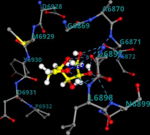
Figure 1. Best docked pose resulted from molecular docking calculation for a. 3CL pro-2-
DG(6lu7-2-DG) b. PLpro-2-DG(6wuu-2-DG) c. NSp-16-2-DG(6wkq-2-DG) d. RdRp-2-DG(7bv1-2-
DG) from PRinMTML-ESS platform.

Gly-271, Tyr-268) obtained from a study that revealed the inhibitory
mechanisms and determined the crystal structure of inhibitor VIR250 and
VIR251 in complex with SARS-CoV-2 PLpro.9 A study by Yang et al. determined the
crystal structure of 3CLpro of SARS-CoV-2 in complex with the inhibitor (N3).10
They have predicted the bonding site of inhibitors (binding region residues: Cys-
145 to His-41 cavity) which we have used for the grid generation. Yin et al.
determined the crystal structure of polymerase bound to RNA and to the drug
Remdesivir. The binding site information of RdRP (Gly-683, Ser-682, Asp-684,
Asp-760, Ser-759, Ala-685, Tyr-689, Ser-814, Gln-815) has been obtained from
this study and used for grid generation.11 Rosas-Lemus et al. unravelled the
crystal structures 2’-O-methyltransferase, the nsp16-nsp10 heterodimer, in
complex with an inhibitor Sinefungin.12 Sinefungin binds to Nsp-16 at the binding
site (ASN-6899, ASP-6897, ASP-6928, ASN-6841, CYS-6913, TYR-6930) which has
been used for grid generation. In all the protein ligand combination the best
docked pose was found be residing at that particular binding site, whose
information we have obtained from crystal structure (Figure 1). The binding
energy of the best docked pose for 3CLpro-2-DG(6lu7-2-DG), NSp-16-2-DG(6wkq-
2-DG), PLpro-2-DG(6wuu-2-DG) and RdRp-2-DG(7bv1-2-DG) are listed in Table 1.
Docking scores were found to be -2.35 kcal/mol for 3CLpro-2-DG(6lu7-2-DG)
a

 b

 c

 d

Figure 2. The histogram plot of conformers after molecular docking for a. 3CLpro-2-DG(6lu7-2-
DG) b. PLpro-2-DG(6wuu-2-DG) c. NSp-16-2-DG(6wkq-2-DG) d. RdRp-2-DG(7bv1-2-DG)
generated from PRinMTML-ESS platform.

combination, -1.7 kcal/mol for NSp-16-2-DG(6wkq-2-DG) combination, -1.77
kcal/mol for PLpro-2-DG(6wuu-2-DG) and -1.26 kcal/mol for RdRp-2-DG(7bv1-2-
DG).
Docking Energy and Docking Score are plotted for all the protein-ligand
combination in the Docking Analysis part of PRinMTML-ESS platform. The
histogram plots for all the combinations indicates that the lowest energy
configuration is the highest in number in the clusters (Figures 2). The
combination 3CLpro-2-DG have the lowest docking energy and docking score
having three hydrogen bonding interaction of the ligand, two between the OH
group of 2-DG with the carbonyl group of the surrounding residue Phe-140 and
one between OH group of 2-DG with the amine nitrogen of surrounding residue
Gly-143.
The hydrogen bonding and hydrophobic interactions of all 2-DG with the four
viral proteins at their lowest energy configuration are shown in Figures 3. To
select the combinations for molecular dynamic simulation in PRinMTML-ESS a
screening is done based on the docking energy. Since the lowest energy
configuration for all the protein-ligand combination, we have set a cutoff score
of -6.00 kcal/mol which selects all the four protein-ligand combinations for
further MD simulations. The lowest energy configuration of the docked
structure was used as the starting structure for performing MD simulation for
apprehending the stability of the interaction of the ligand with respect to the
protein binding site in explicit water. In the PRinMTML-ESS platform for
quantified the interactions between the amino acids in the binding pocket and
the ligand several analysis has been carried out. The average root mean square

 a b

 c d

Figure 3. The ligand interaction showing hydrogen bonding and hydrophobic interaction at
the best docked pose a. 3CLpro-2-DG(6lu7-2-DG) b. PLpro-2-DG(6wuu-2-DG) c. NSp-16-2-
DG(6wkq-2-DG) d. RdRp-2-DG(7bv1-2-DG) generated from PRinMTML-ESS platform.
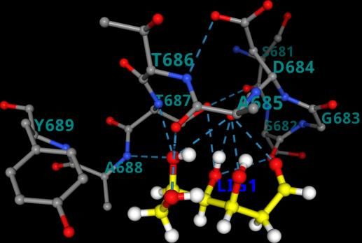
Table 1. The docking energy, docking score, MD non-bonded total energy, free energy barriers
for dissociation, average RMSD values, Centre of Mass(COM) distance

 Target Docking Docking MD non- Free Average COM Interact Residue-
 Energy Score bonded Energy RMSD Distance ing ligand
 (kcal/mol) (kcal/mol) total barrier (nm) (nm) Residue interacting
 energy (kJ/mol) groups
 (kJ/mol)
 NSp16 -7.71 -1.7 -197.7 29.2 0.13 0.47 Asp- OH-C=O
 6897 OH-C=O
 Asp- OH-C=O
 6928
 Gly-
 6871
 RdRp -6.79 -1.26 -66.2 26.4 0.14 0.16 Ser-682 OH-C=O
 3CLPro -7.75 -2.35 -97.1 --- 0.11 1.27 --- ---

 PLPro -6.97 -1.77 -10.3 --- 0.16 1.08 --- ---

deviation (RMSD) of the target protein binding pocket residues is calculated
between the initial MD configuration (docked structure) and the MD
equilibrated structure (final frame of MD simulation). Average RMSD of the final
frame of MD simulated structure in the four combinations is found to vary
between 0.11 nm to 0.16 nm (Table 1). Average RMSD value lower than 0.2 nm
clearly shows that all the protein ligand combinations are stable. The hydrogen
bonding section plots the number of hydrogen bonds between the ligand and
the residues at the binding site of the target. NSp16-2-DG(6wkq-2-DG) is found
to have the highest number of hydrogen bonding interaction, six followed by
two hydrogen bonding in RdRp-2-DG(7bv1-2-DG) and no hydrogen bonding in
PLpro- 2-DG(6wuu-2-DG), and 3CLpro-2-DG(6lu7-2-DG) combination (Figure 4). In
the Interaction Energy section, the non-bonded interaction energies, coulomb
and van der Waals interaction energies, between the ligand and protein are
plotted for each combination in the platform.
The total non-bonded interaction energy was found to -197.7 kJ/mol for NSp16-
2-DG(6wkq-2-DG), hence the most favourable interaction of ligand 2-DG occurs
with O-methyl transferase (NSp-16) of SARS-CoV-2. Total non-bonded
interaction energy for 3CLpro-2-DG(6lu7-2-DG), RdRp-2-DG(7bv1-2-DG) and
PLpro-2-DG(6wuu-2-DG) are found to be -97.1 kJ/mol, -66.2 kJ/mol and -10.3
kJ/mol respectively (Table 1). These energy values predict that only in
NSp16(6wkq) the ligand 2-DG is stable and tends to be in the binding site.
However, for the other three viral proteins 3CLpro, RdRp and PLpro the interaction
energy is much lower to stabilise the ligand in the binding cavity.
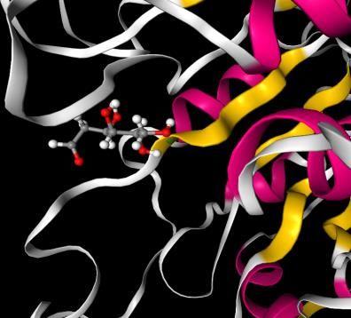
The next section of MD analysis is the centre of mass distance which calculates
the distance between the centre of mass of the residues in the binding site with
the centre of mass of the ligand. Average centre of mass distance if larger than
1 nm would predict the ligand has escaped the binding pocket after MD
simulation.
The average centre of mass (COM) distance plot shows that the combinations
NSp16-2-DG(6wkq-2-DG) and RdRp-2-DG(7bv1-2-DG) have value 0.47 nm and
0.16 nm respectively, hence lies within the binding site cavity at the equilibrated
structure after MD simulation. (Table 1) However, in both the combination
3CLpro-2-DG(6lu7-2-DG) and PLpro-2-DG(6wuu-2-DG) the average COM distance
are 1.27 nm and 1.08 nm respectively, hence here the ligand 2-DG have escaped
the binding sites because of less interaction strength with the binding site
residues of protein. The final equilibrated structure of the four combinations
and visualization of the trajectory is consistent with the COM distance results.

 a b a

 c b
 d

Figure 4. [A] The equilibrated structure after MD simulation for the combinations a. 3CLpro-2-
DG(6lu7-2-DG) b. PLpro-2-DG(6wuu-2-DG) c. NSp-16-2-DG(6wkq-2-DG) d. RdRp-2-DG(7bv1-2-
DG) generated from PRinMTML-ESS platform (Left). [B] The ligand interaction with the
binding residues showing hydrogen bonding and hydrophobic interaction in a. NSp-16-2-
DG(6wkq-2-DG) and b. RdRp-2-DG(7bv1-2-DG) (Right).
The equilibrated structure of NSp16-2-DG(6wkq-2-DG) have three hydrogen
bonding interaction between 2-DG and the binding residues Asp-6897, Asp-6928
and Gly-6871. (Figure 4). The equilibrated structure of RdRp-2-DG(7bv1-2-DG)
has two hydrogen bonds with Ser-682. Our platform generates a combination
score based on the average RMSD, internal energy and hydrogen bonding data
obtained from analysis of MD simulation. Based on this score the selection of
combinations for running the enhanced sampling free energy simulation is done.
Among the four combination we have selected NSp16-2-DG(6wkq-2-DG) and
RdRp-2-DG(7bv1-2-DG) combination based on their combination score 0.51 and
0.55 respectively, since only in these two combinations the ligand is still in the
binding cavity after MD simulation. The sampling around the binding sites in
instance of an MD simulation is not sufficient to predict the stability of the ligand
in the binding cavity of protein, as conformations might get stuck in local
minima. Therefore, meta-dynamics an enhanced sampling method of
simulation19 for quantitatively predicting ligand binding energy and analysis of
changes in the conformation of ligands is important to ascertain the most stable
(bound) protein-ligand complex and predicting the inhibition property of the
ligand. The equilibrium structures obtained from MD simulations were used as
the starting configurations in the enhanced sampling free energy simulations
(FES). The average free energy of dissociation obtained from 5 independent
dissociation simulations for the combination NSp16-2-DG(6wkq-2-DG) and
RdRp-2-DG(7bv1-2-DG) are 29.1 kJ/mol and 26.4 kJ/mol (Table 1). For clarity and
understanding of the free energy surfaces the individual free energy surfaces of
all the independent runs and the average surface is shown in Figure 5. The free
energy surfaces predict that the surfaces have multiple local minima and one
global minimum for the ligand bound at the binding cavity. This structural
characteristic represents multiple interactions between the ligands and residues
of the binding site. The free energy of dissociation of the ligand 2-DG is of same
order for both the viral protein NSp16(6wkq) and RdRp(7bv1), however the
combination NSp16-2-DG(6wkq-2-DG) has the maximum ligand dissociation free
energy. However previous studies show that antiviral drugs like Carfilzomib,
Eravacycline, Valrubicin, Lopinavir, Elbasvir and Ritonavir interacts with SARS-
COV-2 viral proteins 3CLpro, PLpro, RdRp and spike protein where total binding
free energy ranges from -79.4 kJ/mol to -168.1 kJ/mol with the van der Waal
a

 b

 c

 d

Figure 5. a. Free energy surface obtained from five Individual dissociation simulations for the
combination NSp16-2-DG(6wkq-2-DG) b. Average free energy surface for the combination
NSp16-2-DG(6wkq-2-DG) c. Free energy surface obtained from five Individual dissociation
simulations for the combination RdRp-2-DG(7bv1-2-DG) b. Average free energy surface for
the combination NSp16-2-DG(6wkq-2-DG) d. Average free energy surface for the combination
RdRp-2-DG(7bv1-2-DG)
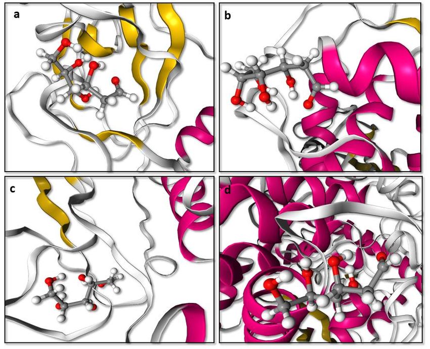
Scheme 2. a. Inhibition of viral proteins taking part in replication and transcription of SARS-
COV 2 b. Glycolytic inhibition via 2-deoxy-D-glucose (2-DG)

energy components ranging from -133 kJ/mol to -255.1 kJ/mol when calculated
using molecular dynamics and MM-GBSA based scoring function.20 Since 2-DG
shows a lower value of binding energy to all the viral proteins compared to most
of the antiviral drugs it is challenging to predict if 2-DG plays a role of antiviral
drug for inhibiting the SARS-CoV-2 viral proteins. The free energy barrier of
dissociation of the 2-DG from both the viral proteins Nsp16 and RdRp is
extremely low and can hardly predict any inhibition property of 2-DG. We also
carried a 50 ns simulation for all the four combination and aligned with the
enhanced free energy sampling results we found in all the case 2-DG have
escaped the binding cavity at the final equilibrated structure.
On the other hand, several studies have shown glucose plays a major role in
proliferation of SARS-CoV-2.21 This is one of the reasons why obese and diabetic
patients with uncontrolled blood glucose level are more prone to develop a
severe form of COVID-19. Due to the structural similarity between 2-DG and
glucose (Scheme 1), it is expected that 2-DG would act as the competitive
inhibitor of glucose metabolism and would strongly effect the metabolic
processes like glycolysis and glycosylation which are dependent on glucose.
When SARS-CoV-2 attacks our cells, they co-opt both of these metabolic
Table 2. The docking energy, MD non-bonded total energy, free energy barriers for
dissociation, average RMSD values, Centre of Mass(COM) distance
 Target Ligand Docking MD Free Average COM Interacting Residue-
 Energy non- Energy RMSD Distance Residue ligand
 (kcal/mol) bonded barrier (nm) (nm) interacting
 total (kJ/mol) groups
 energy
 (kJ/mol)
 Hexokinase Glucose -7.72 -330.2 66.3 3.7 0.50 Gln-291 NH2-OH
 Lys-173 N-OH
 Asn-235 NH2-O
 Thr-172 OH-OH
 Asn-208 NH2-OH
 Glu-260 C=O-OH
 Asp-209 C=O-OH
 Glu-294 C=O-OH
 Hexokinase 2-DG -7.33 -257.2 52.4 3.6 0.38 Glu-260 C=O-OH
 Asn-235 NH2-O
 Asp-209 C=O-OH
 Thr-172 OH-OH

processes to increase its replication and transcription. SARS-CoV-2 induces an
anabolic state in their host cell which causes these infected cells to upregulate
their production of energy using glycolysis as compared with their healthy
neighbours.22 2-DG differs from the glucose by removal of an oxygen atom from
the 2-position (Scheme 1). In the glycolysis 2-DG is absorbed by the cell and
undergoes phosphorylation at the 6 position to generate 2-DG-6P, in the next
step unlike glucose-6-phosphate, 2-DG-6P cannot undergo isomerisation by
glucose-6-phosphate isomerase hence the glycolytic flux is reduced (Scheme 2).
Therefore, in presence of 2-DG, the rate limiting reaction of glycolysis is ATP-
dependent phosphorylation of glucose to form glucose-6-phosphate (G-6-P) and
is catalysed by tissue-specific isoenzymes known as hexokinases. A recent study
showed that SARS-COV-2 infection induces higher amount of glucose influx and
glycolysis in the infected cells, resulting in selective high accumulation of the
fluorescent glucose/2-DG analogue in the viral infected cells. They further
observed that mannose inhibit the entry of 2-DG analogue at a very low
concentration hence predicted that 2-DG entry in virus-infected cells might be
manipulating specific mannose transporter or high-affinity glucose transporter,
GLUT3, which was found to be increased on SARS-CoV-2 infection.23 In our study
we have tried to explore the competitive inhibition property of 2-DG on the first
step of phosphorylation with the aid of hexokinase enzyme after entering the
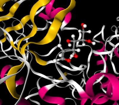
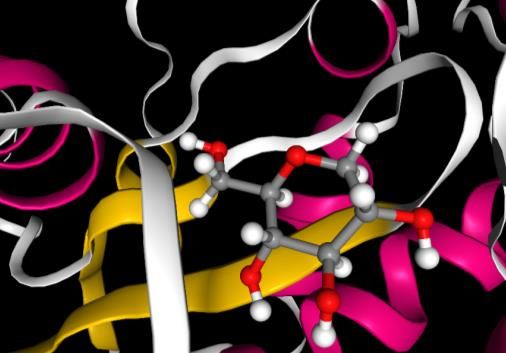
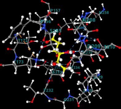
a b

 c d

Figure 6. [A] The equilibrated structure after MD simulation for the combinations a.
Hexokinase-Glucose(1qha-glucose) c. Hexokinase-2-deoxy-D-glucose(1qha-2-DG) (Left) [B]
The ligand interaction with the binding residues showing hydrogen bonding and hydrophobic
interaction in b. Hexokinase-Glucose(1qha-glucose) and d. Hexokinase-2-deoxy-D-
glucose(1qha-2-DG) (Right).

 viral infected cell. The 2-DG and glucose cyclic conformers were docked at the
binding site of hexokinase (Thr-210, Thr-232, Ser-155, Lys-173).The binding
energy was found to be -7.72 kcal/mol and -7.33 kcal/mol for glucose and 2-DG
respectively. The entropic contribution associated with the solvent effect and
conformational changes in the docked ligand is not accounted in the docking.
The best docked pose and MD simulation is carried out in the next step. The
non-bonded total interaction energy in the equilibrated structure were found to
be slightly lower in glucose, -330.2 kJ/mol than 2-DG, -257.2 kJ/mol. The
Coulomb energy and Van der Walls energy was also found to follow the same
trend (Table 2). RMSD was computed for the target binding pocket residues is
the initial MD configuration and the MD equilibrated structure for both the 2-
DG-hexokinase and glucose-hexokinase structure (Table 2). The Centre of mass
distance 0.50nm and 0.38 nm for glucose and 2-DG indicated that the ligands
are stable in the binding cavity. Number of hydrogen bonding interaction in the
equilibrated structure in glucose and 2-DG are ten and seven respectively, the
interacting residues are indicated in Table 2 and Figure 6. To increase the
sampling around the binding site of the protein and calculate the more accurate
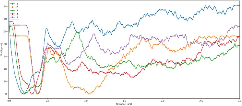
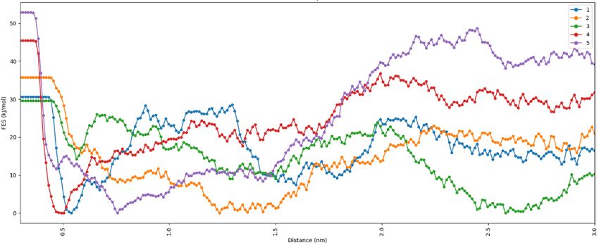
a

 b

 c

 d

Figure 7. a. Free energy surface obtained from five Individual dissociation simulations for the
combination Hexokinase-Glucose(1qha-glucose) b. Average free energy surface for the
combination Hexokinase-Glucose(1qha-glucose) c. Free energy surface obtained from five
Individual dissociation simulations for the combination Hexokinase-2-deoxy-D-glucose(1qha-
2-DG) d. Average free energy surface for the combination Hexokinase-2-deoxy-D-
glucose(1qha-2-DG)
binding energy of the ligand considering the changes in conformation of the
ligand the equilibrium structure obtained from MD simulations was used as the
starting configuration in the enhanced wt-metaD simulation. The average free
energy barrier of dissociation obtained from enhanced wt-metaD simulations
was found to be 66.3 kJ/mol and 52.4 kJ/mol for glucose and 2-DG (Table 2).
Hence, it can be predicted that since the free energy barrier is comparable and
of same order in both glucose and 2-DG, they have the similar binding affinity to
hexokinase enzyme. Therefore, the phosphorylation step of conversion of
glucose to glucose-6-phosphate is inhibited in presence of 2-DG at a certain
concentration. Thus, competitive inhibition is caused by 2-DG, which structurally
similar to glucose and can combine at the same binding site of hexokinase. For
clearly understanding the free energy surface the average and the individual
surfaces are plotted in PRinMTML-ESS platform as shown in Figure 7. The free
energy surfaces show jagged and uneven surfaces arising due to the multiple
interaction of the ligand at the binding site. The MD simulation was extended
to 50 ns keeping all other parameters same as the 2 ns simulation. However
negligible change in the ligand configuration and interaction of the ligand with
the binding residues at the final equilibrated structure.

Computational Method
 In this study for carrying out all the calculations and generating the figure the
Prescience in silico Multi Target Multi Ligand Enhanced Sampling Screening
(PRinMTML-ESS) has been used. In PRinMTML-ESS the process flow is in-build
and fully automated, so no manual intervention is required from the user-side.
The process flow here consists of molecular docking for docking the ligands in
by automated grid generation at the target binding site. To explore the stability
of the target-ligand complex all-atom molecular dynamic simulation is carried
out in the next step and enhanced free energy sampling at the last stage for final
analysis of the binding property of the ligand with the target
(protein/enzyme/DNA-RNA). Efficient scoring algorithms are used after Docking
and MD simulation for screening at each stages. All molecular docking
calculation and MD simulations are performed using Autodock 4 and GROMACS-
5.1.4 simulation package.24-25 The enhanced sampling using metadynamics19
(metaD) and its variant well-tempered metadynamics26 (wt-metaD) using
Scheme 2. PRinMTML-ESS platform structure of integrated computational methods and
analysis

Plumed 2.3.027 patched with MD engine GROMACS 5.1.4 integrated within the
PRinMTML-ESS platform. Enhanced sampling-based method metadynamics
developed by Laio et al.19, is widely used in simple and complex molecules due
to its advantage over free energy perturbation and thermodynamic integration
methods which are computationally more expensive for larger complex system
like protein-ligand, protein-protein binding. A time-dependent bias is included
to the system in a metadynamic simulation along with some deposited bias like
a suitably chosen reaction coordinate(s) which will eventually push the complex
away from the minimum energy state, so that the system doesn’t get trapped
at a local minima for sufficiently long time. Since this method is independent on
the choice of reaction coordinate and not very sensitive to the precise choice of
biasing parameters (except in the case where the parameters are chosen to be
too high or low), any rudimentary reaction coordinate can bias the system
helping it to escape the local minima within a small time and generate a
qualitative free energy surface (FES). Metadynamics simulation uses a history-
dependent bias which inhibits the system from repeated revisiting previously
visited regions of the phase space. Another, widely used scoring method over
molecular dynamic simulation is the molecular mechanics Poisson-Boltzmann
surface area (MMPBSA) which uses approximations to calculate enthalpic and
entropic contributions using implicit continuum solvent hence less accurate in
comparison to enhanced free energy sampling method used in our study.
Additionally, as an alternative to MMPBSA we have performed all-atom
simulation in explicit solvent medium including dynamics of solvent, solute and
ions. Considering the effectiveness and computational viability of enhanced free
energy sampling method, it has been used as the final scoring method for
calculating free energy barrier for dissociation in PRinMTML-ESS platform. In-
house codes have been used for generating the analysis in each section of
computational methods used. A scheme of the methodologies and analysis used
in PRinMTML-ESS platform are shown in Scheme 2. The structure of ligands used
in this study has been downloaded from PubChem
(https://pubchem.ncbi.nlm.nih.gov/). The crystal structure of the viral proteins
of SAR-COV2 PLpro (PDB ID: 6wuu), 3CLpro (PDB ID: 6lu7), RNA-dependent RNA
polymerase (PDB ID: 7bv1) and the Hexokinase enzyme (PDB ID: 1QHA) are
obtained from RCSB Protein Data Bank(https://www.rcsb.org/). All the protein
and enzyme structures were cleaned (removing ligands, ions, water molecules)
and missing loop was modelled using our PRinBio platform. A ligand
conformational search is carried out generating an automated grid box of the
dimension 60*60*60 Å by taking the binding site centroid as a grid center with
a spacing of 0.375 Å. The docking was conducted the Lamarckian genetic
algorithm (LGA), and a total of 100 GA-LA hybrid runs for performing the
conformational search. In the MD simulation the proteins and enzyme were
modelled using the CHARMM27 force field28 parameters. For generating the
CHARMM27 force field for all the ligands, SwissParam29 was used and the force
field generation is automated in PRinMTML-ESS platform. The target-ligand
systems were solvated in water and equilibrated using MD simulations at room
temperature. The systems were first equilibrated using an NVT ensemble at 300
K for 1 ns and extended to the NPT ensemble at 300 K and 1 atm for another 2
ns. The temperature and pressure during the simulations were maintained using
a velocity rescaling thermostat and Parrinello-Rahman barostat, respectively. A
time step of 1 fs was used to integrate the equation of motion, and a non-
bonded cutoff of 10Å was used to perform the MD simulations. We have used a
bias V(s,t) in the form of Gaussians with every 500 steps (1 ps) deposition pace
with a Gaussian hill-height of 2.0 kJ/mol, width of σ (0.1 nm), bias factor of 15,
and temperature (T) of 300 K. For smooth convergence of the system in a wt-
metaD the amplitude of the bias is tuned accordingly. Here a tempering factor
ΔT (Equation 1) is used to adjust the height of the hills, and henceforth a smooth
convergence of the free energy landscape is attained.

 + ∆
 ( ) = − ( , ) + ( ) (1)
Once the system converges, free energy F(s) (Eq. (1))30 can be extracted by
adding the deposited hills along the biased reaction coordinates. The centre of
mass distance between the heavy atoms of the ligands and the protein
backbone in the surrounding area of the binding pocket is considered as the
reaction coordinates in the free energy sampling, since the aim of the calculation
is taking into account the ligand dissociation from the binding site. In this study
5 independent simulations are done and an averaging is done for each
combination to obtain better sampling and statistically reliable results.

Conclusion
This computational study aims to understand the role of 2-deoxy-D-glucose as
an antiviral and glycolysis pathway inhibitor in SAR-CoV-2 affected human body
using PRinMTML-ESS platform. Using this platform molecular docking, all-atom
molecular dynamics simulations with enhanced free energy simulation was
performed and screening was done in each stage using effective scoring
algorithm. Here we have explored the inhibition property of 2-DG with the four
SAR-CoV-2 viral proteins 3CLpro, PLpro, NSp-16 and RdRp which are important for
transmission and replication of SARS-CoV-2. 2-DG has very low dissociation
barrier with O-methyl-transferase (Nsp16) and RdRp in free energy simulation.
Hence it can be predicted that 2-DG can barely inhibit any viral protein taking
part in replication and translation of SARS-CoV-2. On the other hand,
PRinMTML-ESS platform predicts 2-DG binds strongly to hexokinase, the enzyme
responsible for phosphorylation of glucose to glucose-6-phosphate in glycolysis.
Though glucose has higher binding affinity to hexokinase, comparable binding
affinity of 2-DG may lead to competitive inhibition of the glycolysis, hence the
glycolytic flux is reduced which in turn reduces the replication and transcription
of SARS-CoV-2 virus in human cells. Therefore, our study predicts that 2-DG
though doesn’t show any comparable inhibition for non-structured protein
associated with translation and replication of SAR-CoV-2 can be used as a clinical
therapy in COVID-19 infected patient for competitive inhibition of glycolysis
process in virus infected cells, thereby reducing the rate the replication of virus.
Reference
1. Hoffmann, M.; Kleine-Weber, H.; Schroeder, S.; Krüger, N.; Herrler, T.; Erichsen, S.; Schiergens,
T. S.; Herrler, G.; Wu, N.-H.; Nitsche, A., SARS-CoV-2 cell entry depends on ACE2 and TMPRSS2 and is
blocked by a clinically proven protease inhibitor. cell 2020, 181 (2), 271-280. e8.
2. Lan, J.; Ge, J.; Yu, J.; Shan, S.; Zhou, H.; Fan, S.; Zhang, Q.; Shi, X.; Wang, Q.; Zhang, L., Structure
of the SARS-CoV-2 spike receptor-binding domain bound to the ACE2 receptor. Nature 2020, 581
(7807), 215-220.
3. Koopmans, M.; Haagmans, B., Assessing the extent of SARS-CoV-2 circulation through
serological studies. Nat Med 2020, 26 (8), 1171-2.
4. Woo, P. C.; Huang, Y.; Lau, S. K.; Yuen, K.-Y., Coronavirus genomics and bioinformatics analysis.
Viruses 2010, 2 (8), 1804-1820.
5. Sadee, W., Genomics and personalized medicine. Elsevier: 2011.
6. Helmy, Y. A.; Fawzy, M.; Elaswad, A.; Sobieh, A.; Kenney, S. P.; Shehata, A. A., The COVID-19
pandemic: a comprehensive review of taxonomy, genetics, epidemiology, diagnosis, treatment, and
control. Journal of clinical medicine 2020, 9 (4), 1225.
7. Yuan, M.; Wu, N. C.; Zhu, X.; Lee, C.-C. D.; So, R. T.; Lv, H.; Mok, C. K.; Wilson, I. A., A highly
conserved cryptic epitope in the receptor binding domains of SARS-CoV-2 and SARS-CoV. Science
2020, 368 (6491), 630-633.
8. Bagdonaite, I.; Wandall, H. H., Global aspects of viral glycosylation. Glycobiology 2018, 28 (7),
443-467.
9. Rut, W.; Lv, Z.; Zmudzinski, M.; Patchett, S.; Nayak, D.; Snipas, S. J.; El Oualid, F.; Huang, T. T.;
Bekes, M.; Drag, M., Activity profiling and crystal structures of inhibitor-bound SARS-CoV-2 papain-like
protease: A framework for anti–COVID-19 drug design. Science advances 2020, 6 (42), eabd4596.
10. Jin, Z.; Du, X.; Xu, Y.; Deng, Y.; Liu, M.; Zhao, Y.; Zhang, B.; Li, X.; Zhang, L.; Peng, C., Structure
of M pro from SARS-CoV-2 and discovery of its inhibitors. Nature 2020, 582 (7811), 289-293.
11. Yin, W.; Mao, C.; Luan, X.; Shen, D.-D.; Shen, Q.; Su, H.; Wang, X.; Zhou, F.; Zhao, W.; Gao, M.,
Structural basis for inhibition of the RNA-dependent RNA polymerase from SARS-CoV-2 by remdesivir.
Science 2020, 368 (6498), 1499-1504.
12. Rosas-Lemus, M.; Minasov, G.; Shuvalova, L.; Inniss, N. L.; Kiryukhina, O.; Brunzelle, J.; Satchell,
K. J., High-resolution structures of the SARS-CoV-2 2′-O-methyltransferase reveal strategies for
structure-based inhibitor design. Science Signaling 2020, 13 (651).
13. Namsani, S.; Pramanik, D.; Khan, M. A.; Roy, S.; Singh, J. K., Metadynamics-based enhanced
sampling protocol for virtual screening: case study for 3CLpro protein for SARS-CoV-2. Journal of
Biomolecular Structure and Dynamics 2021, 1-16.
14. Kumawat, A.; Namsani, S.; Pramanik, D.; Roy, S.; Singh, J. K., Integrated docking and enhanced
sampling based selection of repurposing drugs for SARS-CoV-2 by targeting host dependent factors.
2020.
15. Gorbalenya, A. E.; Enjuanes, L.; Ziebuhr, J.; Snijder, E. J., Nidovirales: evolving the largest RNA
virus genome. Virus research 2006, 117 (1), 17-37.
16. V’kovski, P.; Kratzel, A.; Steiner, S.; Stalder, H.; Thiel, V., Coronavirus biology and replication:
implications for SARS-CoV-2. Nature Reviews Microbiology 2020, 1-16.
17. Báez-Santos, Y. M.; John, S. E. S.; Mesecar, A. D., The SARS-coronavirus papain-like protease:
structure, function and inhibition by designed antiviral compounds. Antiviral research 2015, 115, 21-
38.
18. Vithani, N.; Ward, M. D.; Zimmerman, M. I.; Novak, B.; Borowsky, J. H.; Singh, S.; Bowman, G.
R., SARS-CoV-2 Nsp16 activation mechanism and a cryptic pocket with pan-coronavirus antiviral
potential. Biophysical Journal 2021.
19. Laio, A.; Parrinello, M., Escaping free-energy minima. Proceedings of the National Academy of
Sciences 2002, 99 (20), 12562-12566.
20. Murugan, N. A.; Kumar, S.; Jeyakanthan, J.; Srivastava, V., Searching for target-specific and
multi-targeting organics for Covid-19 in the Drugbank database with a double scoring approach.
Scientific reports 2020, 10 (1), 1-16.
21. Codo, A. C.; Davanzo, G. G.; de Brito Monteiro, L.; de Souza, G. F.; Muraro, S. P.; Virgilio-da-
Silva, J. V.; Prodonoff, J. S.; Carregari, V. C.; de Biagi Junior, C. A. O.; Crunfli, F., Elevated glucose levels
favor SARS-CoV-2 infection and monocyte response through a HIF-1α/glycolysis-dependent axis. Cell
metabolism 2020, 32 (3), 437-446. e5.
22. Gualdoni, G. A.; Mayer, K. A.; Kapsch, A.-M.; Kreuzberg, K.; Puck, A.; Kienzl, P.; Oberndorfer,
F.; Frühwirth, K.; Winkler, S.; Blaas, D., Rhinovirus induces an anabolic reprogramming in host cell
metabolism essential for viral replication. Proceedings of the National Academy of Sciences 2018, 115
(30), E7158-E7165.
23. Bhatt, A. N.; Kumar, A.; Rai, Y.; Kumari, N.; Vedagiri, D.; Harshan, K. H.; Chinnadurai, V.;
Chandna, S., Glycolytic inhibitor 2-Deoxy-D-glucose attenuates SARS-CoV-2 multiplication in host cells
and weakens the infective potential of progeny virions. bioRxiv 2021.
24. Abraham, M. J.; Murtola, T.; Schulz, R.; Páll, S.; Smith, J. C.; Hess, B.; Lindahl, E., GROMACS:
High performance molecular simulations through multi-level parallelism from laptops to
supercomputers. SoftwareX 2015, 1, 19-25.
25. Pronk, S.; Páll, S.; Schulz, R.; Larsson, P.; Bjelkmar, P.; Apostolov, R.; Shirts, M. R.; Smith, J. C.;
Kasson, P. M.; van der Spoel, D., GROMACS 4.5: a high-throughput and highly parallel open source
molecular simulation toolkit. Bioinformatics 2013, 29 (7), 845-854.
26. Barducci, A.; Bussi, G.; Parrinello, M., Well-tempered metadynamics: a smoothly converging
and tunable free-energy method. Physical review letters 2008, 100 (2), 020603.
27. Bonomi, M.; Branduardi, D.; Bussi, G.; Camilloni, C.; Provasi, D.; Raiteri, P.; Donadio, D.;
Marinelli, F.; Pietrucci, F.; Broglia, R. A., PLUMED: A portable plugin for free-energy calculations with
molecular dynamics. Computer Physics Communications 2009, 180 (10), 1961-1972.
28. MacKerell Jr, A. D.; Banavali, N.; Foloppe, N., Development and current status of the CHARMM
force field for nucleic acids. Biopolymers: Original Research on Biomolecules 2000, 56 (4), 257-265.
29. Zoete, V.; Cuendet, M. A.; Grosdidier, A.; Michielin, O., SwissParam: a fast force field
generation tool for small organic molecules. Journal of computational chemistry 2011, 32 (11), 2359-
2368.
30. Clark, A. J.; Tiwary, P.; Borrelli, K.; Feng, S.; Miller, E. B.; Abel, R.; Friesner, R. A.; Berne, B. J.,
Prediction of protein–ligand binding poses via a combination of induced fit docking and metadynamics
simulations. Journal of chemical theory and computation 2016, 12 (6), 2990-2998.
You can also read