The Effects of Intraoperative Hypothermia on Postoperative Cognitive Function in the Rat Hippocampus and Its Possible Mechanisms
←
→
Page content transcription
If your browser does not render page correctly, please read the page content below
brain
              sciences
Article
The Effects of Intraoperative Hypothermia on Postoperative
Cognitive Function in the Rat Hippocampus and Its
Possible Mechanisms
Guangyan Xu                 , Tianjia Li *     and Yuguang Huang *

                                          Department of Anesthesiology, Peking Union Medical College Hospital, Chinese Academy of Medical Sciences
                                          and Peking Union Medical College, Beijing 100730, China; bonnie_xgy08@163.com
                                          * Correspondence: litianjia@pumch.cn (T.L.); garypumch@163.com (Y.H.)

                                          Abstract: Intraoperative hypothermia is a common complication during operations and is asso-
                                          ciated with several adverse events. Postoperative cognitive dysfunction (POCD) and its adverse
                                          consequences have drawn increasing attention in recent years. There are currently no relevant stud-
                                          ies investigating the correlation between intraoperative hypothermia and POCD. The aim of this
                                          study was to assess the effects of intraoperative hypothermia on postoperative cognitive function
                                          in rats undergoing exploratory laparotomies and to investigate the possible related mechanisms.
                                          We used the Y-maze and Morris Water Maze (MWM) tests to assess the rats’ postoperative spa-
                                          tial working memory, spatial learning, and memory. The morphological changes in hippocampal
                                          neurons were examined by haematoxylin-eosin (HE) staining and hippocampal synaptic plasticity-
                                          related protein expression. Activity-regulated cytoskeletal-associated protein (Arc), cyclic adenosine
                                          monophosphate-response element-binding protein (CREB), S133-phosphorylated CREB (p-CREB

                                   [S133]), α-amino-3-hydroxy-5-methyl-4-isoxazole propionic acid receptor 1 (AMPAR1), and S831-
Citation: Xu, G.; Li, T.; Huang, Y. The   phosphorylated AMPAR1 (p-AMPAR1 [S831]) were evaluated by Western blotting. Our results
Effects of Intraoperative                 suggest a correlation between intraoperative hypothermia and POCD in rats and that intraoperative
Hypothermia on Postoperative              hypothermia may lead to POCD regarding impairments in spatial working memory, spatial learning,
Cognitive Function in the Rat             and memory. POCD induced by intraoperative hypothermia might be due to hippocampal neurons
Hippocampus and Its Possible              damage and decreased expression of synaptic plasticity-related proteins Arc, p-CREB (S133), and
Mechanisms. Brain Sci. 2022, 12, 96.      p-AMPAR1 (S831).
https://doi.org/10.3390/
brainsci12010096                             Keywords: intraoperative hypothermia; postoperative cognitive dysfunction; hippocampus; synaptic
Academic Editors: Luigi De Gennaro           plasticity; synaptic plasticity-related protein
and Dona E. Locke

Received: 22 November 2021
Accepted: 8 January 2022
                                             1. Introduction
Published: 12 January 2022
                                                  Intraoperative hypothermia (defined as a core temperature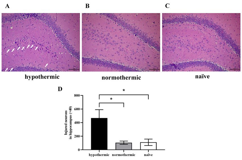
Brain Sci. 2022, 12, 96                                                                                            2 of 14

                          and societal burdens [11,12]. Although many mechanisms, including apoptosis [13], neu-
                          roinflammation and oxidative stress [11,14,15], autophagy [16], and synaptic plasticity dys-
                          function [16,17] are involved in POCD, the exact molecular mechanism remains unknown.
                               The hippocampus, which is integral to the formation and retention of episodic and
                          spatial memory, is a brain region that is responsible for cognitive functions, such as learning,
                          memory, and spatial navigation [18,19]. Several studies have demonstrated that amygdala–
                          hippocampus–prefrontal cortex neural network is widely involved in emotion regulatory
                          and memory consolidation processes [20,21]. Moreover, the neural circuitry of fear acquisi-
                          tion and extinction includes the amygdala, hippocampus, ventromedial prefrontal cortex
                          (vmPFC) and dorsolateral prefrontal cortex (dlPFC), which is well established in rodent
                          and humans [22,23].
                               Synaptic plasticity, the ability of synapses to modulate their strength or efficacy of
                          synaptic transmission, underlies learning, memory, and information processing in the
                          brain [24]. Non-invasive brain stimulation (NIBS), an innovative set of technologies and
                          techniques, was used to stimulate or alter synaptic plasticity, modify and enhance cognitive,
                          behavioural, social, and emotional processes [25]. Recently, the effects of NIBS on attention
                          and memory functions were widely examined [26,27].
                               Activity-regulated cytoskeletal-associated protein (Arc) is involved in multiple forms
                          of synaptic plasticity, including long-term potentiation (LTP) and long-term depression
                          (LTD) [28]. Previous findings have implicated that Arc promotes the hippocampal neuronal
                          maturation critical for learning, memory storage, and memory consolidation [29,30].
                               Cyclic adenosine monophosphate-response element-binding protein (CREB) is widely
                          involved in neuronal development, plasticity, survival and neuroprotection in the central
                          nervous system, and participates in LTP formation [17,31]. Several studies have confirmed
                          that CREB exhibits vital biological functions, including learning ability, memory formation
                          and cognition regulation [31,32]. CREB activation is mainly regulated by its phosphoryla-
                          tion at S133. CREB phosphorylation has also been shown to mediate neuronal proliferation,
                          plasticity, survival and differentiation [33,34].
                               Postsynaptic α-amino-3-hydroxy-5-methyl-4-isoxazole propionic acid receptors (AM-
                          PARs) are responsible for the majority of fast excitatory synaptic transmission in the brain.
                          Previous studies have reported that AMPARs are assembled from four distinct subunits
                          (GluA1-4), which are involved in several forms of neuronal plasticity [35]. AMPARs play
                          an important role in synapse formation and stabilisation, and the regulation of functional
                          AMPARs is the principal mechanism underlying synaptic plasticity [36]. It has also been
                          suggested that proper synaptic localisation and dynamic trafficking of AMPARs play a
                          crucial role in synaptic plasticity [37,38]. S831-phosphorylated AMPAR1 regulates AM-
                          PAR functions, which are necessary for synaptic plasticity, spatial learning, and memory
                          functions [39]. Hence, Arc, CREB, and AMPAR1 are functional synaptic proteins related to
                          synaptic plasticity.
                               Currently, there are few specific studies addressing the correlation between intraoper-
                          ative hypothermia and POCD. Based on the above clinical and basic studies, we sought to
                          further explore the relationship between intraoperative hypothermia and postoperative
                          cognitive function in rodents. The purpose of this study was to explore the effects of intra-
                          operative hypothermia on postoperative cognitive function in rats undergoing exploratory
                          laparotomies and to explore the possible molecular mechanisms.

                          2. Materials and Methods
                          2.1. Animals
                               Adult male Sprague Dawley rats (300–400 g, aged 10–12 weeks) were purchased from
                          HFK Bioscience Co., Ltd., Beijing, China. All rats were housed in a specific pathogen-free
                          environment (room temperature 23 ± 1 ◦ C, standard 12 h light/dark cycle, two rats per
                          cage) with food and water available ad libitum. All animal procedures were approved by
                          the Ethical Committee for Animal Experimentation of the Peking Union Medical College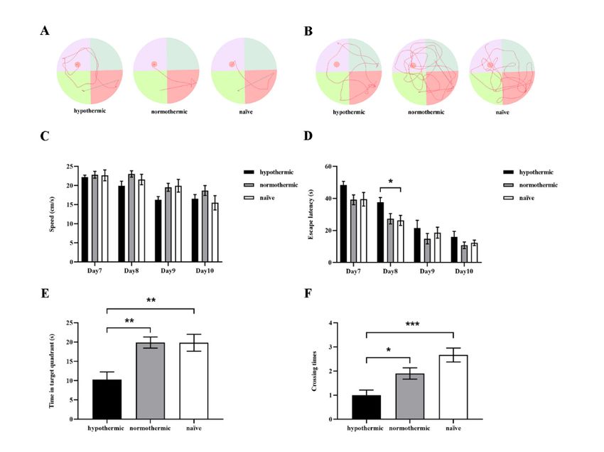
Brain Sci. 2022, 12, x FOR PEER REVIEW 3 of 14
Brain Sci. 2022, 12, 96 environment (room temperature 23 ± 1 °C, standard 12 h light/dark cycle, two rats3 ofper
14
cage) with food and water available ad libitum. All animal procedures were approved by
the Ethical Committee for Animal Experimentation of the Peking Union Medical College
Hospital (XHDW-2019-003) and conformed to the Guide for the Care and Use of Labora-
Hospital (XHDW-2019-003) and conformed to the Guide for the Care and Use of Laboratory
tory Animals
Animals by thebyNational
the National Institutes
Institutes of Health.
of Health.
2.2. Protocols
2.2. Protocols
Theanimals
The animalswere
wereallowed
allowedto toacclimate
acclimateto tothe
theenvironment
environmentfor foraaminimum
minimumof of33days
days
priorto
prior tothe
thebeginning
beginningof ofexploratory
exploratorylaparotomies.
laparotomies.Rats Ratswere
wererandomly
randomlyassigned
assignedto tothree
three
groups as
groups asfollows
follows(n(n== 10 10per
pergroup):
group): thethe hypothermic
hypothermic group,
group, thethe normothermic
normothermic group group
and the naïve group. Postoperatively, the rats were allowed to recover
and the naïve group. Postoperatively, the rats were allowed to recover for 5 days without for 5 days without
any manipulation.
any manipulation. The The rats
rats then
then underwent
underwent behavioural
behavioural tests
tests starting
starting with
with the
the Y-maze
Y-maze
spontaneous alteration
spontaneous alteration test
test on
on day
day 6,6, followed
followed by the the Morris
Morris Water
Water Maze
Maze (MWM)
(MWM) test.test.
TheMWM
The MWMtest testwas
wascarried
carriedout outfor
forfive
fivesuccessive
successivedaysdaysasasfollows:
follows: training
training on on days
days7–10
7–10
followed by the probe
followed probetrialtrialon
onday
day11.11.
UponUponcompleting
completingthe the
behavioural
behavioural tests,tests,
all rats
all were
rats
euthanised
were by intraperitoneal
euthanised by intraperitonealinjection of pentobarbital
injection sodiumsodium
of pentobarbital to collect
toblood
collectsamples
blood
samples and hippocampal
and hippocampal tissues fortissues for biochemical
biochemical and molecular
and molecular studies.studies. A schematic
A schematic of
of the ex-
the experimental
perimental protocol
protocol is summarised
is summarised in Figure
in Figure 1A. 1A.
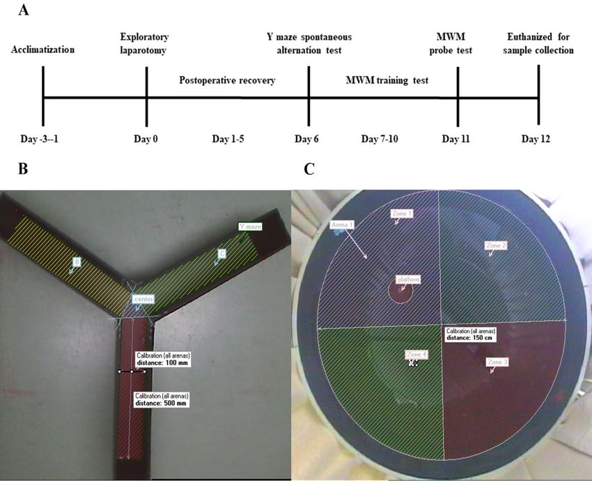
Figure 1.
Figure 1. Study
Studydesign
designtotoevaluate
evaluatethe effects
the of of
effects intraoperative hypothermia
intraoperative on postoperative
hypothermia cog-
on postoperative
nitive function in rats. (A) Schematic timeline of the experimental procedure; (B) representative
cognitive function in rats. (A) Schematic timeline of the experimental procedure; (B) representative im-
age of Y-maze apparatus; (C) representative graph of the MWM apparatus.
image of Y-maze apparatus; (C) representative graph of the MWM apparatus.
2.3.
2.3. Surgery
Surgery
Rats
Rats were
wereanaesthetised
anaesthetisedwith
with4040mg/kg
mg/kg sodium
sodium pentobarbital
pentobarbital (Beijing
(Beijing Sunbiotech
Sunbiotech
Co.,
Co.,Ltd.,
Ltd., Beijing,
Beijing, China) administered intraperitoneally, followed by by exploratory
exploratorylaparot-
laparo-
tomies. Briefly, the rats in
omies. Briefly, in the
the hypothermic
hypothermic and and normothermic
normothermic groups
groups were
were shaved,
shaved,and
and
an
an abdominal
abdominal median
median incision
incision of
of approximately
approximately 33cm cm was
was made
made totoallow
allowpenetration
penetration of
of
the
theperitoneal
peritoneal cavity.
cavity.The
Theoperator
operator then
then explored
explored the
the small
small intestine,
intestine, and
and 5–10
5–10 cm
cm of
ofthe
the
small
smallintestine
intestinewas
wasexteriorised
exteriorisedand
andleft
leftin
inair
airfor
forat
at60
60min.
min.The
Theintestine
intestinewas
wasthen
thenplaced
placed
inside the peritoneal cavity, and the wound was sutured with 4-0 non-absorbable sutures
in three layers consisting of the peritoneal lining, abdominal muscles, and skin. Core
body temperatures were measured every 5 min throughout the operation using a rectal
temperature probe. The entire surgical procedures lasted for at least 60 min. The body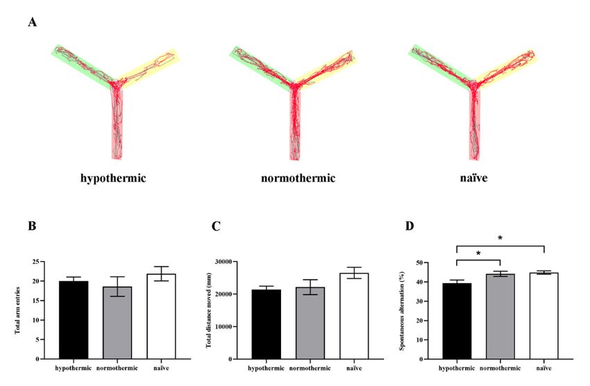
Brain Sci. 2022, 12, 96                                                                                             4 of 14

                              temperatures in hypothermic rats were maintained at 33 ± 0.5 ◦ C by spraying 75% alcohol
                              onto the rat’s body, while body temperatures of rats in the normothermic group were
                              maintained at 37 ± 0.5 ◦ C with a heating pad during operation. After the wounds were
                              sutured, the rats were immediately intraperitoneally injected with penicillin (60,000 U) to
                              prevent postoperative infections, and the body temperatures of those in the hypothermic
                              group were raised to 37 ± 0.5 ◦ C using a heating pad. For the rats in the naïve group, no
                              operation was performed.

                              2.4. Behavioural Tests
                                    According to the experimental protocol, the postoperative cognitive function of the
                              rats was evaluated by the Y-maze and MWM tests from postoperative days 6–11. All the
                              behavioural experiments were conducted each day, and the camera was tracked using
                              the EthoVision tracking system (EthoVision XT9, Noldus). The MWM test is the gold
                              standard for assessing cognitive function in rodents, especially for testing hippocampus-
                              related spatial learning and memory [40,41]. The Y-maze spontaneous alteration test is a
                              hippocampal-dependent spatial working memory test used to measure the willingness of
                              rodents to explore new environments [42,43]. In our experiment, the MWM and Y-maze
                              tests were performed as previously reported with minor modifications [41,43].

                              2.5. Y-Maze Test
                                   The Y-maze apparatus, made of grey plastic, has three identical arms separated at
                              120◦ angles (arm dimensions: 50 × 10 × 40 cm). The apparatus was used to evaluate the
                              spontaneous alteration performance of rats at 6 days postoperatively. A representative
                              image of the Y-maze apparatus is shown in Figure 1B. Each rat was placed in the centre
                              of the Y-maze and was free to explore the three different arms of the maze for 8 min.
                              After each test, the apparatus was thoroughly cleaned with 75% ethanol. Arm entry was
                              recorded when the rat placed all four paws within the arm. The sequence and the total
                              number of arms entered were recorded using a digital camera. Spontaneous alteration
                              behaviour was determined from successive consecutive entries to the three different arms
                              on overlapping triads in which all arms were represented. For example, a sequence of
                              entries to the three arms A, B, and C, ACBABACABA, would generate four “successful”
                              spontaneous alterations: ACB, BAC, and CAB. The percentage of spontaneous alterations
                              was determined using the following equation:

        Spontaneous alteration percentage = ((number of spontaneous alterations)/(total arm entries − 2)) × 100%

                              2.6. MWM Test
                                    The MWM consisted of a circular pool (diameter: 150 cm, height: 50 cm; black interior
                              wall) filled with water (depth: 25 cm; temperature: 22 ± 1.0 ◦ C) and a circular platform
                              (diameter: 10 cm) for the rats to escape. The pool was divided into four equal-sized virtual
                              quadrants (1, 2, 3, and 4), and the removable escape platform was placed 2 cm below the
                              water’s surface in the first quadrant (Figure 1C). Furthermore, the water was made opaque
                              by adding nontoxic black ink to prevent the rats from seeing the submerged platform. In
                              each training trial, the rat was gently placed into the water facing the tank wall at the
                              central point of the quadrant. Then, each rat was allowed to swim for 60 s to find the
                              hidden platform and was subjected to three consecutive trials from quadrants 2–4 per day.
                              When successful, the rats were allowed a 15 s rest period on the platform. If unsuccessful
                              within the allotted time, the rat was guided to the platform and allowed to rest for 15 s
                              on the platform. For all training trials, the average swimming speed and the mean time
                              to reach the platform (escape latency) across three trials were noted as the daily result
                              of learning ability for the rats, which were recorded and analysed by the computerised
                              tracking system.
                                    On the 11th day, the rats were additionally tested for spatial learning and memory
                              abilities by removing the underwater platform from the pool. The rat was gently placed in
Brain Sci. 2022, 12, 96 5 of 14
the water at the central point of the quadrant 3, opposite the platform location, and had
60 s to search for the platform’s original location. The time that each rat spent searching for
the platform in the target quadrant and the number of platform crossings were recorded.
2.7. Haematoxylin-Eosin (HE) Staining
To assess the morphological changes in the rats’ hippocampal neurons, brain tissues
were harvested after the rats were deeply anaesthetised with intraperitoneal pentobarbital
sodium injections (40 mg/kg). The rats were transcardially perfused with sterile phosphate-
buffered saline (PBS), followed by 4 ◦ C paraformaldehyde (Sigma, St. Louis, MO, USA).
Air bubbles were avoided throughout the perfusion. The rats’ brains were then dissected
and post-fixed for at least 24 h in 4% paraformaldehyde at 4 ◦ C. Subsequently, the tissue
samples were dehydrated using graded alcohol steps, immersed in xylene, embedded in
paraffin, sectioned along the coronal plane at 5 µm thickness (Leica 2000, Leica Microsys-
tems, Wetzlar, Germany), and stained with HE. Images were examined under bright-field
illumination using a fluorescence microscopy imaging system (Ti-S, Olympus FluoView
Software, Olympus, Tokyo, Japan). The total number of injured neurons in each image was
counted and analysed by Image-Pro Plus 6.0 Software.
2.8. Western Blot Analysis
After deep anaesthesia with pentobarbital sodium (40 mg/kg) and transcardial perfu-
sion with sterile PBS, the brains were rapidly removed, and bilateral hippocampal tissues
were dissected and flash-frozen in liquid nitrogen. Hippocampal tissues were homogenised
in Tissue Protein Extraction Reagent (Thermo Fisher Scientific, Waltham, MA, USA) with
a protease inhibitor cocktail (CWbio, Beijing, China) and phosphatase inhibitor cocktail
(CWbio, Beijing, China). After centrifugation (12000× g for 15 min at 4 ◦ C), the super-
natants were collected and denatured in sodium dodecyl sulphate-polyacrylamide gel
electrophoresis (SDS-PAGE) loading buffer (Solarbio, Beijing, China) for 10 min at 100 ◦ C.
Protein concentrations were determined using the Pierce BCA Protein Assay (Thermo
Scientific, Rockford, IL, USA).
The protein samples were separated by SDS-PAGE and transferred to a polyvinylidene
fluoride membrane (Millipore, Billerica, MA, USA). The membranes were blocked with
5% non-fat dry milk (Solarbio, Beijing, China) or 5% bovine serum albumin (Solarbio,
Beijing, China) in Tris-buffered saline with 0.5% Tween 20 (TBST, Solarbio, Beijing, China)
for 1 h at room temperature and incubated with primary antibodies overnight at 4 ◦ C.
The following primary antibodies were used: mouse anti-Arc (sc-17839, 1:500, Santa Cruz,
CA, USA), rabbit anti-CREB (ab32515, 1:1000, Abcam, Cambridge, MA, USA), rabbit
anti-S133-phosphorylated CREB (p-CREB [S133]; #9198, 1:1000, Cell Signaling Technology,
Danvers, MA, USA), rabbit anti-AMPAR1 (ab183797, 1:1000, Abcam, Cambridge, MA, USA),
rabbit anti-S831-phosphorylated AMPAR1 (p-AMPAR1 [S831]; ab109464, 1:1000, Abcam,
Cambridge, MA, USA), and mouse anti-β-actin (cat No. 66009-1-lg, 1:5000; Proteintech
Group, Chicago, IL, USA).
The membranes were subsequently washed three times for 5 min each in TBST (Solar-
bio, Beijing, China) and incubated for 1 h at room temperature with the following secondary
antibodies: HRP-conjugated affinipure goat anti-rabbit IgG (H+L; cat No. SA00001-2, 1:5000,
Proteintech Group, Chicago, IL, USA), and HRP-conjugated affinipure goat anti-mouse
IgG (H+L; cat no. SA00001-1, 1:5000, Proteintech Group, Chicago, IL, USA). The bands
were visualised via a Tanon 5800 Luminescent Imaging Workstation (Tanon Science &
Technology Co., Ltd., Shanghai, China) using High-sig ECL Western blotting Substrate (So-
larbio, Beijing, China). The band intensity was measured using ImageJ software (National
Institutes of Health, Bethesda, MD, USA). The ratio of each band/β-actin was considered
as the expression level of the target protein.ageJ software (National Institutes of Health, Bethesda, MD, USA). The ratio of each
band/β-actin was considered as the expression level of the target protein.
2.9. Statistical Analysis
Brain Sci. 2022, 12, 96 6 of 14
All data are presented as mean ± SEM. Statistical analysis was performed using
GraphPad Prism 8 software (version 8.01, GraphPad Software, San Diego, CA, USA). The
normality of the data distribution was determined by visual inspection of quantile-quan-
2.9. Statistical Analysis
tile (Q-Q) plot. Levene’s test was used to assess the homogeneity of variances. The rats’
data are presented as mean ± SEM. Statistical analysis was performed using
escapeAll latency and swimming speed in the MWM were analysed using two-way repeated
GraphPad Prism 8 software (version 8.01, GraphPad Software, San Diego, CA, USA).
measures analysis of variance (ANOVA) followed by Tukey’s post hoc multiple compar-
The normality of the data distribution was determined by visual inspection of quantile-
isons test.(Q-Q)
quantile Otherplot.
results were test
Levene’s analysed using
was used one-way
to assess ANOVA followed
the homogeneity by Tukey’s
of variances. The post
hoc test.
rats’ In this
escape study,
latency pent groups; (B) the number of total arm entries of rats from different groups; (C) the total distan
moved during 8 min free exploration in the Y-maze; (D) the spontaneous alternation percentage
different groups. Data are expressed as mean ± SEM. * p < 0.05, n = 9–10.
Brain Sci. 2022, 12, 96 3.2. Intraoperative Hypothermia Reduced Rats’ Postoperative Spatial Learning and7 ofMemory
14
Ability
We conducted Hypothermia
3.2. Intraoperative the MWMReduced test toRats’
evaluate hippocampal-dependent
Postoperative Spatial Learning and spatial learni
and memory of rats in each group. The representative swimming trajectories of rats fro
Memory Ability
each group Weon the lastthe
conducted day of the
MWM training
test trials
to evaluate and probe trials arespatial
hippocampal-dependent shown in Figure 3A
learning
and memory of rats in each group. The representative swimming
respectively. During the consecutive 4-day training phase, we found that the trajectories of rats fromescape
each group on the last day of the training trials and probe trials are shown in Figure 3A,B,
tencies were obviously shortened in all groups (F2,26 = 4.669, p = 0.0185; Figure 3D). Spec
respectively. During the consecutive 4-day training phase, we found that the escape
ically,latencies
the ratswere
in the hypothermic
obviously shortenedgroup spent (F
in all groups more time searching for the hidden pl
2,26 = 4.669, p = 0.0185; Figure 3D).
form Specifically,
than thosetheinratstheinnaïve group ongroup
the hypothermic day spent
8 (p =more
0.0415; Figure 3D).
time searching Onhidden
for the day 11, in t
probeplatform
test, the hypothermic
than rats spent
those in the naïve group less time
on day 8 (pin=the target
0.0415; quadrant
Figure 3D). Onthan the norm
day 11,
in the probe test, the hypothermic rats spent less time in the target
thermic (p = 0.0031; Figure 3E) and naïve rats (p = 0.0041; Figure 3E), but there was quadrant than the
normothermic (p = 0.0031; Figure 3E) and naïve rats (p = 0.0041; Figure 3E), but there
significant difference between the normothermic and naïve groups (p = 0.9997; Figure 3
was no significant difference between the normothermic and naïve groups (p = 0.9997;
Figure 3E).
Figure 3. The effects of intraoperative hypothermia on spatial learning and memory of rats were
Figuredetermined by the of
3. The effects MWM. (A) The representative
intraoperative hypothermiaswimming trajectories
on spatial of ratsand
learning frommemory
each group
of rats w
on the last day of training trials; (B) the representative swimming paths of rats from each group in
determined by the MWM. (A) The representative swimming trajectories of rats from each group
the probe test; (C) the average swimming speeds of rats in different groups on successive MWM
the last day of training trials; (B) the representative swimming paths of rats from each group in
training days; (D) the average escape latencies to find the hidden platform across three trials on four
probe test; (C) the average swimming speeds of rats in different groups on successive MWM tra
consecutive training days; (E) the time spent in the target quadrant in the probe test; (F) the number
ing days; (D) the average escape latencies to find the hidden platform across three trials on fo
of platform crossings during the probe test. Data are expressed as mean ± SEM. * p < 0.05, ** p < 0.01
consecutive
and *** ptraining
< 0.001, ndays;
= 9–10.(E) the time spent in the target quadrant in the probe test; (F) the num
of platform crossings during the probe test. Data are expressed as mean ± SEM. * p < 0.05, ** p < 0
and *** pFigure 3C). Taken together, these results suggest that intraoperative hypothermia could
lead to spatial learning memory impairment in rats; however, maintaining the normal
body temperature of rats during operation could ameliorate this cognitive decline.
Brain Sci. 2022, 12, 96 8 of 14
3.3. Intraoperative Hypothermia Increased Hippocampal Neuron Injuries in Postoperative Rats
In this study, we observed postoperative morphological changes of neurons in rats’
learning memory impairment in rats; however, maintaining the normal body temperature
hippocampal dentate gyrus
of rats (DG)
during region
operation by ameliorate
could HE staining (Figuredecline.
this cognitive 4). We found the neurons
in the naïve group were round or oval with rich, large, and lightly stained cytoplasm, and
3.3. Intraoperative Hypothermia Increased Hippocampal Neuron Injuries in Postoperative Rats
orderly arranged nuclei (Figure 4C). Similarly, we observed that the hippocampal neurons
In this study, we observed postoperative morphological changes of neurons in rats’
of rats in the normothermic
hippocampal group showed
dentate normal
gyrus (DG) regionmorphology, similar
by HE staining (Figure 4). to
Wethe
foundnaïve group
the neurons
(p = 0.9983; Figure 4B,D). However,
in the naïve group weredisorderly
round or ovalarranged nuclei,
with rich, large, neuronal
and lightly stainedpyknosis and
cytoplasm, and
orderly arranged nuclei (Figure 4C). Similarly, we observed that the hippocampal neurons
necrosis in the hippocampus were evident in the hypothermic group (Figure 4A, white
of rats in the normothermic group showed normal morphology, similar to the naïve group
arrows) compared to (p =the naïve
0.9983; (p =4B,D).
Figure 0.0222; Figure
However, 4D) and
disorderly normothermic
arranged (p = pyknosis
nuclei, neuronal 0.0241; and
Fig-
ure 4D) groups. Ournecrosis
findings
in theindicate thatwere
hippocampus rats’evident
postoperative cognitive
in the hypothermic groupfunction
(Figure 4A,may be
white
arrows) compared to the naïve (p = 0.0222; Figure 4D) and normothermic (p = 0.0241;
related to the morphological changes of hippocampal neurons, and intraoperative hypo-
Figure 4D) groups. Our findings indicate that rats’ postoperative cognitive function may
thermia could significantly
be relateddamage hippocampal
to the morphological nerve
changes cells. This neurons,
of hippocampal suggests andthat maintain-
intraoperative
ing rats’ body temperature
hypothermia within the physiological
could significantly range could
damage hippocampal alleviate
nerve cells. hippocampal
This suggests that main-
neuronal damage. taining rats’ body temperature within the physiological range could alleviate hippocampal
neuronal damage.
Figure 4. Morphological changes of hippocampal neurons in rats among groups were observed under
Figure 4. Morphological changes of hippocampal neurons in rats among groups were observed un-
bright-field illumination using a fluorescence microscopy imaging system. (A–C) Representative
der bright-field illumination
images ofusing a fluorescence
HE staining in hippocampal microscopy imaging analysis
slices; (D) quantitative system.of(A–C)
injured Representa-
neurons with
tive images of HE staining inpyknosis
neuronal hippocampal slices;
and necrosis in the (D) quantitative
hippocampal analysis
DG region. ofarrow
The white injured neurons
indicates with
a disorderly
neuronal pyknosis and necrosis
nucleus, in the
neuronal hippocampal
pyknosis and necrosis.DG region. The
Magnification: ×40;white arrow
scale bar: indicates
50 µm. a disor-
Data are expressed
as mean ± SEM. * p < 0.05, n = 4.
derly nucleus, neuronal pyknosis and necrosis. Magnification: ×40; scale bar: 50 μm. Data are ex-
pressed as mean ± SEM.3.4.*Intraoperative
p < 0.05, n =Hypothermia
4. Suppressed the Expression of Synaptic Plasticity-Related Proteins
in the Hippocampal Region of Rats
To further
3.4. Intraoperative Hypothermia elucidate thethe
Suppressed underlying molecular
Expression mechanism,
of Synaptic the expression levels of
Plasticity-Related
synaptic plasticity-related proteins such as Arc, CREB, and AMPAR1 in the hippocampi of
Proteins in the Hippocampal Region of Rats
To further elucidate the underlying molecular mechanism, the expression levels of
synaptic plasticity-related proteins such as Arc, CREB, and AMPAR1 in the hippocampi
of rats were examined by Western blotting. We also determined CREB and AMPAR1 ac-OR PEER REVIEW 9 of 14
Brain Sci. 2022, 12, 96 9 of 14
extracts of rats. Therats were examined
hypothermic by Western
group blotting.
exhibited We also determined
significantly lower CREB andof
levels AMPAR1 activities
Arc protein
by measuring p-CREB (S133) and p-AMPAR1 (S831) levels in the hippocampal extracts of
expression than therats.
normothermic (p = 0.0117; Figure 5D) and naïve groups (p = 0.0005;
The hypothermic group exhibited significantly lower levels of Arc protein expression
Figure 5D), although thanthere was no significant
the normothermic difference
(p = 0.0117; between
Figure 5D) and the normothermic
naïve groups and
(p = 0.0005; Figure 5D),
naïve groups (p = 0.1128; Figure 5D). Moreover, no remarkable between-group differences
although there was no significant difference between the normothermic and naïve groups
(p = 0.1128; Figure 5D). Moreover, no remarkable between-group differences were found
were found regarding CREB expression levels (F2,9 = 0.05574, p = 0.9641; Figure 5B) and
regarding CREB expression levels (F2,9 = 0.05574, p = 0.9641; Figure 5B) and AMPAR1
AMPAR1 (F2,9 = 3.147,(F2,9p= =3.147,
0.0920; Figure
p = 0.0920; 5E).5E).
Figure
Figure 5. Expression levels of hippocampal synaptic plasticity-related proteins in rats and the effects
Figure 5. Expression levels of hippocampal synaptic plasticity-related proteins in rats and the effects
of intraoperative hypothermia. (A) The visualisation of protein bands of CREB, p-CREB (S133),
of intraoperative hypothermia. (A) The visualisation of protein bands of CREB, p-CREB (S133), Arc,
Arc, AMPAR1, p-AMPAR1 (S831), and β-actin; (B) quantitative analysis of CREB expression levels
AMPAR1, p-AMPAR1 (S831), and β-actin; (B) quantitative analysis of CREB expression levels nor-
normalised to β-actin band intensities; (C) quantitative analysis of p-CREB (S133) expression levels
malised to β-actin band intensities;
normalised (C)band
to CREB quantitative
intensities;analysis of p-CREB
(D) quantitative analysis(S133) expression
of Arc expression levels
levels nor-
normalised
malised to CREB band to intensities; (D) quantitative
β-actin band intensities; analysis
(E) quantitative of Arc
analysis expression
of AMPAR1 levelslevels
expression normalised
normalised toto
β-actin band intensities; (E) quantitative analysis of AMPAR1 expression levels normalised to β-to
β-actin band intensities; (F) quantitative analysis of p-AMPAR1 (S831) expression levels normalised
actin band intensities;AMPAR1
(F) quantitative analysis
band intensities. ofexpressed
Data are p-AMPAR1 as mean ± SEM.expression
(S831) * p < 0.05, ** plevels *** p < 0.001,
normalised
< 0.01 and
n = 3–4.
to AMPAR1 band intensities. Data are expressed as mean ± SEM. * p < 0.05, ** p < 0.01 and *** p <
0.001, n = 3–4. However, after the band intensities of p-CREB (S133) were normalised to CREB,
we found that hypothermic rats displayed significantly lower p-CREB (S133) expression
However, aftercompared
the band to intensities of p-CREB
the normothermic (S133)
(p = 0.0466; were
Figure 5C)normalised to CREB,
and naïve groups we
(p = 0.0028;
Figure 5C), whereas no significant difference was found between
found that hypothermic rats displayed significantly lower p-CREB (S133) expression com- the normothermic and
naïve groups (p = 0.0819; Figure 5C).
pared to the normothermic (p = 0.0466;
Additionally, Figure a5C)
we performed and naïve
quantitative groups
analysis of the(pexpression
= 0.0028;levels
Figure
of p-
5C), whereas no significant
AMPAR1 (S831)difference was
normalised found between
to AMPAR1 the normothermic
band intensities and showed that theand naïve
hypothermic
group presented
groups (p = 0.0819; Figure 5C). decreased levels of p-AMPAR1 (S831) compared to the normothermic
(p = 0.0470; Figure 5F) and naïve groups (p = 0.0081; Figure 5F). However, there was no
Additionally, we performed a quantitative analysis of the expression levels of p-AM-
significant difference between the normothermic and naïve groups (p = 0.5133; Figure 5F).
PAR1 (S831) normalised to AMPAR1 band intensities and showed that the hypothermic
group presented decreased levels of p-AMPAR1 (S831) compared to the normothermic (p
= 0.0470; Figure 5F) and naïve groups (p = 0.0081; Figure 5F). However, there was no sig-
nificant difference between the normothermic and naïve groups (p = 0.5133; Figure 5F).Brain Sci. 2022, 12, 96 10 of 14
4. Discussion
This study was designed to explore the effects of intraoperative hypothermia on post-
operative cognitive function in rats undergoing exploratory laparotomies and to investigate
the related molecular mechanisms. In our study, hypothermic rats showed impaired spatial
working memory (Figure 2), as demonstrated by the reduced spontaneous alteration per-
centage evaluated by the Y-maze test. Notably, when the rats’ temperature was maintained
within a normal range (37 ± 0.5 ◦ C) during surgical operation, the spontaneous alteration
percentage of rats in the normothermic group was statistically indistinguishable from that
of naïve rats (Figure 2), suggesting that maintaining core temperature in a normal range
could dramatically alleviate the detrimental effects of intraoperative hypothermia on rats’
spatial working memory. Similarly, in the MWM test, rats in the hypothermic group exhib-
ited markedly decreased spatial learning and memory ability, as evidenced by significant
reductions in time spent in target quadrant and number of platform crossings, comparable
to the normothermic and naïve groups (Figure 3). However, no significant differences
were observed in time spent in target quadrant and number of platform crossings between
the normothermic and naïve groups, indicating that intraoperative hypothermia seriously
affects spatial learning and memory in rats.
To further examine the possible mechanisms behind these behavioural phenomena,
we performed HE staining to examine neuronal integrity and orderliness in the rats’ hip-
pocampi. Our histopathological results revealed significant damage in the hippocampi
of hypothermic rats (Figure 4A,D), which showed disorderly nuclei, neuronal pyknosis
and necrosis. However, the hippocampal tissues taken from rats in the normothermic
(Figure 4B,D) and naïve groups (Figure 4C,D) were similar, showing normal pyramidal
nerve cell morphology, confirming that intraoperative hypothermia could induce POCD by
damaging hippocampal neurons, and that maintaining normal body temperatures could
alleviate POCD and hippocampal neuronal damage.
Multiple studies have established that synaptic plasticity plays a critical role in learn-
ing and memory by governing the number and strength of connections within a cell
assembly [44–46]. Although various pathological factors have been demonstrated to be
responsible for POCD, evidence from experimental studies revealed that synaptic dys-
function in the central nervous system, particularly in the hippocampus, may be involved
in POCD’s progression [47,48]. Furthermore, synaptic dysfunction is associated with the
abnormal expression of proteins required for synaptic function [49,50]. Therefore, we
measured the expression levels of synaptic plasticity-associated proteins, including Arc,
CREB, p-CREB (S133), AMPAR1, and p-AMPAR1 (S831) by Western blotting to further
explore the underlying molecular mechanism.
We found that the expression of hippocampal synaptic plasticity-related proteins is
consistent with rats’ behavioural results and histopathological outcomes. We observed
a dramatic decrease in Arc levels in the hippocampi of hypothermic rats compared to
normothermic and naïve rats (Figure 5D), whereas no differences were observed between
the normothermic and naïve rats. It is well recognised that Arc plays a vital role in multiple
forms of learning and memory by regulating synaptic plasticity [29,51]. Specifically, Arc
was found to be strongly related to synaptic plasticity processes dependent on protein
synthesis [52]. Moreover, Plath et al. suggested that Arc-knockout mice show severe
long-term memory impairment [29]. Thus, the lower expression of Arc in the hypothermic
group demonstrated the impairment of synaptic plasticity, learning, and memory in rats,
consistent with the outcomes of the Y-maze spontaneous alteration and MWM tests.
Subsequently, we measured the levels of CREB and phosphorylation of CREB at the
Ser133 site in the rat hippocampus among the three groups and found significant p-CREB
(S133) expression inhibition in hypothermic rats compared to normothermic and naïve rats
(Figure 5C). However, no significant difference was found between normothermic and naïve
rats. In addition, no significant between-group difference was found regarding the level
of CREB expression (Figure 5B). Several studies have illustrated that CREB mobilisation
is dependent on phosphorylation at the Ser133 site, which is widely involved in manyBrain Sci. 2022, 12, 96 11 of 14
forms of neuronal activity: synaptic plasticity, learning, and memory [53,54]. Lv et al.
identified that Arc is the critical downstream molecule of the extracellular signal-related
kinase (ERK)-CREB pathway [55]. Briefly, the above results show that a decrease in p-CREB
(S133) is associated with intraoperative hypothermia-induced POCD, and lower levels of
Arc may be caused by the reduction of p-CREB (S133).
Previous studies have reported that Arc has a close, bidirectional relationship with
postsynaptic glutamate neurotransmission because it is stimulated by numerous gluta-
matergic receptor mechanisms and regulates AMPAR internalisation, trafficking, and
activity-dependent scaling [56,57]. At excitatory synapses in the central nervous system,
AMPARs are responsible for most synaptic transmission [51]. The effects on AMPAR
trafficking are likely associated with Arc’s ability to mediate different forms of synaptic
plasticity, such as LTP, LTD, and synaptic scaling, each of which is essential for normal
cognitive function [52,58]. Therefore, AMPARs are critical downstream molecules of Arc.
In this study, to further explore Arc function, we detected AMPAR1 and p-AMPAR1
(S831) in the hippocampi of rats. We found that AMPAR1 protein expression differed
slightly between the groups (Figure 5E). In addition, p-AMPAR1 (S831) was decreased
in rat hippocampi after intraoperative hypothermia compared with the normothermic
and naïve groups, whereas no differences were found between normothermic and naïve
rats (Figure 5F). Overall, our results show that intraoperative hypothermia can signifi-
cantly inhibit hippocampal synaptic plasticity-related proteins, including Arc, p-CREB
(S133), and p-AMPAR1 (S831), indicating that intraoperative hypothermia impairs the
postoperative cognitive function of rats by reducing the levels of hippocampal synaptic
plasticity-related proteins.
To the best of our knowledge, there are few relevant studies exploring the effects
of intraoperative hypothermia on postoperative cognitive function in rats undergoing
exploratory laparotomies and investigating the related mechanisms. In our research, we
found a correlation between intraoperative hypothermia and POCD in rats. The underlying
mechanism may associate with hippocampal neuron damage and downregulation of
synaptic plasticity-associated proteins Arc, p-CREB (S133), and p-AMPAR1 (S831). When
interpreting these results, it is important to consider some limitations. First, POCD, as
a commonly used clinical term, has some limitations when used to describe impaired
cognitive function in rodents after operations. In clinical research studies, the Montreal
Cognitive Assessment (MoCA) and the Mini-Mental State Examination (MMSE) are the
most commonly used neuropsychological rating scales for the diagnosis of POCD. However,
such behavioural tests as the Y-maze and MWM tests are two commonly used evaluation
methods to examine spatial cognitive functions of rodents in basic research. It is debatable
whether the two types of evaluation methods present concordance both in clinical research
and basic research for a valid diagnosis of POCD. In addition, we are only presenting a
preliminary exploration of the mechanisms underlying intraoperative hypothermia effect
on POCD; the precise mechanism requires further investigation.
5. Conclusions
POCD has been shown to be associated with impaired concentration, memory, and
learning. Intraoperative hypothermia and its adverse consequences have drawn increasing
attention in recent years. We found a correlation between intraoperative hypothermia and
POCD in rats. Specifically, our study indicated that intraoperative hypothermia could
cause POCD, affecting rats’ spatial working memory, spatial learning, and memory. POCD
induced by intraoperative hypothermia might be due to hippocampal neuron damage
and the reduced expression of synaptic plasticity-related proteins, such as Arc, p-CREB
(S133), and p-AMPAR1 (S831). This study was a preliminary exploration of the mechanisms
underlying intraoperative hypothermia effect on POCD; the precise mechanism requires
further investigation.Brain Sci. 2022, 12, 96 12 of 14
Author Contributions: G.X. conducted the experiments, analysed the results and drafted the
manuscript; T.L. and Y.H. conceived and designed the study and helped to edit the manuscript.
All authors have read and agreed to the published version of the manuscript.
Funding: This research was funded by the Post-Doctoral Science Foundation of China (grant number
2019M660550), the National Natural Science Youth Foundation of China (grant number 81900433)
and the National Natural Science Foundation of China (grant number 82071252).
Institutional Review Board Statement: The study was conducted according to the guidelines of the
Declaration of Helsinki and approved by the Ethical Committee for Animal Experimentation of the
Peking Union Medical College Hospital (XHDW-2019-003).
Informed Consent Statement: Not applicable.
Data Availability Statement: All data included in this study are available upon request from the
corresponding authors.
Acknowledgments: We thank Institute of Laboratory Animal Sciences, Chinese Academy of Medical
Sciences and Peking Union Medical College for providing behavioural devices and guidance.
Conflicts of Interest: The authors declare no conflict of interest.
References
1. Sari, S.; Aksoy, S.M.; But, A. The incidence of inadvertent perioperative hypothermia in patients undergoing general anesthesia
and an examination of risk factors. Int. J. Clin. Pract. 2021, 75, e14103. [CrossRef] [PubMed]
2. Yi, J.; Lei, Y.; Xu, S.; Si, Y.; Li, S.; Xia, Z.; Shi, Y.; Gu, X.; Yu, J.; Xu, G.; et al. Intraoperative hypothermia and its clinical outcomes in
patients undergoing general anesthesia: National study in China. PLoS ONE 2017, 12, e0177221. [CrossRef]
3. Sun, Z.; Honar, H.; Sessler, D.I.; Dalton, J.E.; Yang, D.; Panjasawatwong, K.; Deroee, A.F.; Salmasi, V.; Saager, L.; Kurz, A.
Intraoperative core temperature patterns, transfusion requirement, and hospital duration in patients warmed with forced air.
Anesthesiology 2015, 122, 276–285. [CrossRef] [PubMed]
4. Schmied, H.; Kurz, A.; Sessler, D.I.; Kozek, S.R.A. Mild hypothermia increases blood loss and transfusion requirements during
total hip arthroplasty. Lancet 1996, 347, 289–292. [CrossRef]
5. Frank, S.; Fleisher, L.; Breslow, M.; Higgins, M.; Olson, K.; Kelly, S.; Beattie, C. Perioperative maintenance of normothermia
reduces the incidence of morbid cardiac events. A randomized clinical trial. JAMA 1997, 277, 1127–1134. [CrossRef]
6. Kurz, A.; Sessler, D.I.; Lenhardt, R. Perioperative normothermia to reduce the incidence of surgical-wound infection and shorten
hospitalization. Study of wound infection and temperature group. N. Engl. J. Med. 1996, 334, 1209–1215. [CrossRef] [PubMed]
7. Yi, J.; Xiang, Z.; Deng, X.; Fan, T.; Fu, R.; Geng, W.; Guo, R.; He, N.; Li, C.; Li, L.; et al. Incidence of Inadvertent Intraoperative
Hypothermia and Its Risk Factors in Patients Undergoing General Anesthesia in Beijing: A Prospective Regional Survey. PLoS
ONE 2015, 10, e0136136. [CrossRef]
8. Yi, J.; Zhan, L.; Lei, Y.; Xu, S.; Si, Y.; Li, S.; Xia, Z.; Shi, Y.; Gu, X.; Yu, J.; et al. Establishment and Validation of a Prediction Equation
to Estimate Risk of Intraoperative Hypothermia in Patients Receiving General Anesthesia. Sci. Rep. 2017, 7, 13927. [CrossRef]
9. Needham, M.J.; Webb, C.E.; Bryden, D.C. Postoperative cognitive dysfunction and dementia: What we need to know and do. Br.
J. Anaesth. 2017, 119, i115–i125. [CrossRef]
10. Evered, L.; Silbert, B.; Knopman, D.S.; Scott, D.A.; DeKosky, S.T.; Rasmussen, L.S.; Oh, E.S.; Crosby, G.; Berger, M.; Eckenhoff,
R.G.; et al. Recommendations for the nomenclature of cognitive change associated with anaesthesia and surgery-2018. Br. J.
Anaesth. 2018, 121, 1005–1012. [CrossRef]
11. Skvarc, D.R.; Berk, M.; Byrne, L.K.; Dean, O.M.; Dodd, S.; Lewis, M.; Marriott, A.; Moore, E.M.; Morris, G.; Page, R.S.; et al.
Post-Operative Cognitive Dysfunction: An exploration of the inflammatory hypothesis and novel therapies. Neurosci. Biobehav.
Rev. 2018, 84, 116–133. [CrossRef]
12. Androsova, G.; Krause, R.; Winterer, G.; Schneider, R. Biomarkers of postoperative delirium and cognitive dysfunction. Front.
Aging Neurosci. 2015, 7, 112. [CrossRef]
13. Chen, C.; Gao, R.; Li, M.; Wang, Q.; Chen, H.; Zhang, S.; Mao, X.; Behensky, A.; Zhang, Z.; Gan, L.; et al. Extracellular RNAs-TLR3
signaling contributes to cognitive decline in a mouse model of postoperative cognitive dysfunction. Brain Behav. Immun. 2019, 80,
439–451. [CrossRef]
14. Oberman, K.; Hovens, I.; de Haan, J.; Falcao-Salles, J.; van Leeuwen, B.; Schoemaker, R. Acute pre-operative ibuprofen improves
cognition in a rat model for postoperative cognitive dysfunction. J. Neuroinflam. 2021, 18, 156. [CrossRef]
15. Netto, M.B.; de Oliveira Junior, A.N.; Goldim, M.; Mathias, K.; Fileti, M.E.; da Rosa, N.; Laurentino, A.O.; de Farias, B.X.; Costa,
A.B.; Rezin, G.T.; et al. Oxidative stress and mitochondrial dysfunction contributes to postoperative cognitive dysfunction in
elderly rats. Brain Behav. Immun. 2018, 73, 661–669. [CrossRef] [PubMed]
16. Gao, S.; Zhang, S.; Zhou, H.; Tao, X.; Ni, Y.; Pei, D.; Kang, S.; Yan, W.; Lu, J. Role of mTOR-Regulated Autophagy in Synaptic
Plasticity Related Proteins Downregulation and the Reference Memory Deficits Induced by Anesthesia/Surgery in Aged Mice.
Front. Aging Neurosci. 2021, 13, 628541. [CrossRef] [PubMed]Brain Sci. 2022, 12, 96 13 of 14
17. Xiao, J.-Y.; Xiong, B.-R.; Zhang, W.; Zhou, W.-C.; Yang, H.; Gao, F.; Xiang, H.-B.; Manyande, A.; Tian, X.-B.; Tian, Y.-K. PGE2-
EP3 signaling exacerbates hippocampus-dependent cognitive impairment after laparotomy by reducing expression levels of
hippocampal synaptic plasticity-related proteins in aged mice. CNS Neurosci. Ther. 2018, 24, 917–929. [CrossRef]
18. Pacheco Estefan, D.; Sanchez-Fibla, M.; Duff, A.; Principe, A.; Rocamora, R.; Zhang, H.; Axmacher, N.; Verschure, P. Coordinated
representational reinstatement in the human hippocampus and lateral temporal cortex during episodic memory retrieval. Nat.
Commun. 2019, 10, 2255. [CrossRef] [PubMed]
19. Bellmund, J.L.S.; Gardenfors, P.; Moser, E.I.; Doeller, C.F. Navigating cognition: Spatial codes for human thinking. Science 2018,
362, eaat6766. [CrossRef]
20. Borgomaneri, S.; Battaglia, S.; Sciamanna, G.; Tortora, F.; Laricchiuta, D. Memories are not written in stone: Re-writing fear
memories by means of non-invasive brain stimulation and optogenetic manipulations. Neurosci. BioBehav. Rev. 2021, 127, 334–352.
[CrossRef] [PubMed]
21. Battaglia, S.; Garofalo, S.; di Pellegrino, G.; Starita, F. Revaluing the Role of vmPFC in the Acquisition of Pavlovian Threat
Conditioning in Humans. J. Neurosci. 2020, 44, 8491–8500. [CrossRef] [PubMed]
22. Battaglia, S.; Harrison, B.J.; Fullana, M.A. Does the human ventromedial prefrontal cortex support fear learning, fear extinction or
both? A commentary on subregional contributions. Mol. Psychiatry 2021, 1–3. [CrossRef]
23. Borgomaneri, S.; Battaglia, S.; Garofalo, S.; Tortora, F.; Avenanti, A.; di Pellegrino, G. State-Dependent TMS over Prefrontal Cortex
Disrupts Fear-Memory Reconsolidation and Prevents the Return of Fear. Curr. Biol. 2020, 30, 3672–3679.e4. [CrossRef] [PubMed]
24. Mansvelder, H.D.; Verhoog, M.B.; Goriounova, N.A. Synaptic plasticity in human cortical circuits: Cellular mechanisms of
learning and memory in the human brain? Curr. Opin. Neurobiol. 2019, 54, 186–193. [CrossRef] [PubMed]
25. Borgomaneri, S.; Serio, G.; Battaglia, S. Please, don’t do it! Fifteen years of progress of non-invasive brain stimulation in action
inhibition. Cortex 2020, 132, 404–422. [CrossRef]
26. Borgomaneri, S.; Vitale, F.; Battaglia, S.; Avenanti, A. Early Right Motor Cortex Response to Happy and Fearful Facial Expressions:
A TMS Motor-Evoked Potential Study. Brain Sci. 2021, 11, 1203. [CrossRef]
27. Battaglia, S.; Serio, G.; Scarpazza, C.; D’Ausilio, A.; Borgomaneri, S. Frozen in (e)motion: How reactive motor inhibition is
influenced by the emotional content of stimuli in healthy and psychiatric populations. Behav. Res. Ther. 2021, 146, 103963.
[CrossRef]
28. Wall, M.J.; Collins, D.R.; Chery, S.L.; Allen, Z.D.; Pastuzyn, E.D.; George, A.J.; Nikolova, V.D.; Moy, S.S.; Philpot, B.D.;
Shepherd, J.D.; et al. The Temporal Dynamics of Arc Expression Regulate Cognitive Flexibility. Neuron 2018, 98, 1124–1132.e7.
[CrossRef]
29. Plath, N.; Ohana, O.; Dammermann, B.; Errington, M.L.; Schmitz, D.; Gross, C.; Mao, X.; Engelsberg, A.; Mahlke, C.;
Welzl, H.; et al. Arc/Arg3.1 is essential for the consolidation of synaptic plasticity and memories. Neuron 2006, 52, 437–444.
[CrossRef]
30. Gao, X.; Castro-Gomez, S.; Grendel, J.; Graf, S.; Susens, U.; Binkle, L.; Mensching, D.; Isbrandt, D.; Kuhl, D.; Ohana, O. Arc/Arg3.1
mediates a critical period for spatial learning and hippocampal networks. Proc. Natl. Acad. Sci. USA 2018, 115, 12531–12536.
[CrossRef]
31. Benito, E.; Barco, A. CREB’s control of intrinsic and synaptic plasticity: Implications for CREB-dependent memory models. Trends
Neurosci. 2010, 33, 230–240. [CrossRef]
32. Takeo, S.; Niimura, M.; Miyake-Takagi, K.; Nagakura, A.; Fukatsu, T.; Ando, T.; Takagi, N.; Tanonaka, K.; Hara, J. A possible
mechanism for improvement by a cognition-enhancer nefiracetam of spatial memory function and cAMP-mediated signal
transduction system in sustained cerebral ischaemia in rats. Br. J. Pharmacol. 2003, 138, 642–654. [CrossRef]
33. Landeira, B.S.; Santana, T.; Araujo, J.A.M.; Tabet, E.I.; Tannous, B.A.; Schroeder, T.; Costa, M.R. Activity-Independent Effects of
CREB on Neuronal Survival and Differentiation during Mouse Cerebral Cortex Development. Cereb. Cortex 2018, 28, 538–548.
[CrossRef] [PubMed]
34. Sakamoto, K.; Karelina, K.; Obrietan, K. CREB: A multifaceted regulator of neuronal plasticity and protection. J. Neurochem. 2011,
116, 1–9. [CrossRef]
35. Stewart, M.; Lau, P.; Banks, G.; Bains, R.S.; Castroflorio, E.; Oliver, P.L.; Dixon, C.L.; Kruer, M.C.; Kullmann, D.M.; Acevedo-
Arozena, A.; et al. Loss of Frrs1l disrupts synaptic AMPA receptor function, and results in neurodevelopmental, motor, cognitive
and electrographical abnormalities. Dis. Model. Mech. 2019, 12, dmm036806. [CrossRef]
36. Palmer, C.L.; Cotton, L.; Henley, J.M. The molecular pharmacology and cell biology of alpha-amino-3-hydroxy-5-methyl-4-
isoxazolepropionic acid receptors. Pharmacol. Rev. 2005, 57, 253–277. [CrossRef]
37. Zhang, D.; Watson, J.F.; Matthews, P.M.; Cais, O.; Greger, I.H. Gating and modulation of a hetero-octameric AMPA glutamate
receptor. Nature 2021, 594, 454–458. [CrossRef]
38. Diering, G.H.; Huganir, R.L. The AMPA Receptor Code of Synaptic Plasticity. Neuron 2018, 100, 314–329. [CrossRef] [PubMed]
39. Yang, J.H.; Seo, S.Y.; Oh, J.H.; Ryu, I.S.; Kim, J.; Lee, D.K.; Ryu, Y.; Choe, E.S. Activation of Protein Kinase G After Repeated
Cocaine Administration Is Necessary for the Phosphorylation of alpha-Amino-3-Hydroxy-5-Methyl-4-Isoxazolepropionic Acid
Receptor GluA1 at Serine 831 in the Rat Nucleus Accumbens. Front. Mol. Neurosci. 2018, 11, 263. [CrossRef] [PubMed]
40. Meilin, S.; Machicao, F.; Elmlinger, M. Treatment with Actovegin improves spatial learning and memory in rats following transient
forebrain ischaemia. J. Cell. Mol. Med. 2014, 18, 1623–1630. [CrossRef]Brain Sci. 2022, 12, 96 14 of 14
41. Vorhees, C.V.; Williams, M.T. Morris water maze: Procedures for assessing spatial and related forms of learning and memory. Nat.
Protoc. 2006, 1, 848–858. [CrossRef]
42. Qiu, L.L.; Pan, W.; Luo, D.; Zhang, G.F.; Zhou, Z.Q.; Sun, X.Y.; Yang, J.J.; Ji, M.H. Dysregulation of BDNF/TrkB signaling mediated
by NMDAR/Ca(2+)/calpain might contribute to postoperative cognitive dysfunction in aging mice. J. Neuroinflam. 2020, 17, 23.
[CrossRef]
43. Ghafouri, S.; Fathollahi, Y.; Javan, M.; Shojaei, A.; Asgari, A.; Mirnajafi-Zadeh, J. Effect of low frequency stimulation on impaired
spontaneous alternation behavior of kindled rats in Y-maze test. Epilepsy Res. 2016, 126, 37–44. [CrossRef]
44. Neves, G.; Cooke, S.F.; Bliss, T.V.P. Synaptic plasticity, memory and the hippocampus: A neural network approach to causality.
Nat. Rev. Neurosci. 2008, 9, 65–75. [CrossRef]
45. Zenke, F.; Agnes, E.J.; Gerstner, W. Diverse synaptic plasticity mechanisms orchestrated to form and retrieve memories in spiking
neural networks. Nat. Commun. 2015, 6, 6922. [CrossRef] [PubMed]
46. Vutskits, L.; Xie, Z. Lasting impact of general anaesthesia on the brain: Mechanisms and relevance. Nat. Rev. Neurosci. 2016, 17,
705–717. [CrossRef] [PubMed]
47. Alam, A.; Hana, Z.; Jin, Z.; Suen, K.C.; Ma, D. Surgery, neuroinflammation and cognitive impairment. EBioMedicine 2018, 37,
547–556. [CrossRef] [PubMed]
48. Li, L.; Li, Z.; Cao, Y.; Fan, D.; Chui, D.; Guo, X. Increased extrasynaptic GluN2B expression is involved in cognitive impairment
after isoflurane anesthesia. Exp. Ther. Med. 2016, 12, 161–168. [CrossRef] [PubMed]
49. Colbran, R.J. Thematic Minireview Series: Molecular Mechanisms of Synaptic Plasticity. J. Biol. Chem. 2015, 290, 28594–28595.
[CrossRef] [PubMed]
50. Li, Y.; He, Z.; Lv, H.; Chen, W.; Chen, J. Calpain-2 plays a pivotal role in the inhibitory effects of propofol against TNF-alpha-
induced autophagy in mouse hippocampal neurons. J. Cell. Mol. Med. 2020, 24, 9287–9299. [CrossRef]
51. Liu, Y.; Zhou, Q.X.; Hou, Y.Y.; Lu, B.; Yu, C.; Chen, J.; Ling, Q.L.; Cao, J.; Chi, Z.Q.; Xu, L.; et al. Actin polymerization-dependent
increase in synaptic Arc/Arg3.1 expression in the amygdala is crucial for the expression of aversive memory associated with
drug withdrawal. J. Neurosci. 2012, 32, 12005–12017. [CrossRef] [PubMed]
52. Bramham, C.R.; Alme, M.N.; Bittins, M.; Kuipers, S.D.; Nair, R.R.; Pai, B.; Panja, D.; Schubert, M.; Soule, J.; Tiron, A.; et al. The
Arc of synaptic memory. Exp. Brain Res. 2010, 200, 125–140. [CrossRef] [PubMed]
53. Uchida, S.; Teubner, B.J.W.; Hevi, C.; Hara, K.; Kobayashi, A.; Dave, R.M.; Shintaku, T.; Jaikhan, P.; Yamagata, H.; Suzuki, T.; et al.
CRTC1 Nuclear Translocation Following Learning Modulates Memory Strength via Exchange of Chromatin Remodeling Com-
plexes on the Fgf1 Gene. Cell Rep. 2017, 18, 352–366. [CrossRef]
54. Shi, M.; Ding, J.; Li, L.; Bai, H.; Li, X.; Lan, L.; Fan, H.; Gao, L. Effects of Ketamine on Learning and Memory in the Hippocampus
of Rats through ERK, CREB, and Arc. Brain Sci. 2020, 11, 27. [CrossRef]
55. Lv, X.F.; Sun, L.L.; Cui, C.L.; Han, J.S. NAc Shell Arc/Arg3.1 Protein Mediates Reconsolidation of Morphine CPP by Increased
GluR1 Cell Surface Expression: Activation of ERK-Coupled CREB is Required. Int. J. Neuropsychopharmacol. 2015, 18, 1–10.
[CrossRef] [PubMed]
56. Shepherd, J.D.; Rumbaugh, G.; Wu, J.; Chowdhury, S.; Plath, N.; Kuhl, D.; Huganir, R.L.; Worley, P.F. Arc/Arg3.1 mediates
homeostatic synaptic scaling of AMPA receptors. Neuron 2006, 52, 475–484. [CrossRef]
57. Roth, R.H.; Cudmore, R.H.; Tan, H.L.; Hong, I.; Zhang, Y.; Huganir, R.L. Cortical Synaptic AMPA Receptor Plasticity during
Motor Learning. Neuron 2020, 105, 895–908.e5. [CrossRef]
58. Li, Y.; Pehrson, A.L.; Waller, J.A.; Dale, E.; Sanchez, C.; Gulinello, M. A critical evaluation of the activity-regulated cytoskeleton-
associated protein (Arc/Arg3.1)’s putative role in regulating dendritic plasticity, cognitive processes, and mood in animal models
of depression. Front. Neurosci. 2015, 9, 279. [CrossRef]You can also read