The effect of valproic acid on the transcriptional activity of Ngf and Bdnf genes of in vitro cultured neurons under oxidative stress conditions ...
←
→
Page content transcription
If your browser does not render page correctly, please read the page content below
INTERNATIONAL JOURNAL OF BIOLOGY AND BIOMEDICAL ENGINEERING
        DOI: 10.46300/91011.2021.15.45                                                                      Volume 15, 2021

      The effect of valproic acid on the transcriptional
     activity of Ngf and Bdnf genes of in vitro cultured
         neurons under oxidative stress conditions
A.D. Filev1,2, E.S. Ershova1,2, E.A. Savinova1, A.M. Кalakov 4, N.N. Veiko 1, P.E. Umriukhin1,3, S.V. Kostyuk1,2.
                      1
                       Research Centre for Medical Genetics (RCMG), Moscow 115478, Russia.
2
    V. A. Negovsky Research Institute of General Reanimatology, Federal Research and Clinical Centre of Intensive
                             Care Medicine and Rehabilitology, 107031 Moscow Russia;
       3
         I.M. Sechenov First Moscow State Medical University (Sechenov University), 125007 Moscow, Russia.
    4
      N. A. Alexeev Clinical Psychiatric Hospital №1, Moscow Healthcare Department, 117152, Moscow, Russia.

    Received: July 30, 2021. Revised: August 27, 2021. Accepted: August 30, 2021. Published: September 1, 2021.

Abstract – Brain-derived neurotrophic factor (BDNF) is              cultured neurons in vitro. We also found that in the
a secretory molecule that promotes peripheral neurons               presence of valproic acid, the number of neuronal
synaptic transmission and plasticity by TrkB receptor               processes and contacts between them significantly
activation. This is shown in cultured central nervous               increased. However, we have also found that the
system (CNS) neurons, including hippocampal and                     oxidative stress accompanying the schizophrenia can
cortical cholinergic, dopaminergic and serotonergic                 significantly reduce the valproic acid effect on the Ngf
neurons. Hypotheses suggesting that BDNF may play a                 and Bdnf genes expression. The results of the study may
potential role in the pathophysiology of schizophrenia              be potentially useful for new schizophrenia therapy
are based on the key role of BDNF in the synaptic                   strategies development.
plasticity and, consequently, regulation of cognitive
functions. In the schizophrenia treatment valproic acid is          Keywords— BDNF, neurons, oxidative stress, valproic
used in complex combined therapy regimens. Treatment                acid.
of schizophrenia patients with valproate increases the
BDNF level. Since it is not yet clear whether the BDNF                               I. INTRODUCTION
protein levels measured in serum samples and in the                          Complex interactions between BDNF and neural
brain correlate, we investigated valproate effects on the
cultured neurons Bdnf transcription level. The primary              activity may be key components in the control of cognitive
neuron-glia culture was obtained from the cerebellum of             functions in the mammalian brain disrupted in schizophrenia
8-9-day-old Wistar rats. Valproic acid was added to the
neurons (at a concentration of 50 µg/ml), oxidative stress          [1]. Schizophrenia is a multifactorial disease in which both
was stimulated by 40 µMof H2O2, and injury was caused               genetic and environmental factors play a significant role. It
by mechanical damage to the neuron culture. It was
shown that valproic acid in 3-24 hours increases the                is well known that physical and mental disorders can
transcriptional activity of the Bdnf and Ngf                        provoke psychotic behavior in (genetically) vulnerable
(nerve growth factor) genes 2–2.5-fold (p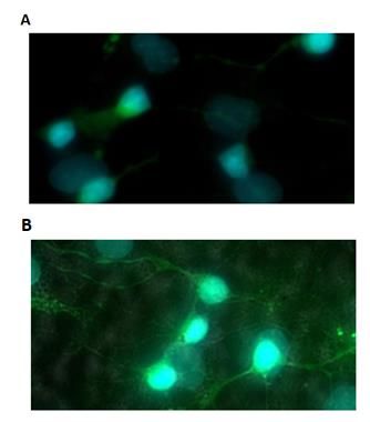
INTERNATIONAL JOURNAL OF BIOLOGY AND BIOMEDICAL ENGINEERING
DOI: 10.46300/91011.2021.15.45 Volume 15, 2021
of schizophrenia with BDNF gene polymorphisms and with ethics committee approval laboratory animals study
changes in BDNF mRNA levels has been reported [7]. It (Protocol No. 14). The cerebellums tissue was lysed in a
was found in the patients with the first schizophrenia solution of 0.25% trypsin solution / Versen-EDTA solution
episode that childhood trauma and stressful situations (in equal volumes) for 15 minutes at 37°C. The supernatant
affected leukocytes BDNF mRNA levels [8]. BDNF levels was removed by centrifugation at 200 g for 30 seconds, the
were also negatively correlated with interleukin 6 (IL-6) tissue was homogenized in a DMEM medium, after
expression, suggesting an inflammation-mediated BDNF precipitation, the upper fraction was passed through a 70
expression decrease [8, 9]. microns filter (SPL Lifesciences), centrifuged at 200 g for 3
BDNF signaling can be regulated or changed at the minutes, the cell precipitate was placed in a Neuromed
different levels. Valproic acid (VPA) is used in the medium (PanEco, RF), in which the cells were cultured. The
schizophrenia complex combined treatment regimens both number of cells from one cerebellum is approximately 15
to reduce inflammation and to prevent the appearance of million, the number of cells per well is 2.2 million. The
catatonic symptoms [9-12]. The use of valproates in the experiment was carried out in 6-well plates with a pre-
treatment of schizophrenia differs in different countries – for applied adhesive material: poly-D-lysine. Valproic acid was
example, in 2018 in Hong Kong, 38.41% of women of added to the neurons (at a concentration of 50 μg / ml),
childbearing age with bipolar disorder were prescribed oxidative stress was created by 40 mM of H2O2, injury was
valproates compared to 8.46% in the UK [11]. Treatment of caused by mechanical damage to the neuron culture. Before
schizophrenia patients with valproate has been shown to exposure to the damaging factors, the growth medium was
increase BDNF level [10]. However, it is still unclear replaced and the restoration of neurons was visually
whether BDNF protein levels measured in blood serum evaluated (AxioVert "CarlZeissMicroscopy" microscope,
samples reflect the level of BDNF in the brain, since animal Germany). To confirm the presence of neurons in the
studies conducted so far yielded contradictory results [6]. culture, a fluorescent analysis was performed using specific
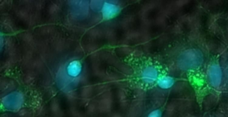
We studied Bdnf gene transcription changes in neuronal antibodies (anti-Map-2, Fig. 1).
neurons in vitro under valproic acid and oxidative stress that
accompanies different diseases including schizophrenia. In
addition, we analyzed Ngf gene transcriptional activity
under valproic acid on cultured neurons under oxidative
stress, since NGF plays an integral role, stimulating the
proliferation, differentiation and development of the central
nervous system neurons, and activates the PI3K, MAPK,
PLC-γ and Ras signaling pathways in a similar way to
Fig. 1 Fluorescence microscopy. Staining of neurons of the
BDNF [13].
primary neuron-glia culture of the rat cerebellum with anti-
II. MATERIALS AND METHODS Map2 antibodies, followed by treatment with secondary
1. Cultivation of primary neuron-glia cell culture antibodies conjugated with FITC (green). The nuclei are
In the present work we used in vitro method, DAPI-colored (blue). Magnification x63.
studying VPA effects on the neuron-glia culture. Such
approach has limitations as cells are grown generally outside
The ability of VPA to activate the Bdnf and Ngf
their natural environment. Meanwhile, such methodology
genes was analyzed in rat neurons.
makes possible to carefully investigate gene expression in
standard laboratory conditions. The primary neuron-glia 2. Determination of the Bdnf and Ngf genes expression.
culture was obtained from the cerebellum of 8-9-day-old Total RNA was isolated with the RNeasy Mini kit
Wistar rats. Research Centre for Medical Genetics (RCMG) (Qiagen, Germany) by the standard method and treated with
Ε-ISSN: 1998-4510 372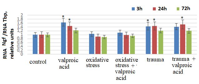
INTERNATIONAL JOURNAL OF BIOLOGY AND BIOMEDICAL ENGINEERING
       DOI: 10.46300/91011.2021.15.45                                                                                Volume 15, 2021

DNase I. The isolated RNA was stored at -80°C. The purity                    studied 3, 24 and 72 hours after its addition into the neuron
of the isolated RNA was determined spectrophotometrically                    culture medium. It was shown that 3 and 24 hours after the
on NanoDrop™ OneC (Thermo Scientific™, USA). The                             valproic acid addition to the primary neuron-glia culture,
optimal values are greater than 1.8. If the sample had                       Bdnf expression increases 2.3-fold (p< 0.01) and 1.5-fold
indicators below 1.8, additional cleaning was performed                      (p< 0.01), respectively (Fig. 2).
with RNA Clean & Concentrator – 5 kits (Zymo Research,                                The schizophrenia is accompanied by oxidative
USA). The concentration of the isolated RNA was                              stress. Oxidative stress in neuron-glia cell culture was
determined using a Qubit 3.0 fluorimeter (Thermo                             modeled by hydrogen peroxide (40 μM) added into the cell
Scientific™, USA). Reverse transcription was performed                       culture medium for 30 min. Mechanical damage to neurons
using "Silex" (Russia) reagents with random hexaprimers.                     disrupts contacts between neurons to a greater extent, but is
For real-time polymerase chain reaction (PCR), an internal                   not accompanied by intense oxidative stress. Hydrogen
standard gene was selected from three genes most                             peroxide in a dose 40 μM in the culture medium does not
commonly used in the literature: GAPDH (glyceraldehyde-                      change Bdnf expression and valproic acid under oxidative
3-phosphate dehydrogenase), TBP (a protein that binds the                    stress causes only slight 1.5-fold (p< 0.01) Bdnf expression
TATA sequence), βACT (β-actin). TBP was chosen as the                        increase after 3 hours (Fig. 2). Both mechanical damage to
gene least susceptible to change during exposure. PCR was                    neurons and the addition of valproic acid after mechanical
performed     with      Synthol      primers       and    SybrGreen          culture damage increased Bdnf expression 2-2.5-fold (p<
intercalating dye (Invitrogen, USA) on a StepOnePlus                         0.01) after 3 to 24 hours. Addition of valproic acid in these
device (Applied Byosystems, USA). Amplification reaction                     conditions did not produce any significant effect (Fig.2).
conditions: 95°C for 5 minutes, then 35 cycles in the mode:
96°C-20 seconds, 57°C-24 seconds, 72°C-24 seconds.
         The level of gene expression was analyzed in
several independent experiments; the results were processed
in the PCR device software. The error was approximately
2%.
Tbp F: GCTCAGGGCTTGGCCTCCCC,                                                 Fig. 2. Bdnf mRNA content in the cells. Bar chart reflects
R: GCTGTCTTTGTTGCTCTTCC                                                      Bdnf expression in the cells of the primary neuron-glia
Bdnf     F: GTGTGGACCCTGAGTTCCAC,                                            culture of the rat cerebellum in 3, 24 and 72 hours. X-axis
R: CTGGGTAGGCCAAGTTGCCT                                                      illustrate condition of experiment; control – the cells
Ngf F: AAGGGGAGCGCATCGCTCTC                                                  cultured without any action. Bdnf mRNA amount is the
R: ATAGAAAGCTGCGTCCTTGG                                                      average value for three experiments relatively to the TBP
                                                                             standard gene expression. (*) data differ from the control
3. Statistics methods                                                        according to the Mann-Whitney criterion; the differences are
The reliability of the results obtained in the study was                     significant, p< 0.01.
analyzed by statistical methods using the nonparametric
Mann-Whitney      criterion       (U-criterion).    Statistics   was                  Another factor necessary for the survival, growth of
calculated   using      the   standard         software    packages          axons and dendrites, specification and formation of synapses
Statgraphics, Statistica 10 and SigmaStat.                                   of sympathetic and sensory neurons is NGF. The signaling
                                                                             pathways and processes that NGF triggers occur mainly in
                                                                             sympathetic and sensory neurons. NGF binds to the TrkA
                         III. RESULTS
                                                                             receptor leading to its phosphorylation. Then, similarly to
The effect of valproic acid on Bdnf expression in the cells of               BDNF effect, PI3K, MAPK, PLC - γ, Ras – signaling
the primary neuron-glia culture of the rat cerebellum was                    pathways in neuronal cells are activated [13].

       Ε-ISSN: 1998-4510                                               373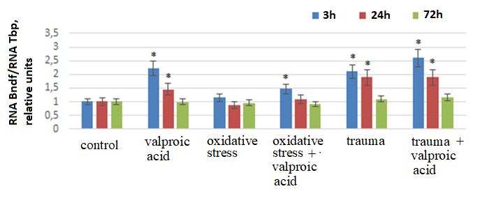
INTERNATIONAL JOURNAL OF BIOLOGY AND BIOMEDICAL ENGINEERING
      DOI: 10.46300/91011.2021.15.45                                                                             Volume 15, 2021

         We studied the valproic acid effect on the Ngf                valproic acid positive effect on the neuroplasticity [4].
expression in the cells of the primary neuron-glia culture of          However, its effect on neurons is, possibly, more complex
the rat cerebellum 3, 24 and 72 hours after its addition into          than simply the effect on the Ngf and Bdnf expression.
the neuron culture medium. It was shown that 3 and 24
hours after valproic acid addition to the primary neuron-glia
culture, Ngf expression increases 1.6-fold (p< 0.01) and 1.5-
fold (p
INTERNATIONAL JOURNAL OF BIOLOGY AND BIOMEDICAL ENGINEERING
DOI: 10.46300/91011.2021.15.45 Volume 15, 2021
activation of the Ngf and Bdnf genes transcription by VPA on Neuroinflammation”, Mol Neurobiol., 56, 2019, pp.
3295-3312. doi: 10.1007/s12035-018-1283-6.
in cultured neurons in vitro. We also have found that in the
[7] B. Lu, K. Lu B. Martinowich et al., “Cell biology of
presence of valproic acid, the number of neuronal processes BDNF and its relevance to schizophrenia”, Novartis
Found Symp, 289, 2008, pp. 119-129; discussion 129-
and contacts between them significantly increased. The
35, 193-5. doi: 10.1002/9780470751251.ch10.
results of the present study show that valproic acid under [8] V. Mondelli, A. Cattaneo, M.B. Murri, et al., “Stress
and inflammation reduce brain-derived neurotrophic
oxidative stress conditions causes only slight Bdnf
factor expression in first-episode psychosis: a pathway
expression increase after 3 hours in comparison with to smaller hippocampal volume”, J Clin Psychiatrym,
72, 2011, pp. 1677–1684. doi: 10.4088/JCP.10m06745.
valproic acid effect in control experiments. Therefore we
[9] Y.K. Kim, H.G. Jung, A.M. Myint, et al., “Imbalance
conclude that oxidative stress accompanying the between pro-inflammatory and anti-inflammatory
cytokines in bipolar disorder”, J Affect Disord, 104,
schizophrenia can significantly reduce the valproic acid
2007, pp. 91–95. doi: 10.1016/j.jad.2007.02.018.
effectiveness on the Ngf and Bdnf genes expression. Further [10] C.A. Zarate, H.K. Manji, “Protein kinase C inhibitors:
rationale for use and potential in the treatment of
studies of the valproic acid effect on the transcriptional
bipolar disorder”, CNS Drugs, 23, 2009, pp. 569–582.
activity of key signaling pathways genes in neurons under doi: 10.2165/00023210-200923070-00003.
[11] V.W.S Ng, K.K.C. Man, L. Gao, E.W. Chan, E.H.M.
oxidative stress are needed. Also more research should be
Lee, J.F. Hayes, I.C.K. Wong, “Bipolar disorder
conducted to determine VPA effects using in vivo conditions prevalence and psychotropic medication utilisation in
Hong Kong and the United Kingdom”,
to justify its effects in living mammalian brain and the
Pharmacoepidemiol Drug Saf, 2021 doi:
oxidative stress role in schizophrenia patients’ therapy 10.1002/pds.5318.
[12] M. Saito, T. Takizawa, H. Miyaoka, “Factors
approaches.
associated with blood carnitine levels in adult epilepsy
patients with chronic valproic acid therapy”, Epilepsy
ACKNOWLEDGMENT Res, 175, 2021, 106697. doi:
10.1016/j.eplepsyres.2021.106697.
Funding: The study was supported by Russian [13] M.C. Marlin, G. Li, “Biogenesis and function of the
Science Foundation, project No. 18-15-00437. NGF/TrkA signaling endosome”, Int Rev Cell Mol
Biol, 314, pp. 239-257.
doi: 10.1016/bs.ircmb.2014.10.002
CONFLICTS OF INTEREST: The authors declare [14] Dedoni S., Marras L., Olianas M.C., Ingianni A., Onali
no conflict of interest. P.J., “Downregulation of TrkB Expression and
Signaling by Valproic Acid and Other Histone
Deacetylase Inhibitors”, Parmacol Exp Ther., 2019
REFERENCES Sep;370(3):490-503. doi: 10.1124/jpet.119.258129
[1] A. Mohammadi, E. Rashidi, V.G. Amooeian, “Brain, [15] T.D. Gould, J.A. Quiroz, J. Singh, et al, “Emerging
blood, cerebrospinal fluid, and serum biomarkers in experimental therapeutics for bipolar disorder: insights
schizophrenia”, Psychiatry Res, 265, 2018; pp. 25-38. from the molecular and cellular actions of current mood
doi: 10.1016/j.psychres.2018.04.036. stabilizers”, Mol Psychiatry, 9, 2004, pp. 734–755. doi:
[2] M.E. Benros, P.R. Nielsen, M. Nordentoft et al., 10.1038/sj.mp.4001518.
“Autoimmune diseases and severe infections as risk
factors for schizophrenia: a 30-year population-based Creative Commons Attribution License 4.0
register study”, Am J Psychiatry, 168, 2011, pp. 1303– (Attribution 4.0 International, CC BY 4.0)
1310. doi: 10.1176/appi.ajp.2011.11030516.
[3] G.M. Khandaker, L. Cousins, J. Deakin, et al., This article is published under the terms of the Creative
“Inflammation and immunity in schizophrenia: Commons Attribution License 4.0
implications for pathophysiology and treatment”, https://creativecommons.org/licenses/by/4.0/deed.en_US
Lancet Psychiatry, 2, 2015, pp. 258–270. doi:
10.1016/S2215-0366(14)00122-9.
[4] G. Leal, D. Comprido, C.B. Duarte, “BDNF-induced
local protein synthesis and synaptic plasticity”,
Neuropharmacology, 2014 Jan;76 Pt C:639-56. doi:
10.1016/j.neuropharm.2013.04.005. Epub 2013 Apr 16.
[5] Leal , C.R. Bramham , C.B. Duarte, “BDNF and
Hippocampal Synaptic Plasticity”, Vitam Horm,
2017;104:153-195. doi:
10.1016/bs.vh.2016.10.004. Epub 2016 Nov 29.
[6] B. Lima Giacobbo, J. Doorduin, H.C. Klein, RAJO
Dierckx, E. Bromberg, EFJ de Vries, et al., “Brain-
Derived Neurotrophic Factor in Brain Disorders: Focus
Ε-ISSN: 1998-4510 375You can also read