Structure of Vibrio collagenase VhaC provides insight into the mechanism of bacterial collagenolysis - Nature
←
→
Page content transcription
If your browser does not render page correctly, please read the page content below
ARTICLE
                  https://doi.org/10.1038/s41467-022-28264-1                 OPEN

                  Structure of Vibrio collagenase VhaC provides
                  insight into the mechanism of bacterial
                  collagenolysis
                  Yan Wang1,2,3,10, Peng Wang2,3,10, Hai-Yan Cao1,3, Hai-Tao Ding4, Hai-Nan Su1,3, Shi-Cheng Liu5,
                  Guangfeng Liu6, Xia Zhang5, Chun-Yang Li2,3, Ming Peng1,3, Fuchuan Li 7, Shengying Li 1,3, Yin Chen                                                       2,8,

                  Xiu-Lan Chen 1,3 ✉ & Yu-Zhong Zhang 2,3,9 ✉
1234567890():,;

                  The collagenases of Vibrio species, many of which are pathogens, have been regarded as an
                  important virulence factor. However, there is little information on the structure and
                  collagenolytic mechanism of Vibrio collagenase. Here, we report the crystal structure of the
                  collagenase module (CM) of Vibrio collagenase VhaC and the conformation of VhaC in
                  solution. Structural and biochemical analyses and molecular dynamics studies reveal that
                  triple-helical collagen is initially recognized by the activator domain, followed by subsequent
                  cleavage by the peptidase domain along with the closing movement of CM. This is different
                  from the peptidolytic mode or the proposed collagenolysis of Clostridium collagenase.
                  We propose a model for the integrated collagenolytic mechanism of VhaC, integrating
                  the functions of VhaC accessory domains and its collagen degradation pattern. This study
                  provides insight into the mechanism of bacterial collagenolysis and helps in structure-based
                  drug design targeting of the Vibrio collagenase.

                  1 State Key Laboratory of Microbial Technology, Shandong University, Qingdao 266237, China. 2 College of Marine Life Sciences, and Frontiers Science Center for

                  Deep Ocean Multispheres and Earth System, Ocean University of China, Qingdao 266003, China. 3 Laboratory for Marine Biology and Biotechnology, Pilot
                  National Laboratory for Marine Science and Technology, Qingdao 266237, China. 4 Antarctic Great Wall Ecology National Observation and Research Station,
                  Polar Research Institute of China, Shanghai 200136, China. 5 Department of Molecular Biology, Qingdao Vland Biotech Inc., Qingdao, China. 6 National Center
                  for Protein Science Shanghai, Shanghai Advanced Research Institute, Chinese Academy of Sciences, Shanghai 201210, China. 7 National Glycoengineering
                  Research Center and Shandong Key Laboratory of Carbohydrate Chemistry and Glycobiology, Shandong University, Qingdao, China. 8 School of Life Sciences,
                  University of Warwick, Coventry, UK. 9 Marine Biotechnology Research Center, State Key Laboratory of Microbial Technology, Shandong University, Qingdao
                  266237, China. 10These authors contributed equally: Yan Wang, Peng Wang. ✉email: cxl0423@sdu.edu.cn; zhangyz@sdu.edu.cn

                  NATURE COMMUNICATIONS | (2022)13:566 | https://doi.org/10.1038/s41467-022-28264-1 | www.nature.com/naturecommunications                                      1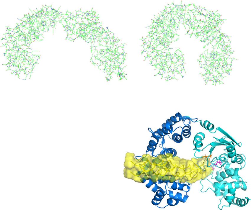
ARTICLE                                                                  NATURE COMMUNICATIONS | https://doi.org/10.1038/s41467-022-28264-1

C
        ollagens are the most abundant proteins in mammals,                   disease-like domain (PKD-like domain) and the bacterial pre-
        mainly in the extracellular matrix (ECM)1. Of the 28 types            peptidase C-terminal domain (PPC domain)8,20. The PPC
        of collagens, type I collagen is the most widely occurring            domain of Vibrio collagenase Ghcol from Grimontia hollisae
and has a hierarchical structure2. Monomeric type I collagen                  (formerly known as Vibrio hollisae) has been demonstrated to
(tropocollagen, ~300 nm in length and ~1.5 nm in diameter) is a               function as a collagen binding domain (CBD)32. Despite these
righthanded triple helix comprising three α chains (two α1 chains             studies, little is known about the mechanism of Vibrio col-
and one α2 chain). Each chain consists of Gly-X-Y repeating tri-              lagenase for collagen recognition and degradation due to the
plets where X and Y are often occupied by proline and hydro-                  lack of structural information.
xyproline, respectively. The N- and C-terminal telopeptides,                     In this work, we report the structure and propose the col-
located on the flanks of the triple-helical region, constitute the             lagenolytic mechanism of a Vibrio collagenase VhaC from
non-triple helical regions of tropocollagen1. Every five tropo-                Vibrio harveyi VHJR7, which is isolated from an infected
collagens form one quasi-hexagonal microfibril unit3,4. Micro-                 fish33,34. Protease VhaC is a multi-domain enzyme composed of
fibrils are interdigitated regularly with each other, forming mature           a CM containing an activator domain and a peptidase domain, a
collagen fibrils. Various covalent cross-links, such as pyridinium             PKD-like domain and a PPC domain. The crystal structure
compounds and pyrroles within or between microfibrils, maintain                of the CM is solved at 1.8 Å resolution and the overall con-
the strength and stability of the fibrils5. Collagen fibrils further            formation and interdomain arrangement of VhaC in solution
aggregate with proteoglycans to form collagen fibers and other                 is determined by small angle x-ray scattering (SAXS). The
super structures in the ECM3,6,7. Due to its tight structure and              mechanism of VhaC for the recognition and catalysis of triple-
hierarchical assembly, collagen can only be degraded by a very few            helical collagen is illustrated based on structural and biochem-
proteases8.                                                                   ical analyses and molecular dynamics studies, and the functions
   Collagenolytic proteases, capable of hydrolyzing native collagen           of the C-terminal accessory PKD-like and PPC domains
under physiological conditions, are found in animals and microbes.            in collagenolysis are analyzed. Finally, combined with its col-
Mammalian matrix metalloproteinases (MMPs), belonging to the                  lagen degradation pattern, a model for collagen fibers degra-
M10A subfamily, play crucial roles in the interstitial collagen cat-          dation by VhaC is proposed. These results reveal the structure
abolism and their structures and collagenolytic mechanisms have               of Vibrio collagenase and its mechanism for collagen recogni-
been extensively studied9–11, especially for MMP-1. MMP-1                     tion and degradation, providing insight into the mechanism
cleaves type I tropocollagen chain at a single Gly-Ile/Leu site               of bacterial collagenolysis and helping in the development of
generating 1/4 and 3/4 length fragments via a complicated mole-               strategies to prevent and treat infection caused by pathogenic
cular mechanism12–18. Bacterial collagenolytic proteases, that are            Vibrio species.
capable of hydrolyzing native collagen at multiple sites, are dis-
tributed in the M9, S1, and S8 MEROPS peptidase families, some
of which are strongly linked to bacterial pathogenesis19–21. Most             Results
known bacterial collagenases belong to the M9 family, which                   Characterization and structural analysis of VhaC. The vhaC
is subdivided into the M9A (Vibrio collagenases) and M9B                      gene from Vibrio harveyi VHJR7 was predicted to encode a putative
(Clostridium collagenases) subfamilies. Up to now, information on             M9A collagenase (GenBank accession No. WP_047516938.1) of
the three-dimensional structure of Vibrio collagenase is still lack-          814 amino acid residues. Mature VhaC protein contains 745 resi-
ing. As for Clostridium collagenases, only the crystal structure of           dues from Ser70 to Gln814 with the N-terminal signal peptide and
the collagenase module (CM) of ColG22 and those of the peptidase              propeptide being removed, which presented as a monomer in
domains of ColH and ColT have been solved23. Eckhard et al.22                 solution (Supplementary Fig. 1a). VhaC had activity toward various
proposed a “chew and digest” collagenolytic model based on the                types of collagens (Supplementary Table 1), showing that it is a
saddle-shaped architecture of the CM of ColG. In this model,                  collagenase. With type I collagen as the substrate, VhaC has optimal
triple-helical collagen initially contacts the peptidase domain in the        enzymatic reaction conditions at 40 °C and pH 8.0 (Supplementary
open state; in the closed state, triple-helical collagen interacts with       Fig. 1b, c).
both the activator and the peptidase domains in the CM and then                  Sequence analysis using Conserved Domain Search showed
is unwound and degraded progressively. However, due to the lack               that VhaC is a multi-domain protein containing an activator
of sufficient experimental evidence for this model, how triple-                domain (Ser70-Tyr346), a peptidase domain (Gly347-Val605),
helical collagen is recognized by the activator and the peptidase             a PKD-like domain (Ser606-Ala698), and a PPC domain
domains is still largely unclear. Thus, the mechanism of collagen             (Leu699-Gln814) (Fig. 1a). The activator domain and the
recognition by M9 bacterial collagenases warrants further study in            peptidase domain form the CM. To obtain the structure of
order to better understand its role in pathogenesis.                          VhaC, the full-length VhaC (Met1-Gln814) and its CM (Met1-
   Collagenases of certain pathogenic Vibrio species accelerate               Val605) were expressed, and an attempt was made to crystallize
bacterial dissemination and facilitate diffusion of other toxins              the recombinant proteins. However, only the crystal structure
through hydrolysis of the collagenous components of the ECM,                  of CM was solved to 1.8 Å resolution. Data collection statistics
they are therefore regarded as an important virulence factor24.               are shown in Supplementary Table 2. The CM crystal belongs to
So far, studies have largely been focused on the physicochemical              the P 21 space group with one molecule per asymmetric unit.
properties25–28, cytotoxicity, and fish pathogenicity29 of Vibrio              The CM structure contains residues from Cys78 to His609,
collagenases. Except for an early report that Vibrio collagenase              suggesting that the N-terminal signal peptide and propeptide in
attacked the collagen molecule at 3/4 from the N-terminus by                  the CM precursor are cleaved off during enzyme maturation.
digesting the preferential peptide bond, Y-Gly, in the first step              The eight N-terminal residues from Ser70 to Val77 were not
of collagen degradation30, information on the collagen cleavage               observed in the CM structure due to the ambiguous electron
pattern of Vibrio collagenase is scarce. Like Clostridium col-                density. The CM structure presents a saddle-shaped architec-
lagenases, the characteristic domain architecture of Vibrio                   ture (Fig. 1b), which is similar to that of ColG (PDB: 2Y50) but
collagenase consists of a peptidase M9N domain (activator                     with a more contracted catalytic cavity (Fig. 1c). The activator
domain) and a peptidase M9 domain (peptidase domain),                         domain of VhaC is similar to that of ColG with a root mean
forming a CM26,31. Some Vibrio collagenases contain additional                square deviation (RMSD) of 3.861 Å (Fig. 1d). The peptidase
C-terminal accessory domains, such as the polycystic kidney                   domain of VhaC is also similar to that of ColG with an RMSD

2                 NATURE COMMUNICATIONS | (2022)13:566 | https://doi.org/10.1038/s41467-022-28264-1 | www.nature.com/naturecommunications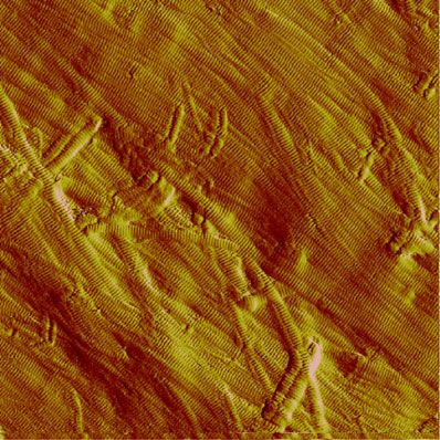
NATURE COMMUNICATIONS | https://doi.org/10.1038/s41467-022-28264-1                                                                                                           ARTICLE

    a                                                                                      b

               ep ide
                     e
                   pt
                 tid
                pe
              al
                   op
          gn
               Pr
         Si

                     Peptidase_M9_N         Peptidase_M9         PKD PPC

        M1 A21 L22 S70               Y346 G347           V605 S606 A698 L699 Q814                             Peptide P1
                   Protein crystallized, collagenase module (CM)                                       Peptide P2                                               Peptide P1
                                             Mature enzyme
                                                                                                                                                  Peptide P2

        Peptidase_M9_N   Activator domain   PKD   Polycystic kidney disease domain (PKD)

         Peptidase_M9    Peptidase domain   PPC   Prepeptidase C-terminal domain (PPC)

                                                                                               Activator domain            Peptidase domain

    c                                                                                                   d                                     e

                                                                                Catalytic
                                                                                helper
                                                                                subdomain

    f
                                                                                    h

                                                                                                                                                               90o

    g                                                                                                                                               90o

Fig. 1 Crystal structure of the CM and overall conformation of VhaC in solution. a Schematic diagram of the domain organization of VhaC precursor.
b The overall structure of CM. The activator domain is shown in marine, and the peptidase domain in cyan. Zn2+ (gray) and Ca2+ (wheat) are shown as
spheres. Peptides P1 and P2 derived from the propeptide are colored in orange and magenta, respectively. A close-up view of the 2Fo-Fc omit electron
density map of the peptide P1 (orange sticks), peptide P2 (magenta sticks) and Zn2+ ion (gray sphere) contoured at 1.0 σ (gray) is shown. c A comparison
of the CM structures between VhaC and ColG (golden) (PDB: 2Y50). The catalytic helper subdomain of ColG is indicated by dotted ellipse. d Structural
superimposition of the activator domains of VhaC (marine) and ColG (golden). e Structural superimposition of the peptidase domains of VhaC (cyan) and
ColG (golden). f Structural superposition of the catalytic centers of CM and 2Y6I. Peptide P1 in CM is colored in orange and isoamyl-phosphonyl-Gly-Pro-
Ala in 2Y6I is colored in blue. The double-Gly motif representing the S1′ recognition site is shown in red (CM) and magenta (2Y6I). The edge strand
representing the non-primed substrate-recognition site is shown in green (CM) and marine (2Y6I). g Ribbon representation of the catalytic center of
CM. Residues His477, His481, Glu505 (shown in yellow), and histidine from peptide P1 (shown in orange) tetrahedrally coordinate the catalytic Zn2+
(gray sphere). The catalytic Glu478 is shown in magenta. The Ca2+ is coordinated by Gly485, Leu489, Gly491, and Glu446 (shown in salmon).
h Superimposition between the ab initio beads model and the rigid body model of VhaC. The beads model is represented as grey spheres. The rigid body
model is drawn as a ribbon representation and colored marine (the activator domain), cyan (the peptidase domain), yellow (the PKD-like domain), and
magenta (the PPC domain). The poly-glycine linker added by CORAL is shown in green.

value of 1.720 Å (Fig. 1e). The CM of ColG contains an                                                       collagen degradation mechanisms of the M9A and M9B
additional catalytic helper subdomain (Fig. 1c), which is a part                                             subfamilies.
of the peptidase domain and indispensable for the full                                                          Unexpectedly, the structure data show that the CM molecule
enzymatic activity of ColG22,35. However, this domain is absent                                              binds two peptides (peptide P1: 24-EPSQQVTEIYQHHA-37;
from VhaC, suggesting that differences may exist in the                                                      peptide P2: 48-DYAPTKLLPQQP-59) derived from the propeptide

NATURE COMMUNICATIONS | (2022)13:566 | https://doi.org/10.1038/s41467-022-28264-1 | www.nature.com/naturecommunications                                                            3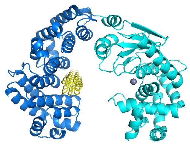
ARTICLE                                                                 NATURE COMMUNICATIONS | https://doi.org/10.1038/s41467-022-28264-1

of VhaC via hydrogen bonds and hydrophobic interactions (Fig. 1b).           [(POG)10]3 and only 27% activity against type I collagen fibers
While peptide P2 entirely binds to the activator domain, the                 (Fig. 2a), EGFP-peptidase domain showed approximately 70%
N-terminus of peptide P1 binds to the activator domain and its               activity against Pz peptide but no activity against [(POG)10]3 or
C-terminus extends and binds to the catalytic center of the                  type I collagen fibers (Fig. 2a). Consistently, the inactive mutant of
peptidase domain (Fig. 1b). As shown in Fig. 1f, the complex                 the peptidase domain, peptidase domain-E478A, had no detectable
structure of the CM of ColG with isoamyl-phosphonyl-Gly-Pro-Ala              [(POG)10]3-binding ability (Fig. 2b) and the inactive mutant
(PDB: 2Y6I) defines the S1′ peptide recognition site formed by a              EGFP-peptidase domain-E478A, had little collagen fiber-binding
double-Gly motif (Gly493-Gly494)22. Correspondingly, in the CM               ability (Fig. 2c). These results suggest that the activator domain
structure of VhaC, the C-terminal residues of peptide P1 also bind           plays an indispensable role in the degradation of triple-helical
to the conserved Gly441-Gly442 motif, representing the S1′ peptide           collagen and collagen fibers, likely involved in substrate binding.
recognition site (Fig. 1f). Therefore, peptides P1 and P2 can be             Indeed, isothermal titration calorimetry (ITC) measurements
regarded as substrate analogues in the CM structure, which may               showed that the recombinant activator domain had [(POG)10]3-
provide clues for the recognition pattern of collagen substrates in          binding ability (Fig. 2d) and fluorescence detection showed that the
CM. In addition, as in ColG (495-LYIE-498), an edge strand (443-             activator domain-EGFP protein had collagen fiber-binding ability
MYIE-446) of VhaC forms the canonical non-primed substrate-                  (Fig. 2c).
recognition site with the double-Gly motif as the entry (Fig. 1f). In           To investigate the triple-helical collagen-binding mode in CM, an
the CM structure, a catalytic Zn2+ is tetrahedrally coordinated by           attempt was made to obtain the crystal structure of the inactive
the side chains of His477, His481, and Glu505 from the peptidase             mutant CM-E478A binding [(POG)10]3, but this failed. Instead, the
domain, as well as the side chain of a histidine from peptide P1             structure of unbound-CM (the structure of CM with peptides P1 and
(Fig. 1g) instead of the characteristic water molecule observed in           P2 being removed) in complex with a THP (PDB: 1K6F) was
2Y5022. In addition, a Ca2+ ion, located on the left rim of the              modeled by molecular docking. According to the binding mode with
active-site cleft and 14.2 Å away from the catalytic Zn2+ ion, is            the highest score, THP is bound to the potential substrate-recognition
coordinated by the backbone oxygens of Gly485, Leu489, and                   interface of the activator domain by hydrophobic interactions
Gly491 and the side chain of Glu446 (Fig. 1g). The Ca2+ ion is               (Fig. 2e), away from the site responsible for peptide recognition in
known to stabilize the Zn2+ ion, and as such it is essential for the         the catalytic center of the peptidase domain. This is consistent with
enzymatic activity of M9B collagenase23,36.                                  the afore mentioned ITC data showing that THP [(POG)10]3 can
   Because the structure of full-length VhaC could not be                    bind to the activator domain (Fig. 2d) but not to the peptidase
obtained via crystallization, VhaC was alternatively subjected to            domain (Fig. 2b). The key amino acid residues in the activator
SAXS measurement to analyze its overall conformation and                     domain for collagen binding were further investigated by site-directed
interdomain arrangement in solution. Related SAXS-derived                    mutagenesis. Ten residues (Phe107, Arg153, Tyr157, Phe160,
molecular parameters are shown in Supplementary Table 3 and                  Asp207, Trp234, Phe286, Asn290, Arg293, and Glu294) interacting
Supplementary Fig. 2a, b. The ab initio beads model shows that               with peptides P1 and P2 in the CM structure and/or with THP in the
the overall shape of VhaC is long and flat, with a wide head and a            modeled complex structure were mutated to Ala (Fig. 2f), and the
short tail; dimensions are estimated to be ~176 Å × 63 Å × 28 Å              mutants were expressed as EGFP-fused proteins for detecting their
(Fig. 1h). To estimate the interdomain arrangement of VhaC, a                collagen fiber-binding ability. These residues are strictly or highly
rigid body model was built using the crystal structure of CM and             conserved among M9 collagenases (Supplementary Fig. 3). Among
homology models of PKD-like and PPC domains. The resulting                   the mutants, the collagen fiber-binding ability of mutants F107A,
rigid body model fits well to the ab initio beads model and                   R153A, and Y157A was severely reduced compared with that of the
suggests a side-by-side assembly of each domain (Fig. 1h). The               wild type (Fig. 2g), indicating that these residues are likely the key
theoretical scattering curve of the rigid body model fitted to the            residues involved in collagen binding. Consistently, single mutation
experimental curve is shown in Supplementary Fig. 2c. Accord-                of these amino acid residues (Phe107, Arg153, and Tyr157) in CM to
ing to the elongated conformation of VhaC (Fig. 1h), the PPC                 Ala resulted in noticeable decrease in the activity of CM to
domain (magenta), connected by a flexible linker, extends                     [(POG)10]3 and an increase in its Km value to [(POG)10]3 (Fig. 2h and
outside the core region, self-consistent with the flexibility of              Supplementary Fig. 4) and the triple mutant F107A/R153A/Y157A
VhaC observed in the Kratky plot (Supplementary Fig. 2d).                    almost completely lost its enzymatic activity towards [(POG)10]3
Notably, the solution structure of VhaC is significantly different            (Fig. 2h). These results indicate that Phe107, Arg153 and Tyr157 are
from that of the M9B collagenase ColH determined by SAXS                     key residues involved in triple-helical collagen binding in the catalysis
analysis, which adopts a tapered shape with a swollen head and               of CM. In contrast, these mutations (F107A, R153A, Y157A, and
an elongated tail37.                                                         F107A/R153A/Y157A) had little effect on the enzymatic activity of
                                                                             CM toward Pz peptide and caused no change in the Km value
                                                                             (Fig. 2h and Supplementary Fig. 4), suggesting that the activator
The triple-helical collagen recognition mechanism of CM. The                 domain in CM is not involved in the recognition of peptide
triple-helical collagen recognition and degradation mechanisms of            substrates, consistent with the result that EGFP-peptidase domain
CM were then investigated. Firstly, the activities of VhaC, CM, and          had activity against Pz peptide (Fig. 2a). Circular dichroism (CD)
EGFP-peptidase domain (the peptidase domain fused to the                     spectroscopy assays showed that the secondary structures of all the
enhanced green fluorescent protein, since the recombinant pepti-              site-directed mutants are similar to those of the wild types (Fig. 2i, j),
dase domain alone underwent severe autolysis during purification)             suggesting that the mutations caused little structural alteration.
against insoluble type I collagen fibers, triple-helical peptide (THP)           Together, these results indicate that VhaC adopts different
[(POG)10]3, and 4-phenylazobenzyloixycarbonyl-Pro-Leu-Gly-Pro-               strategies for the recognition of triple-helical collagen and peptide
o-Arg (Pz peptide) were compared. VhaC showed evident activity               substrates. Triple-helical collagen is initially recognized by the
against all three substrates, whereas trypsin (control) showed               activator domain, whereas peptide is directly recognized by the
negligible activity against collagen fibers and no activity against           peptidase domain.
[(POG)10]3 or Pz peptide (Fig. 2a). The slight activity of trypsin on           Since the above results are significantly different from the
collagen fibers may be due to its degradation of proteoglycans or             chew-and-digest model of ColG22, in which the triple-helical
telopeptides within collagen fibers38. Compared to VhaC, CM                   collagen is initially recognized by the peptidase domain, the
showed approximately 100% activity against Pz peptide and                    activator domain, and the peptidase domain of ColG were

4                NATURE COMMUNICATIONS | (2022)13:566 | https://doi.org/10.1038/s41467-022-28264-1 | www.nature.com/naturecommunications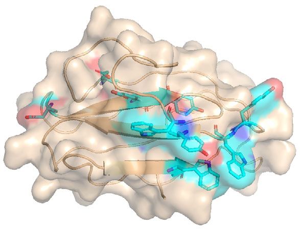
NATURE COMMUNICATIONS | https://doi.org/10.1038/s41467-022-28264-1 ARTICLE
a 120
Collagen fibers [(POG)10]3 Pz peptide b d
ns ns
0.0 0.0
100
Relative activity (%)
-0.1 -0.1
80
-0.2 -0.2
**
DP (μcal/s)
DP (μcal/s)
60 -0.3 -0.3
-0.4 -0.4
40
** -0.5 -0.5
20 -0.6 -0.6
# # ** # # 0 5 10 15 20 25 30 35 0 5 10 15 20 25 30 35
0 Time (min)
ain Time (min)
) CM dom sin
C (WT se Tryp
Vha tida
P -pep
c EGF 0.0
-0.4
450 -0.6
-0.4 -0.8
ΔH (kcal/mol)
400
ΔH (kcal/mol)
Fluorescent intensity
-1.0
-0.8
350 -1.2
300 -1.2 Protein: Peptidase domain-E478A -1.4 Protein: Activator domain
Binding: ND Binding: Yes
-1.6 Kd (μM): 82.80 ± 46.50
250 -1.6
-1.8 ΔH (kcal/mol): -3.90 ± 1.94
EGFP
200 Activator domain-EGFP -2.0 -2.0
0.0 0.2 0.4 0.6 0.8 1.0 1.2 1.4 1.6 1.8 2.0 0.0 0.2 0.4 0.6 0.8 1.0 1.2 1.4 1.6 1.8 2.0
EGFP-peptidase domain-E478A
150 Molar Ratio Molar Ratio
0 2 4 6 8
The amount of collagen (mg)
e f g
120
ns
100 ns
ns ns
Relative binding affinity (%)
80
* *
60 **
**
40 **
**
20
0
Activator domain Peptidase domain WT
3A
0A
3A
A
A
4A
A
A
A
7A
57
94
07
60
86
15
29
29
23
20
Y1
E2
F1
F1
F2
R
N
R
W
D
h
Relative activity towards [(POG)10]3
** Relative Km value to [(POG)10]3
i j
250
Relative activity towards Pz peptide
Relative Km value to Pz peptide 30 20
WT
200 F107A WT
** 20 R153A 10 F107A
Ellipticity (mdeg)
Y157A
Relative value (%)
Ellipticity (mdeg)
R153A
F160A
150 * 10
D207A 0
Y157A
W234A F107A/R153A/Y157A
ns ns 0 F286A
ns ns ns ns ns N290A
ns -10
100 100% R293A
-10 E294A
** -20
-20
50 **
-30 -30
** 200 210 220 230 240 250 200 210 220 230 240 250
0
** Wavelength (nm) Wavelength (nm)
CM (WT) F107A R153A Y157A F107A/R153A/Y157A
expressed in E. coli, and the collagen-binding ability of the activator domain rather than the peptidase domain, it can be
recombinant proteins was analyzed. Similarly, the activator speculated that the activator domain is responsible for collagen
domain of ColG showed evident binding ability towards collagen recognition while the peptidase domain is responsible for catalysis
fibers (Supplementary Fig. 5a) and [(POG)10]3 (Supplementary in CM. To investigate the catalytic mechanism of CM towards
Fig. 5b) but the peptidase domain did not (Supplementary Fig. 5a, triple-helical collagen, unbound-CM and unbound-CM: THP
c), suggesting that in ColG the activator domain rather than the binary complex were considered for performing molecular
peptidase domain, is likely responsible for the initial collagen dynamics simulation (MDS) for 1000 ns. The RMSD profiles of the
recognition, just as in the case of VhaC. backbone atoms of the unbound-CM structure and its complex
structure with THP show that all simulations generated stable
trajectories (Fig. 3a), indicating that the systems reached the
The catalytic mechanism of CM for triple-helical collagen. equilibrium state. Following equilibrium, unbound-CM showed
Considering that triple-helical collagen preferentially binds to the continuous opening and closing changes (Supplementary Movie 1).
NATURE COMMUNICATIONS | (2022)13:566 | https://doi.org/10.1038/s41467-022-28264-1 | www.nature.com/naturecommunications 5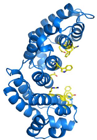
ARTICLE                                                                     NATURE COMMUNICATIONS | https://doi.org/10.1038/s41467-022-28264-1

Fig. 2 The triple-helical collagen recognition mechanism of CM. a The activities of VhaC, CM, and EGFP-peptidase domain towards collagen fibers,
[(POG)10]3 and Pz peptide. The specific activities of VhaC (WT) towards collagen fibers, [(POG)10]3 and Pz peptide were taken as 100%. Trypsin was
served as a negative control to show that collagen fibers were not denatured under the experimental conditions. b ITC analysis of the ability of peptidase
domain-E478A to bind [(POG)10]3. Data representative of the results of triplicate experiments are shown. c Fluorescence analysis of the collagen fiber-
binding ability of EGFP, activator domain-EGFP, and EGFP-peptidase domain-E478A. d ITC analysis of the [(POG)10]3-binding ability of the activator
domain. Data representative of the results of triplicate experiments are shown. e Molecular docking of CM with THP (PDB: 1K6F). THP docked into the
activator domain is shown in yellow. The catalytic center of the peptidase domain is circled. f The residues (yellow sticks) in the activator domain selected
for site-directed mutation. g The collagen-binding ability of activator domain-EGFP (WT) and its mutants. h The activities and Km values of CM and its
mutants towards [(POG)10]3 and Pz peptide. The specific activities and Km values of CM (WT) towards [(POG)10]3 and Pz peptide were taken as 100%,
respectively. i CD spectra of activator domain-EGFP (WT) and its mutants. j CD spectra of CM (WT) and its mutants. The data shown in figures i and j are
representatives of triplicate experiments. Data shown in figures a, c, g, and h are means ± standard deviations (SD) (n = 3 independent experiments). For
comparison of the statistical differences between two groups, a two-tailed t-test was used in statistical analysis. * p < 0.01. ** p < 0.001. ns, no significant
difference (P ≥ 0.01). # The significance was not compared due to the activity of the enzyme was undetectable. The p-values in all cases were compared
with the corresponding wild type, and provided in the source data. Source data are provided as a Source Data file.

Figure 3b shows the conformational opening and closing changes                   structure to that of the wild-type VhaC (Fig. 3g), suggesting that
of the first principal component by principal component analysis,                 the activity loss of the mutants is caused by amino acid
whose movement mode contributes more than 45% to the con-                        replacement rather than structural alteration. Together, these
formational change of CM. Cluster analysis also elucidates the                   results help to identify the key residues involved in collagen
open and closed states of unbound-CM (Supplementary Fig. 6).                     catalysis in the peptidase domain of VhaC.
The root mean-square fluctuation (RMSF) of the N-terminal
and C-terminal residues is greater than that of the middle residues              Functional analysis of the PKD-like and the PPC domains. As
(Fig. 3c), suggesting the open-closed movement of the activator                  shown in Fig. 1a, VhaC contains two C-terminal accessory
domain and the peptidase domain. The radius of gyration (Rg)                     domains, the PKD-like domain, and the PPC domain. Com-
of enzyme molecule (Fig. 3d) and the distances between the Cα                    pared to VhaC, CM had only 27% activity towards type I col-
atoms of the terminal residues in both domains (Supplementary                    lagen fibers (Fig. 2a), suggesting that the two C-terminal
Fig. 7) also show periodic changes. Supplementary Movie 2 shows                  accessory domains play an important role in the degradation of
the 1000 ns-MDS process of unbound-CM: THP binary complex.                       insoluble collagen fibers by VhaC. To reveal the functions of the
As the system gradually reached equilibrium, the THP bound                       PKD-like and the PPC domains, mutants ΔPKD (lacking only
to the activator domain approached the catalytic center of                       the PKD-like domain) and ΔPPC (lacking only the PPC
the peptidase domain with the conformation closing of CM. In                     domain) were constructed. Compared to VhaC, ΔPKD retained
addition, the RMSF of the N-terminal and C-terminal residues                     more than 88% activity towards Pz peptide or [(POG)10]3 but
(Fig. 3c) and the Rg of the enzyme molecule (Fig. 3d) decrease                   only 40% towards collagen fibers. ΔPPC retained more than
compared with those in the unbound-CM MDS. These results                         90% activity towards Pz peptide or [(POG)10]3 but only 35%
potentially suggest that CM maintained an open-closed movement                   towards collagen fibers (Fig. 4a). Therefore, the absence of
in the unbound-CM MDS, and adopted a closed state after binding                  either accessory domain severely affected the activity of VhaC
to the triple-helical collagen in the unbound-CM: THP complex                    towards collagen fibers. It has been reported that the PKD-like
MDS. The most populated cluster in the MDS of the unbound-                       and the PPC domains of certain S8 and M4 proteases are able to
CM: THP complex is shown in Fig. 3e, and the distance between                    bind and swell collagen to facilitate the enzymatic hydrolysis of
the catalytic residue and the nearest peptide bond of the THP                    collagen40–42. The PPC domains of some M9 collagenases have
in this closed state is approximately 6 Å. Since the peptidase                   also been identified as CBD32,43–46. Therefore, the ability of the
domain had activity against the peptide substrate (Fig. 2a), an                  PKD-like domain and the PPC domain of VhaC to bind and
unwinding transition of triple helix molecule may occur, allowing a              swell collagen fibers was investigated. The EGFP-PKD protein
peptide chain and the catalytic residue to reach a distance for                  had neither collagen-binding (Fig. 4b) nor collagen-swelling
interaction (≤3 Å).                                                              ability. Because the PKD-like domain is an intermediate
   Further site-directed mutations also demonstrated the catalysis               domain in VhaC, its absence will affect the spatial arrangement
of the peptidase domain on collagen substrates. Glu478, which is                 of upstream (CM) and downstream (PPC) domains, thereby
strictly conserved among the M9 collagenases, is the general base                affecting the activity of the full-length enzyme against collagen
(Supplementary Fig. 8). Mutation of Glu478 to Ala completely                     fibers. Thus, it is likely that the PKD-like domain functions as a
abolished the activity of VhaC towards type I collagen fibers,                    linker between the CM and the PPC domain.
[(POG)10]3, or Pz peptide (Fig. 3f). Site-directed mutations of the                 The EGFP-PPC protein showed noticeable collagen-binding
ligands of the catalytic Zn2+ (His477, His481, and Glu505) to Ala                ability (Fig. 4b), but no collagen-swelling ability, suggesting that
also completely abolished the activity of VhaC towards these three               the PPC domain likely functions as a CBD. Moreover, based on
substrates (Fig. 3f). The conserved double-Gly motif (Gly441 and                 the elongated conformation of VhaC determined by SAXS
Gly442) (Supplementary Fig. 8) is the S1′ peptide recognition site               analysis, the PPC domain is connected to the core region by a
and its amides form a secondary oxyanion pocket that assists in                  flexible linker, suitable for expressing the collagen-binding
substrate hydrolysis22,39. Mutations of Gly441 and Gly442 to Ala                 function. The amino acid residues involved in binding collagen
significantly reduced the activities of VhaC towards collagen                     in the PPC domain were further investigated. Because aromatic
fibers, [(POG)10]3 and Pz peptide. Furthermore, mutations of                      residues and charged residues usually play key roles in insoluble
Gly441 and Gly442 to Val almost completely abolished the activity                collagen binding40,47, strictly or highly conserved aromatic
of VhaC against the three substrates (Fig. 3f), indicating that the              residues (Trp729, Tyr756, Trp761, Trp770, Tyr788, Trp789,
increase of steric hindrance caused by side-chain affects the                    and Tyr791) and charged residues (Asp751, Lys753, Glu755,
function of the amides in the double-Gly motif. CD spectroscopy                  and Glu778) in the PPC domain were selected based on
analysis showed that all mutants have a similar secondary                        sequence alignment (Supplementary Fig. 9) and mutated to Ala,

6                  NATURE COMMUNICATIONS | (2022)13:566 | https://doi.org/10.1038/s41467-022-28264-1 | www.nature.com/naturecommunications
NATURE COMMUNICATIONS | https://doi.org/10.1038/s41467-022-28264-1 ARTICLE
a b
1.4 Unbound-CM
Unbound-CM: THP
1.2
1.0
RMSD (nm)
0.8
0.6
0.4
0.2
0.0
200 400 600 800 1000
0
Time (ns)
c d e
3.6 Unbound-CM
Unbound-CM
Unbound-CM: THP Unbound-CM: THP
0.8
3.4
0.6
Rg (nm) 3.2
RMSF (nm)
0.4 3.0
2.8
0.2
2.6
0.0
100 200 300 400 500 600 0 200 400 600 800 1000 Activator domain Peptidase domain
residue Time (ns)
f g
120
Collagen fibers 20
100 [(POG)10]3
ns 15 WT
Pz peptide E478A
10
Relative activity (%)
80 H477A
Ellipticity (mdeg)
H481A
* 5 E505A
60 G441V
** 0 G442V
G441A
** -5 G442A
40
** **
-10
20 -15
### ### ### ### ## ** ## ** -20
0 200 210 220 230 240 250
1A
A
A
7A
1A
2A
2V
1V
Wavelength (nm)
78
05
T
48
47
44
44
44
44
W
E4
E5
H
H
G
G
G
G
Fig. 3 The catalytic mechanism of CM for triple-helical collagen. a RMSD of the backbone atoms of the unbound-CM and unbound-CM: THP complex
structures. b Analysis of the conformational change of the first principal component in the unbound-CM MDS. c RMSF of the CM residues in the MDS of
the unbound-CM and unbound-CM: THP structures. d Radius of gyration (Rg) of the unbound-CM and unbound-CM: THP structures over simulated time.
e The most populated cluster during the MDS of the unbound-CM: THP binary complex. The catalytic Glu478 (magenta) and double-Gly motif (orange)
are labeled. The THP is indicated as surface and colored in yellow. f The activities of VhaC (WT) and its mutants towards type I collagen fibers, [(POG)10]3,
and Pz peptide. The specific activities of VhaC towards collagen fibers, [(POG)10]3 and Pz peptide were taken as 100%. Data shown are mean ± SD (n = 3
independent experiments). For comparison of the statistical differences between two groups, a two-tailed t-test was used in statistical analysis. * p < 0.01.
** p < 0.001. ns, no significant difference (P ≥ 0.01). # The significance was not compared due to the activity of the enzyme was undetectable. The p-values
in all cases were compared with the corresponding wild type, and provided in the source data. g CD spectra of VhaC (WT) and its mutants. The data
shown are representatives of triplicate experiments. Source data are provided as a Source Data file.
and all the mutants were expressed as EGFP fusion proteins. type EGFP-PPC (Fig. 4e), suggesting that the decreased
Fluorescence detection indicated that the collagen-binding collagen-binding ability of the mutants is caused by amino
ability of mutants W729A, D751A, E755A, Y756A, W761A, acid replacement rather than structural alteration.
E778A, Y788A, W789A, and Y791A were reduced compared to
that of the wild type EGFP-PPC (Fig. 4c), indicating that these
residues may be essential for the PPC domain to bind to A model for collagen fibers degradation by VhaC. Vibrio col-
collagen. Coincidentally, all of these residues (except Tyr756) lagenase is one of the few enzymes capable of digesting native
are located on one face of the homology-modeled PPC domain, collagen with hierarchical structure. In addition to studying the
with their side chains exposed to the solvent (Fig. 4d), and mechanism of VhaC for triple-helical collagen recognition and
therefore, likely constitute the interface with collagen in the catalysis, its degradation pattern on insoluble type I collagen
PPC domain, which, however, needs further study based on fibers was also investigated. Compared with the compact struc-
crystal structure. CD spectroscopy analysis showed that all ture of collagen fibers treated with 50 mM Tris-HCl (pH 8.0) at
mutants have a similar secondary structure to that of the wild- 30 °C for 5 h (Fig. 5a), collagen fibers treated with 0.1 μM VhaC
NATURE COMMUNICATIONS | (2022)13:566 | https://doi.org/10.1038/s41467-022-28264-1 | www.nature.com/naturecommunications 7ARTICLE NATURE COMMUNICATIONS | https://doi.org/10.1038/s41467-022-28264-1
a b c
120 Collagen fibers [(POG)10]3 Pz peptide
120 ns
500
ns
ns ns
100
ns ns 100
450
Relative binding affinity (%)
Relative activity (%)
Fluorescent intensity
80
400
80 ns
ns ns
60 60
350
40
** ** 300 40
EGFP
EGFP-PKD
20 250 20
EGFP-PPC
0 200 0
0 2 4 6 8
9A
W A
0A
9A
K7 A
A
Y7 A
A
A
A
A
VhaC (WT) ΔPKD ΔPPC WT
1
1
53
55
56
78
88
91
72
76
77
78
75
The amount of collagen (mg)
E7
E7
Y7
Y7
W
W
W
D
d e
WT
10 W729A
D751A
E755A
Y756A
Ellipticity (mdeg)
0 W761A
E778A
Y788A
W789A
-10 Y791A
-20
-30
200 210 220 230 240 250
Wavelength (nm)
Fig. 4 Functional analysis of the PKD-like and the PPC domains. a The activities of VhaC, ΔPKD, and ΔPPC towards collagen fibers, [(POG)10]3, and Pz
peptide. The specific activities of VhaC (WT) towards type I collagen fibers, [(POG)10]3, and Pz peptide were taken as 100%. b Fluorescence analysis of
the collagen-binding ability of EGFP, EGFP-PKD, and EGFP-PPC. c The collagen-binding ability of EGFP-PPC (WT) and its mutants. d Surface representation
of the interface with collagen in the homology-modeled PPC domain. Residues involved in interacting with collagen are shown as cyan sticks. e CD spectra
of EGFP-PPC (WT) and its mutants. The data shown are representatives of triplicate experiments. Data shown in figures a–c are means ± SD (n = 3
independent experiments). For comparison of the statistical differences between two groups, a two-tailed t-test was used in statistical analysis. * p < 0.01.
** p < 0.001. ns, no significant difference (P ≥ 0.01). The p-values in all cases were compared with the corresponding wild type, and provided in the source
data. Source data are provided as a Source Data file.
were cut into fibril fragments (Fig. 5b) and the cleavage was Table 4) and amino acids (Fig. 5h). In contrast, compared with
clearly observed on the surface of the released fibril fragments VhaC, trypsin, as a control, released less pyridinolines (Supple-
(Fig. 5c) using atomic force microscopy (AFM). The broken fibril mentary Fig. 11a) and slight free amino acids (Supplementary
fragments were further digested into microfibrils (Fig. 5d) and Fig. 11b) from collagen fibers, because trypsin is capable of
tropocollagen fragments (Fig. 5e). Further nano LC-MS analysis digesting the non-helical telopeptides and proteoglycans in col-
indicated that VhaC has multiple cleavage sites on the tropo- lagen fibers, but cannot degrade the triple helix structure of
collagen α1 and α2 chains (Supplementary Table 4 and Supple- tropocollagen38.
mentary Fig. 10). The P1 position is often occupied by Arg, Ala, Based on all the above data, a stepwise collagenolysis model of
Lys, or Pro, and Gly dominates the P1′ position, indicating that the Vibrio collagenase VhaC was proposed in five main steps
VhaC prefers to cleave the Y-Gly bonds in the repeating Gly-X-Y (Fig. 6): (I) VhaC molecules bind on collagen fibers via their PPC
triplets in the tropocollagen peptide chains (Fig. 5f). It is worth domains; (II) VhaC molecules then initiate the enzymatic
noting that nearly 93% of the separated peptides are located on hydrolysis of the C-telopeptide exposed on the fibril surface.
the C-telopeptide of tropocollagen (Supplementary Table 4), With the degradation of C-telopeptide, the compact structure of
leading to an extremely high frequency of the enzyme cleavage the collagen fibrils disintegrates and triple-helical tropocollagen
sites in this region (Supplementary Fig. 10). The C-telopeptide is fragments are dissociated from the fibrils; (III) The released
non-helical and exposed on the fibril surface3, thereby vulnerable triple-helical tropocollagen fragment enters the open catalytic
for collagenase assaults4. The high frequency of cleavage sites on cavity and is recognized by the activator domain; (IV) The
the non-helical C-telopeptide indicates that it is the most likely catalytic cavity is closed, making the tropocollagen fragment close
region on tropocollagen that VhaC first assaults. This is sup- to the catalytic center of the peptidase domain to initiate catalysis;
ported by the biochemical results showing that the amount of (V) Tropocollagen fragments are subsequently unwound and
pyridinolines released from VhaC-treated type I collagen fibers progressively cleaved into peptides and amino acids via
increased with treatment time (Fig. 5g). Because pyridinolines are preferential attacks on the Y-Gly bonds in the repeating Gly-X-
involved in the covalent cross-linking between telopeptides and Y triplets.
the helical part of adjacent collagen molecules48,49, the release of
pyridinolines suggests that the telopeptides within collagen fibrils
are degraded, leading to the dissociation of microfibrils fragments Discussion
and tropocollagen fragments, as observed in Fig. 5d and Fig. 5e. Although many bacterial collagenases of the M9 family have been
Further degradation of the released tropocollagen fragments identified, structural information on the M9 bacterial collagenases
resulted in the release of multiple peptides (Supplementary is still limited. For Vibrio collagenases of the M9A subfamily,
8 NATURE COMMUNICATIONS | (2022)13:566 | https://doi.org/10.1038/s41467-022-28264-1 | www.nature.com/naturecommunicationsNATURE COMMUNICATIONS | https://doi.org/10.1038/s41467-022-28264-1 ARTICLE
a b c d e
f g h
Concentration of free amino acids (mM)
120 14
50 mM Tris-HCl (pH 8.0) 50 mM Tris-HCl (pH 8.0)
12
Amino Amino 100 VhaC VhaC
P1 (α1) P1 (α2) Total P1′ (α1) P1′ (α2) Total
Fluorescence Intensity
acid acid
10
Arg 15 2 17 Arg - - - 80
Ala 3 5 8 Ala - - - 8
Lys 4 3 7 Lys - 1 1 60
Pro 2 3 5 Pro 1 - 1 6
Gln 2 1 3 Gln - - -
40
Ser 2 1 3 Ser - - - 4
Val 1 - 1 Val - - -
20 2
Leu - 1 1 Leu - - -
Gly 1 - 1 Gly 29 15 44
0 0
0 10 20 30 40 50 60 0 10 20 30 40 50 60
Time (min) Time (min)
Fig. 5 Degradation pattern of VhaC on type I collagen fibers. AFM observation of collagen fibers degradation by VhaC. Collagen fibers were treated with
50 mM Tris-HCl (pH 8.0) (a) or 0.1 μM VhaC (b–e) at 30 °C for 5 h with continuous stirring and then observed by AFM. Tight collagen fibers were cut into
fibrils fragments (b) and the cleavage (arrow) was clearly observed (c). The broken fibrils fragments were further degraded into microfibrils (d) and
tropocollagen fragments (e). a–c are peak force error images and d, e are height images. Bars, 1 μm. Representative AFM imaging was derived from at
least three biologically independent preparations. f Residue frequency at P1 and P1′ sites in the tropocollagen triple-helical region degradation by VhaC.
g, h Content of pyridinolines (g) and amino acids (h) released from collagen fibers by VhaC. Collagen fibers were treated with 50 mM Tris-HCl (pH 8.0) or
0.5 μM VhaC at 37 °C for 1 h with continuous stirring. Content of pyridinolines is indicated by the fluorescence intensity in the supernatant of the digested
mixture detected on an FP-6500 spectrofluorometer (Jasco, Japan) at an excitation wavelength of 325 nm and an emission wavelength of 400 nm. Data
shown in figures g and h are means ± SD (n = 3 independent experiments). Source data are provided as a Source Data file.
90o
Open state
I II III
Catalytic center Peptidase domain
PKD
90o Activator domain PPC Open state
90o
Closed state A possible
unwinding
transition of triple Closed state
IV V helix α-chains
Cross-links
90o
-(G-X-Y)n-
Tropocollagen
Triple-helical region
N-terminal C-terminal
telopeptide telopeptide
Peptides/Amino acids
Fig. 6 Schematic model of collagen fibers degradation by VhaC. Collagen fibers degradation by VhaC is processed in five steps: (I) VhaC molecules bind
on collagen fibers via their PPC domains; (II) VhaC initiates the enzymatic hydrolysis of the C-telopeptide; (III) Triple-helical tropocollagen fragment enters
the catalytic cavity and is recognized by the activator domain in the open state; (IV) Tropocollagen fragment is close to the catalytic center of the peptidase
domain to initiate catalysis via conformational closing change of CM; (V) Tropocollagen fragments are subsequently unwound and progressively cleaved
into peptides and amino acids by VhaC mainly at the Y-Gly bonds in the repeating Gly-X-Y triplets.
neither a domain nor an intact enzyme has three-dimensional Clostridium collagenase ColG, the only CM structure in the M9
structure information. VhaC from Vibrio harveyi VHJR7 is a true family22. Moreover, the overall conformation and interdomain
Vibrio collagenase composed of a CM containing an activator arrangement of the full-length VhaC in solution was analyzed by
domain and a peptidase domain, a PKD-like domain, and a PPC SAXS. The side-by-side arrangement of the CM, the PKD-like
domain. In this study, the crystal structure of the CM was solved, domain, and the PPC domain results in a long and flat overall
it having a more contracted saddle-shaped architecture and conformation in solution, which is significantly different from the
lacking the catalytic helper subdomain, when compared to that of tapered form of the Clostridium collagenase, ColH (the only
NATURE COMMUNICATIONS | (2022)13:566 | https://doi.org/10.1038/s41467-022-28264-1 | www.nature.com/naturecommunications 9ARTICLE NATURE COMMUNICATIONS | https://doi.org/10.1038/s41467-022-28264-1
structural conformation in solution of an intact M9 collagenase the triple-helical collagen via the HPX domain and the guidance of
determined by SAXS). For ColH, the compact arrangement of the the flexible linker positions the CAT domain toward the intertwined
CM and the PKD-like domain 1 constitutes its swollen head, and cleavage site17. Back-rotation of the CAT and HPX domains to the
the PKD-like domain 2 and the PPC domain extending to the closed conformation leads to the local unwinding of the triple helix,
C-terminus form its tapered tail37. Among MMPs, the structure allowing a single-chain to enter the catalytic cleft and be cleaved17.
of MMP-1 and its complex with triple-helical collagen have been The unwinding of triple-helical collagen (diameter, ~15 Å), which is
extensively studied by X-ray diffraction12,50 and nuclear magnetic stimulated by heat and compensated by entropy54, is a prerequisite
resonance (NMR)17. The mature enzyme structure of MMP-1 for a single chain to enter the catalytic cleft (~5 Å) of MMP-1.
shows a compact arrangement of the catalytic (CAT) domain and In contrast, the hydrolysis of triple-helical collagen by VhaC is per-
the C-terminal hemopexin (HPX)-like domain connected by a formed only by its CM that easily accommodates a triple-helical
linker12, and the catalytic and structural Zn2+ ions play an collagen monomer in its large catalytic cavity (~40 Å) and either the
important role in the structural stability and the dynamics of the PKD-like domain or the PPC domain is not necessary. The activator
MMP-1: THP complex51. There is no sequence or structural domain in the CM initially binds the triple-helical collagen and the
similarity between MMP-1 and VhaC. subsequent closing movement of the CM suggested by MDS studies
Based on structural and biochemical analyses, the function of enables the triple helix close to the catalytic center of the peptidase
each domain of VhaC in collagenolysis was investigated and the domain for hydrolysis. During this process, a local unwinding of
mechanism of VhaC for triple-helical collagen recognition and the triple-helical collagen may occur. Eckhard and coworkers
catalysis was proposed. Presently, our understanding of collagen speculated that the final hydrolysis of triple-helical collagen by the
recognition and degradation by M9 collagenases largely comes CM of Clostridium collagenase ColG depends on an entropy-driven
from the chew-and-digest model of the Clostridium collagenase unwinding and the mechanical energy may come from the release of
ColG proposed by Eckhard et al.22. In this model, the CM of stored ordered water for substrate hydrolysis22. Santra et al.55,56
ColG composed of the activator domain and the peptidase reported that Vibrio collagenase VMC that contains only a CM is
domain is suggested to switch between open and closed states for likely to cause unwinding and structural destabilization of the triple-
collagen hydrolysis. In the open state, triple-helical collagen helical collagen molecule via its C-terminal FAXWXXT motif prior
initially docks to the peptidase domain. Followed by closing of the to hydrolysis. The key residues in this motif are conserved in Vibrio
CM, triple-helical collagen is able to interact with both the acti- collagenases, providing information for revealing the underlying tri-
vator domain and the peptidase domain. Subsequently, an ple helix unwinding mechanism of Vibrio collagenases. Even so, how
entropy-driven unwinding of triple-helical collagen occurs, and the triple-helical collagen is unwound before hydrolysis in the CM of
the cleavage of the peptide chain follows22. Based on this model, the M9 collagenase still needs further study.
only in the closed state can the activator domain interact with the Vibrio collagenase of the M9A subfamily has been identified as
triple-helical collagen bound to the peptidase domain and this an important virulence factor due to its ability to digest native
assists the peptidase domain to complete the substrate collagen of the ECM24. This study reveals the structure of a Vibrio
unwinding22. In this study, molecular docking on the CM of collagenase, VhaC, and its integrated mechanism of collagen
Vibrio collagenase VhaC with a THP (PDB: 1K6F) suggested that recognition and degradation. This information will be crucial for
triple-helical collagen is bound to the activator domain by future development of Vibrio collagenase as a drug target. In
hydrophobic interactions, but not the peptidase domain. Con- addition, functional domains that effectively bind to collagen have
sistently, biochemical analysis showed that the activator domain broader biomedical and clinical applications, such as developing
of VhaC had binding ability to both THP [(POG)10]3 and col- collagen-targeted therapy. For example, CBDs of Clostridium
lagen fibers, but its peptidase domain had neither. Moreover, it collagenases are tailored for collagen-targeted drug delivery by
was also found that the activator domain of ColG showed evident fusing to growth factors57,58. The activator domain of VhaC
binding ability towards [(POG)10]3 and collagen fibers, but its consisting of an array of tandem α-helices has such a potential for
peptidase domain did not. Therefore, during collagen degradation its ability to target and bind collagen, which may be a promising
by the M9 bacterial collagenases, such as VhaC and ColG, it is target for designing drug delivery models. Sequence alignment
most likely that the activator domain is responsible for the initial shows that the key residues responsible for collagen binding are
recognition of triple-helical collagen, rather than the peptidase highly conserved in the M9 family, suggesting that the activator
domain. This is different from the peptide recognition mode of domains of other M9 collagenases, just like that of ColG, may also
the M9 collagenases. Both the results of this study and those have collagen-binding function and act as such collagen-binding
reported on the M9B collagenases suggest that, for peptide moiety in the drug delivery model.
degradation, the peptidase domain of the M9 collagenases alone
can finish the recognition and catalysis of peptides22,23. However, Methods
for collagen degradation, the initial recognition of triple-helical Materials. Fish collagen was extracted from codfish skin using the method
collagen by the activator domain is indispensable. described by Chen et al.59. Briefly, shredded codfish skins bought from Shandong
Oriental Ocean Co., Ltd. (China) were soaked with 0.1 M NaOH for 1 d, and then
Mammalian collagenase MMP-1 initially hydrolyzes the
were treated with acetone by stirring for 3 h. The samples were then washed with
C-telopeptide on the outer edge of the collagen fibril to expose the distilled water and dissolved in 0.5 M acetic acid for 3 d with continuous stirring.
cleavage site and permit interactions of the enzyme with the adjacent After centrifugation, NaCl powder was added to the supernatant to a final con-
tropocollagen triple helix4. Similarly, Vibrio collagenase VhaC is centration of 0.5 M. The precipitated collagen was dialyzed against distilled water
likely to initially attack the C-telopeptide region to release tropo- and then freeze-dried. Type I collagen fibers from bovine achilles tendon were
purchased from Worthington Biochemical Co., (USA). Collagens of types II-V,
collagen fragments for further hydrolysis. However, the recognition gelatin, casein, and Pz peptide were purchased from Sigma–Aldrich (USA). Col-
and catalytic mechanisms of MMP-1 and VhaC on the triple-helical lagenous peptide (POG)10 was purchased from Peptide Institute, Inc (Japan).
collagen monomer are significantly different. In addition to the CAT Trypsin was purchased from Shanghai aladdin Biochemical Technology Co., Ltd.
domain, the hydrolysis of triple-helical collagen by MMP-1 requires (China).
the exosite interaction of the HPX domain with the substrate13,50 and
the interdomain flexibility modulated by the linker17,18,52. MMP-1 Gene synthesis, mutagenesis, and protein expression and purification. The
genes encoding VhaC (WP_047516938.1) and ColG (D87215.1) were synthesized
exhibits an equilibrium between open and closed conformations in by the BGI Tech Solutions Co., Ltd. (China) and cloned into the pET-22b vector
solution14–16, and the binding of triple-helical collagen in the open (Novagen, USA) for protein expression. Using the recombinant plasmid pET-22b-
conformation is productive17,53. MMP-1 in the open state binds on vhaC and pET-22b-colG as templates, truncation mutations of VhaC and ColG
10 NATURE COMMUNICATIONS | (2022)13:566 | https://doi.org/10.1038/s41467-022-28264-1 | www.nature.com/naturecommunicationsNATURE COMMUNICATIONS | https://doi.org/10.1038/s41467-022-28264-1 ARTICLE
were generated by PCR amplification or overlapping extension PCR60. The gene SAXS measurement. SAXS data from solutions of VhaC were collected on the
encoding EGFP amplified from the vector pEGFP-N1 (Clontech, USA) was used to BL19U2 beamline at Shanghai Synchrotron Radiation Facility, equipped with a
fuse with DNA fragments for expressing EGFP fusion proteins. Site-directed PILATUS 1 M detector (DECTRIS, Switzerland) (Supplementary Table 3). The
mutations were performed using a modified QuikChange kit (Agilent Technolo- enzyme solutions ranging from 0.5 to 9 mg/mL in 10 mM Tris-HCl (pH 8.0)
gies, USA). The plasmids and primers used in this study are listed in Supple- containing 100 mM NaCl at 10 °C were loaded by a robotic system into a 1.5-mm
mentary Data 1 and Supplementary Data 2, respectively. All mutants were verified quartz capillary and 20 independent 0.5 s exposures were collected. All SAXS data
by DNA sequencing. processing was accomplished using BioXTAS RAW67 and ATSAS 3.0.3 software
The E. coli strains used in this study are listed in Supplementary Data 1. All of package68. The scattering profile of the protein was obtained after subtracting the
the plasmids for recombinant protein expression were transformed into E. coli buffer profile. The forward scattering, I(0), and the radius of gyration, Rg, were
BL21 (DE3) which were induced by the addition of 0.35 mM isopropyl β-D-1- evaluated using the Guinier approximation. The maximum dimension, Dmax, and
thiogalactopyranoside (IPTG) at 15 °C for 16 h in the Lysogeny-Broth (LB) the interatomic distance distribution function (P(r)) were calculated using the
medium. The selenomethionine (SeMet) derivative of CM was over-expressed in program GNOM69. The Kratky plot was used to analyze for protein flexibility70.
E. coli BL21 (DE3) under the induction of 0.35 mM IPTG in the M9 minimal Ab initio modeling of the molecular envelope was calculated and optimized by the
medium supplemented with selenomethionine, lysine, valine, threonine, leucine, program DAMMIF71 and DAMMIN72. Rigid body model construction to the
isoleucine, and phenylalanine61. After cultivation, E. coli cells were collected and experimental scattering data was performed using CORAL73, using the individual
disrupted by high-pressure cell cracker in the buffer containing 50 mM Tris-HCl domain models of VhaC. Except for the crystal structure of CM, the PKD-like
(pH 8.0), 100 mM NaCl, and 0.5% glycerol. The recombinant proteins were first domain model was prepared by SWISS-MODEL74, using the crystal structure of
purified by affinity chromatography with nickel-nitrilotriacetic acid resin (GE the PKD-like domain from ColG (PDB: 4TN9) as the template. For the model of
Healthcare, USA) and then by anion exchange chromatography using a Source the PPC domain, solution structure of the PPC domain derived from the M4
15Q column (GE Healthcare, USA). The target fractions were further purified by metalloprotease vEP (PDB: 2LUW) was used. The modeled structures lacked
gel filtration chromatography using a Superdex 200 column (GE Healthcare, USA) structural information of the linker region between the PKD-like and the PPC
eluted in the buffer containing 10 mM Tris-HCl (pH 8.0) and 100 mM NaCl. domains (Thr691 to Gly712). Therefore, a poly-glycine segment was involved in
Protein concentration was determined using a BCA protein assay kit (Thermo the rigid body modeling process using CORAL, replacing this missing linker. The
Scientific, USA). Sedimentation velocity of the purified VhaC was performed on an theoretical scattering curve from the atomic model was calculated and compared
XL-I analytical ultracentrifuge (Beckman Coulter, USA) at a speed of 210,000 × g. with the experimental curve by CRYSOL75. Atomic model was docked into ab
The sedimentation coefficients and molecular weight were calculated by SEDFIT initio envelope with the program SUBCOMB76.
V14.4 f software.
Collagen binding and swelling assay and CD spectroscopy assay. Collagenous
peptide (POG)10 was incubated in the buffer of 10 mM Tris-HCl (pH 8.0) con-
Biochemical characterization of VhaC. Fish collagen, collagens of types I-V,
taining 100 mM NaCl at 4 °C for 24 h before ITC measurement. Titrations were
gelatin, and casein were used as substrates for the enzyme specificity assays. The
performed by adding 0.4 μL of [(POG)10]3 solution for the first injection and 3 μL
enzyme activities against type I collagen (fish collagen) and gelatin were deter-
for the following 12 injections to the protein solution with stirring using a
mined using the colorimetric ninhydrin method with L-leucine as the standard47.
MicroCal PEAQ-ITC system (Malvern, United Kingdom) at 25 °C. The con-
For collagens of types II-V, the mixture of 0.05 mL enzyme solution at appropriate
centrations of [(POG)10]3 were 1000 μM for the activator domain and the peptidase
concentration and 0.25 mg substrate was incubated at 37 °C in 50 mM Tris-HCl
domain-E478A proteins (100 μM) and 262 μM for the ColG-activator domain and
(pH 8.0) for 0.5 h with continuous stirring. According to the method used by
the ColG-peptidase domain-E524A proteins (150 μM). The experimental data were
Philominathan et al.45, collagenous peptide (POG)10 was incubated with 50 mM
analyzed using Microcal PEAQ-ITC analysis software. For the binding of insoluble
Tris-HCl (pH 8.0) at 4 °C for 24 h before used for enzyme assays. The mixture of
type I collagen fibers, the individual domains fused with EGFP at a concentration
5 μL enzyme solution at appropriate concentration and 5 μL [(POG)10]3 solution
of 12.5 μM were incubated with various amounts of collagen fibers (0, 2, 4, 6, and
(1000 μM) was incubated at 25 °C for 30 min, and then 50% (v/v) of 1.25 M tri-
8 mg) in 50 mM Tris-HCl (pH 8.0) at 30 °C for 2 h with stirring. The free fluor-
chloroacetic acid (TCA) was added into the mixture to stop the reaction. The
escence intensity in the supernatant was detected on an FP-6500 spectro-
released amino acids were quantified using the colorimetric ninhydrin method with
fluorometer (Jasco, Japan) at an excitation wavelength of 489 nm and an emission
L-leucine as the standard. One unit of enzyme activity is defined as the amount of
wavelength of 510 nm. The collagen-binding ability of fusion proteins and their
enzyme that releases 1 μmol leucine from types I-V collagens (fish collagen) in 1 h
site-directed mutants was determined with the free fluorescence intensity in the
or [(POG)10]3 (gelatin) in 1 min. The activity toward casein was determined at
supernatant before and after incubation with 5 mg collagen fibers. Binding affinity
40 °C using the Folin-Ciocalteu method62. The activity toward Pz peptide was
(%)=(X-Y)/X*100%, where X and Y represent the free fluorescence intensity in the
measured at 40 °C with the method described by Wuensch and Heidrich63 with
solution before and after incubation, respectively.
modifications. Briefly, the mixture of 50 μL enzyme solution at appropriate con-
As for collagen-swelling assay, insoluble type I collagen fibers (5 mg) were
centration and 50 μL Pz peptide solution (1 mg/ml) was incubated at 40 °C for
incubated with 12.5 μM EGFP-PKD or EGFP-PPC in 1 mL 50 mM Tris-HCl
30 min, and then 100 μL 1.25% (w/v) citric acid was added into the mixture to stop
(pH 8.0) at 30 °C for 12 h with continuous stirring. Then, the collagen-swelling
the reaction. After an addition of 1 mL ethyl acetate, the sample was mixed and
effects of the two fusion proteins were checked based on visual inspection.
centrifuged at 15,000 × g for 10 min. The absorbance of the upper phase was
CD spectra of the purified VhaC and its mutants (0.5 mg/mL) in the buffer
determined at 320 nm. One unit of enzyme activity is defined as the enzyme
containing 10 mM Tris-HCl (pH 8.0) and 100 mM NaCl were recorded from
amount that increases 0.1 unit of absorbance at 320 nm per min. With type I
250 nm to 200 nm at a scanning rate of 200 nm/min with a bandwidth of 1 nm on a
collagen as the substrate, the optimum temperature for VhaC activity was deter-
J1500 spectropolarimeter (Jasco, Japan) at 25 °C. JASCO Spectra Manager was used
mined in a range of 0-70 °C at pH 8.0. The optimum pH for VhaC activity was
for data collection and analysis.
determined at 37 °C in Britton-Robinson buffers ranging from pH 3.0 to pH 12.0.
Enzyme kinetic assays of wild-type CM and its mutants against [(POG)10]3 and Pz
peptide were determined by nonlinear analysis. The initial rates were determined
Molecular dynamics simulation. Unbound-CM was constructed by manually
with 0 to 1500 μΜ [(POG)10]3 and 0 to 10 mg/ml Pz peptide in 50 mM Tris-HCl
removing peptide P1 and P2 from the structure of CM with PyMOL. The structure
(pH 8.0). The Km values were calculated by the Michaelis-Menten equation using
of a THP was obtained from the Protein Data Bank (ID: 1K6F). Unbound-CM and
the OriginPro 8.5 software.
THP were subjected to Z-Dock (http://zdock.umassmed.edu/) to generate probable
binding modes with default parameters. Unbound-CM and the most reasonable
complex structure of unbound-CM with THP were subjected to the software
Crystallization, data collection, structure determination, and refinement. package GROMACS 201977 for a 1000-ns MDS, respectively, with the
The protein concentration of CM for crystallization was 10 mg/mL in the buffer AMBER99SB-ILDN78 force field adopted. All simulations were performed under
containing 10 mM Tris-HCl (pH 8.0) and 100 mM NaCl. Crystals of CM were the NPT ensemble with periodic boundary conditions and a time step of 2 fs.
obtained at 4 °C using the sitting drop method in the buffer containing 0.2 M The temperature of the system was kept at 298 K using the v-rescale method,
sodium acetate trihydrate, 0.1 M Tris-HCl (pH 8.5), and 30% (w/v) polyethylene and the pressure was kept at 1 bar using the Parrinello-Rahman method. According
glycol 4000 after two weeks. Crystals of SeMet-CM were obtained at 4 °C using to the backbone-atom RMSD plot, trajectories that reached the equilibrium state
the sitting drop method in the buffer containing 0.2 M calcium acetate, 0.1 M (500-1000 ns) were used for further principal component and cluster analyses.
imidazole/HCl (pH 8.0), and 10% (w/v) polyethylene glycol 8000 after two
weeks. All the x-ray diffraction data were collected on the BL18U1 beamline
at Shanghai Synchrotron Radiation Facility. The initial diffraction data sets The degradation pattern of VhaC on type I collagen fibers. AFM was used to
were processed by the HKL3000 program64. The structure of CM was deter- observe the collagen fibers before and after enzymatic treatment. Insoluble bovine
mined by single-wavelength anomalous dispersion phasing using a seleno- type I collagen fibers (5 mg) were treated with 0.1 μM VhaC or 50 mM Tris-HCl
methionine derivative. Experimental phases were solved using AutoSol in the (pH 8.0) (control) at 30 °C for 5 h with continuous stirring. After treatment, the
Phenix program65. Initial mode was built using Phenix program AutoBuild. samples were centrifuged at 10,000 × g, rinsed with distilled water, and spread onto
Refinement of the structure of CM was done by Phenix program Refine and freshly cleaved mica. After drying in the air, imaging was carried out in ScanAsyst
Coot66 alternately. mode using a Multimode Nanoscope VIII AFM (Bruker AXS, Germany) with a
NATURE COMMUNICATIONS | (2022)13:566 | https://doi.org/10.1038/s41467-022-28264-1 | www.nature.com/naturecommunications 11You can also read