Shape effect of cerium oxide nanoparticles on mild traumatic brain injury
←
→
Page content transcription
If your browser does not render page correctly, please read the page content below
www.nature.com/scientificreports
OPEN Shape effect of cerium oxide
nanoparticles on mild traumatic
brain injury
Dong Hyuk Youn1,5, Ngoc Minh Tran2,5, Bong Jun Kim1, Youngmi Kim1,
Jin Pyeong Jeon1,3,4* & Hyojong Yoo2*
The catalytic performance and therapeutic effect of nanoparticles varies with shape. Here, we
investigated and compared the therapeutic outcomes of ceria nanospheres (Ceria NSs) and ceria
nanorods (Ceria NRs) in an in vivo study of mild traumatic brain injury (mTBI). In vivo TBI was induced
in a mouse model of open head injury using a stereotaxic impactor. Outcomes including cytoprotective
effects, cognitive function, and cerebral edema were investigated after retro-orbital injection of
11.6 mM of ceria nanoparticles. Ceria nanoparticles significantly reduced fluoro-jade B (FJB)-positive
cells and terminal deoxynucleotidyl transferase dUTP nick-end labeling (TUNEL)-positive cells,
and restored mRNA levels of superoxide dismutase 1 (SOD1) and SOD2. They also decreased the
cyclooxygenase-2 (COX-2) expression compared with the untreated control group. Comparing the two
nanomaterials, Ceria NRs showed less stable and high-energy (100) and (110) planes, which increased
the number of active sites. The Ce3+/Ce4+ molar ratio of Ceria NRs (0.40) was greater than that of Ceria
NSs (0.27). Ceria NRs (0.059 ± 0.021) appeared to exhibit better anti-inflammatory effect than Ceria
NSs (0.133 ± 0.024), but the effect was statistically insignificant (p = 0.190). Ceria nanoparticles also
improved cognitive impairment following mTBI compared with the control group, but the effect did
not differ significantly according to the nanoshape. However, Ceria NRs (70.1 ± 0.5%) significantly
decreased brain water content compared with Ceria NSs (73.7 ± 0.4%; p = 0.0015), indicating a more
effective reduction in brain edema (p = 0.0015). Compared with Ceria NSs, the Ceria NRs are more
effective in alleviating cerebral edema following in vivo mTBI.
Traumatic brain injury (TBI) remains a major health concern resulting in death or disability. It accounts for a
huge economic and health burden because the disease is relatively frequent at a young age. Approximately, 69
million individuals are estimated to sustain TBI each y ear1. In particular, the Southeast Asian and Western Pacific
regions exhibit the greatest burden of T BI1. TBI can lead to neurologic sequelae via two different mechanisms
including direct primary mechanical damage and secondary biochemical dysfunction involving acute to chronic
phases2,3. Based on disease severity, TBI is categorized into mild, moderate and severe groups4. Generally, patients
with mild TBI (mTBI) experience transient or no focal neurologic deficits including brief loss of consciousness
without definite abnormalities based on initial radiological investigations such as computed tomography (CT)
or magnetic resonance imaging (MRI)4. However, approximately 10% to 15% of patients with mTBI may con-
tinue to suffer from memory impairment with decreased attention and awareness, which is a major challenge
clinically5,6. Although minimal or no cell death occurs from primary brain injury in such cases, secondary brain
injury due to cellular dysfunction due to imbalance between increased oxidative stress and endogenous anti-
oxidants and radical scavengers can lead to persistent cellular injury and cognitive dysfunction eventually7–9.
Antioxidant drugs have been investigated in clinical studies to ameliorate oxidative damage, although effective
drug treatment has yet to be r eported7.
Cerium oxides (ceria) have attracted substantial attention in the field of environmental applications and
catalysis10. Ceria nanoparticles have demonstrated satisfactory antioxidant, antibacterial, and anticancer func-
tions as well as high resistance to cytotoxicity and n eurotoxicity11,12. The widespread use of ceria nanoparticles
1
Institute of New Frontier Research, Hallym University College of Medicine, Chuncheon, Republic
of Korea. 2Department of Materials Science and Chemical Engineering, Hanyang University, Ansan,
Gyeonggi‑do 15588, Republic of Korea. 3Genetic and Research Inc., Chuncheon, Republic of Korea. 4Department
of Neurosurgery, Hallym University College of Medicine, 77 Sakju‑ro, Chuncheon 24253, Republic of Korea. 5These
authors contributed equally: Dong Hyuk Youn and Ngoc Minh Tran. *email: jjs6553@daum.net; hjhaha73@
hanyang.ac.kr
Scientific Reports | (2021) 11:15571 | https://doi.org/10.1038/s41598-021-95057-9 1
Vol.:(0123456789)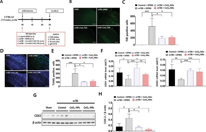
www.nature.com/scientificreports/
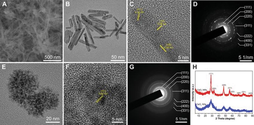
Figure 1. (A). SEM image of Ceria (CeO2) NRs. (B, E) TEM images, (C, F) High-resolution TEM images, and
(D, G) SAED patterns of Ceria NRs and Ceria NSs, respectively; and (H) PXRD patterns of Ceria NRs (in red)
and Ceria NSs (in blue).
is attributed to their high catalytic activities owing to rapid transformation of the oxidative states of Ce3+ and
Ce4+ (so called oxygen storage capacity)13. In addition, ceria nanoparticles exhibit oxygen vacancies in the lat-
tice structure due to the loss of electrons or oxygen atoms, leading to a switch between C eO2 and C eO2-x during
redox processes14. Controlling the morphology is critical to the catalytic performance of ceria nanoparticles, since
the selective exposure of reactive crystal planes on the surface can enrich the catalytic s ites15,16. Recent studies
reported that ceria nanoparticles with a spherical shape decreased neuronal cell death and calcium dysregula-
tion by preserving the antioxidant system in the mTBI model7. However, no comparison with other nanodrugs
was made, and the studies merely demonstrated better therapeutic efficacy of ceria nanoparticle in injured mice
compared with untreated animals7. Therefore, we compared the possible differences in therapeutic effects of ceria
nanoparticles (nanospheres vs. nanorods) in a mouse model of mTBI.
Results
Characterization of ceria nanoparticles. Scanning electron microscope (SEM) and transmission elec-
tron microscope (TEM) images (Fig. 1A,B) of the obtained products revealed that Ceria nanorods (Ceria NRs)
had a rod-like morphology measuring 130.1 ± 42.1 (nm) in length and 9.4 ± 2.1 (nm) in diameter (Fig. S1 A
and B), while Ceria nanospheres (Ceria NSs) (Fig. 1E) were uniform and spherical with a mean particle size of
approximately 3.5 ± 0.5 nm (Fig. S1C). High-resolution TEM images (Fig. 1C,F) revealed the exposed crystal
planes on the surface of Ceria NRs and Ceria NSs, demonstrating the highly crystalline nature of the resulting
ceria nanoparticles. Ceria NRs contained exposed (100), (110), and (111) planes, whereas only (111) plane was
exposed on the surface of Ceria NSs. The selected area electron diffraction (SAED) (Fig. 1D,G) and powder
X-ray diffraction (PXRD) patterns (Fig. 1H) of the two ceria nanoparticles were similar. The predominant peaks
were indexed to (111), (200), (220), (311), (222), (400), and (311) planes indicated that both ceria nanoparticles
were assigned to the pure fluorite cubic structures of CeO2 (JCPDS 34-0394, space group Fm-3m). No diffrac-
tion peaks belonging to other foreign components were detected, indicating the purity of the obtained products.
The composition of the ceria nanoparticles was further established using EDX elemental mapping data (Fig. S2).
The specific surface area (SBET) of Ceria NRs and Ceria NSs were 76 and 230 m2 g−1, respectively (Fig. S3). The
characterization of ceria nanoparticles were summarized in Table 1. In contrast to the white color of pure C eO2,
the resulted ceria nanoparticles exhibited a yellowish color, which demonstrated the coexistence of not only Ce4+
e3+ in the structure. X-ray photoelectron spectroscopy (XPS) analysis revealed a mixed valence state
but also C
4+
of Ce and C e3+ (Fig. 2). Figure 2A and D showed the XPS survey spectra of Ceria NRs and Ceria NSs. Ceria
NRs exhibited characteristic peaks of Ce4+ with binding energy of 882.9, 889.4, 897.6, 900.9, 907.5, and 916.3 eV,
while the peaks at the binding energy of 880.7, 886.4, 898.6, and 904.2 eV corresponded to Ce3+ (Fig. 2B). In the
case of Ceria NSs (Fig. 2E), the peaks were observed at the binding energy of 882.3, 888.7, 898.2, 900.9, 907.1,
916.1 eV in C e3+. The molar ratio between fitted peak areas of C
e4+ and 885.4, 903.7 eV in C e4+ and Ce3+ was used
to estimate their concentrations on the surface of ceria nanoparticles. As shown in Table S1, the Ce3+/Ce4+ molar
ratio of Ceria NRs and Ceria NSs are 0.40 and 0.27, respectively. This data indicate that Ceria NRs contained
more Ce3+ on the surface than Ceria NSs. It is well-known that the biomedical activity of ceria nanoparticles is
enhanced by higher levels of Ce3+ on the surface. As shown in Fig. 2C and F, in the XPS high-resolution binding
Scientific Reports | (2021) 11:15571 | https://doi.org/10.1038/s41598-021-95057-9 2
Vol:.(1234567890)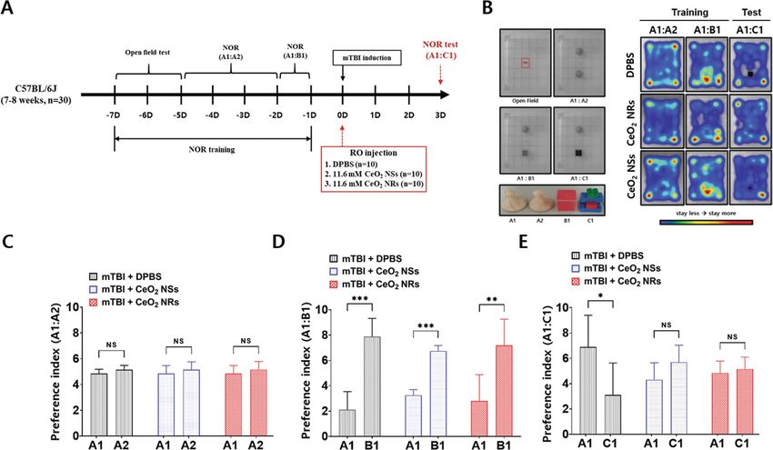
www.nature.com/scientificreports/
Particle characteristic Ceria NRs Ceria NSs
Morphology Nanorod Nanosphere
Length: 130.1 ± 42.1
Size (nm) Diameter: 3.5 ± 0.5
Diameter: 9.4 ± 2.1
3+ 4+
Surface Ce /Ce ratio 0.40 0.27
SBET (m2 g−1) 76 230
Table 1. Characterization of ceria nanoparticles. SBET Brunauer–Emmett–Teller (BET) surface area of ceria
nanoparticles.
Figure 2. XPS survey spectra (A, D), XPS high-resolution binding energy spectra of Ce 3d (B, E) and O 1s (C,
F) of Ceria NRs and Ceria NSs, respectively. Ce4+ and Ce3+ were coexistence in Ceria NRs and Ceria NSs.
energy spectra of O 1 s of Ceria NRs and Ceria NSs the first peak at around 529 eV was associated with lattice
oxygen. A higher binding energy shoulder at around 532 eV was assigned to oxygen vacancies or a mixture of
surface adsorbed oxygen, hydroxyl, and carbonate groups.
Cytoprotective effect in in vivo mild TBI. Fluoro-Jade B (FJB) staining and terminal deoxynucleotidyl
transferase dUTP nick end labeling (TUNEL) assay were performed to further evaluate the role of nanoparticle
shape in the anti-inflammatory activity and subsequent protection of neuronal cell death in mTBI in vivo. As
shown in the schematic diagram (Fig. 3A), mice were sacrificed 3 days after mTBI with or without injection
with Ceria NRs or Ceria NSs. No FJB-positive cells were detected in the control group. FJB-positive neuronal
cell injury was observed in the cortex area after mTBI (Fig. 3B). Compared with DPBS-treated mTBI group
(519.552 ± 76.185), the ceria nanoparticle-injected (11.6 mM/mice) group showed a significant reduction in
the number of FJB-positive cells (188.981 ± 16.329 in the Ceria NSs and 209.205 ± 12.785 in the Ceria NRs)
(Fig. 3B,C). Consistent with FJB staining, both Ceria NRs and Ceria NSs significantly decreased the number of
TUNEL-positive cells compared to the mTBI group (Fig. 3D,E). Treatment with ceria nanoparticles restored the
decreased mRNA levels of SOD1 and SOD2 following mTBI (Fig. 3F). Furthermore, COX-2 expression was also
remarkably reduced in the groups treated with ceria nanoparticles compared with the control group (Fig. 3G,H,
and Supplemental Fig. S4). These results suggest the cytoprotective effects of ceria nanoparticles via suppression
of inflammation and oxidative stress in injured neuronal cells, but the anti-oxidative activities did not differ sig-
nificantly according to the nanoshape. Ceria NRs (0.059 ± 0.021) appeared to exhibit better anti-inflammatory
effect than Ceria NSs (0.133 ± 0.024), although the effects were statistically insignificant (p = 0.190).
Cognitive function in in vivo mild TBI. A schematic representation of in vivo study of novel object rec-
ognition (NOR) and its explanation are presented in Fig. 4 and Supplemental data, respectively. The average
preference index for each group in Fig. 4B (right) is summarized in Figs. 4C–E. Each groups showed intact
NOR memory before mTBI (Fig. 4C,D). Following mTBI, mice showed memory impairment (Fig. 4B, right
upper) and the corresponding preference index was approximately equal to 3.0 (Fig. 4E, gray bar). Both ceria
nanoparticles improved NOR memory in mTBI-induced mice (Fig. 4E; preference index for the novel object of
Scientific Reports | (2021) 11:15571 | https://doi.org/10.1038/s41598-021-95057-9 3
Vol.:(0123456789)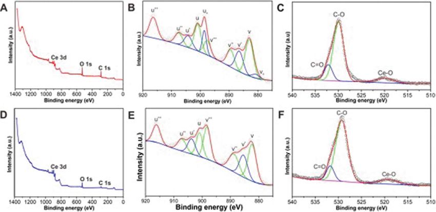
www.nature.com/scientificreports/
Figure 3. Schematic diagram of in vivo study (A). Fluoro-Jade B (FJB) staining was used to detect the neuronal
death (B, C). Fluorescence images represent the relative degree of neuronal death in the cortex. The number
of FJB-positive cells were decreased in Ceria NRs and Ceria NSs than in DBPS group. TUNEL assay in mTBI
in vivo (D, E). Representative images of TUNEL-positive green cells and its quantification. DAPI was used
to counterstaining (blue). The level of SOD1 and SOD2 mRNA expression was analyzed using qRT-PCR (F).
Western blot analysis of COX-2 expression (G) and quantification of blots is based on relative optical densities
of COX-2 and β-actin protein (H). Scale bar = 200 μm. Error bars, mean SEM, *P < 0.05, ** P < 0.01, and
***P < 0.001.
C1, shown in blue and red bars). Thus, ceria nanoparticles improved cognitive function, but the effect was not
different significantly according to the shapes of NRs and NSs.
Cerebral edema in in vivo mild TBI. The cerebral edema was compared additionally via estimation of
reviously17. A schematic diagram of the meas-
changes in overall water content of the brain tissues as described p
urement of cerebral edema is provided in Fig. 5. Ceria nanoparticles significantly decreased the cerebral edema
than in the control group (DPBS-treated mTBI group). Compared with Ceria NSs (73.7 ± 0.4%), Ceria NRs
(70.1 ± 0.5%) significantly decreased brain water content, demonstrating a more effective decline in brain edema
(P = 0.0015).
Discussion
Neuroprotective effects of ceria nanoparticles have been mainly investigated in neurodegenerative and cerebro-
vascular diseases associated with defective antioxidant mechanisms and subsequent susceptibility to oxidative
stress18. Dowding et al.19 reported that ceria nanoparticles reduced the levels of Aβ-induced mitochondrial
fragmentation, endogenous peroxynitrite and neuronal cell death. Treatment with 10 nM ceria nanoparticles
provided confluent astrocyte monolayers with multiple processed n eurons20. In addition, cultures treated with
ceria nanoparticles exhibited higher catalase levels and decreased glutathione compared with untreated controls
during the life s pan20. Treatment with ceria nanoparticles at doses ranging from 0.5 to 0.7 mg k g−1, also reduced
the infarct volume following acute ischemic s troke21. However, only a few studies have focused on the efficacy
of ceria nanoparticles in TBI, although excessive oxidative stress is a major damage mechanism in the brain
after TBI22. In rat models with moderate TBI, pretreatment with ceria nanoparticles showed improved task
performance and recovery than in control g roups22. Bailey et al.7 investigated treatment outcomes of ceria nano-
particles in rats with mild lateral fluid percussion brain injury and showed that ceria nanoparticles significantly
improved catalase activity in the injured brain. During the cellular uptake of ceria nanoparticles, the shape can
affect endocytosis and determine the efficacy of these agents. The shape effect of nanoparticles is mainly deter-
mined by different membrane-bending energies during e ndocytosis23. Spherical ceria nanoparticles required a
Scientific Reports | (2021) 11:15571 | https://doi.org/10.1038/s41598-021-95057-9 4
Vol:.(1234567890)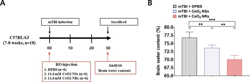
www.nature.com/scientificreports/
Figure 4. Experimental design of the Novel Object Recognition (NOR) test (A). Altered object exploration
ration in the NOR test. Heat map analysis of animal tracking following NOR test (B). Each group shows
similar preferential investigation of the novel objects before mTBI (C, D). Differences in the degree of impaired
discrimination index following mTBI between DPBS and Ceria NSs or Ceria NRs (E). Mice treated with cerium
oxide nanoparticles showed better preference for novel objects than DPBS, indicating cognitive improvement
after injury.
Figure 5. Schematic diagram depicts brain water content analysis of the degree of cerebral edema (A).
Comparison of brain water content in the three groups: DPBS, Ceria NSs, and Ceria NRs. Error bars, mean
SEM, **P < 0.01, and ***P < 0.001.
minimal membrane-bending energy barrier than the non-spherical counterparts. Li et al.23 concluded that the
shape of the nanomaterial only affects the free energy change of grafted polyethylene glycol (PEG) polymers
during internalization.
In brief, we first tested the ability of ceria nanoparticles based on their shape in an in vivo mTBI model.
Theoretically, Ceria NRs exhibit better antioxidant activity and cytotoxicity than Ceria NSs for the following
reasons. First, the difference in reactive crystal planes is attributed to the difference in therapeutic effects. As
shown by high-resolution TEM analysis (Fig. 1), the less stable and high-energy (100) and (110) planes were
predominantly exposed in Ceria NRs. However, the (111) plane on the surface was mainly exposed in Ceria NSs,
which is consistent with previous r eports24. It is believed that the improved biomedical activities of Ceria NRs
originate in the abundance of (100) and (110) terminated planes compared with Ceria NSs, since the crystalline
planes with high surface energy provide more active sites. The effects of structural features in ceria nanoparticles
Scientific Reports | (2021) 11:15571 | https://doi.org/10.1038/s41598-021-95057-9 5
Vol.:(0123456789)www.nature.com/scientificreports/
on the catalysis such as CO oxidation have been investigated comprehensively16,25. For example, Ceria NRs were
found to be more active in CO oxidation than nanocubes, nanowires, and nanoparticles16,26. Similarly, Co3O4
NR with a higher exposure of the reactive crystal planes also exhibited particularly high activity towards CO
oxidation at low temperature27. Second, the Ce3+/Ce4+ molar ratio on the surface of ceria nanoparticles also
affects the biomedical a ctivities28,29. By changing the oxidation state from Ce3+ to Ce4+, ceria nanoparticles can
scavenge the free radicals or reactive oxygen s pecies11. Meanwhile, changing the oxidation state from C e4+ to Ce3+
in ceria nanoparticles completes the autoregenerative reaction c ycle . As shown in Fig. 5, the Ce /Ce4+ molar
30 3+
ratio of Ceria NRs (0.40) is greater than that of Ceria NSs (0.27) (calculated from the XPS data), which indicates
the concentration of C e3+ on the surface of Ceria NRs is higher than Ceria NSs. Third, the oxygen vacancies in
the lattice structure of ceria nanoparticles, which are the most active on the surface, also play an essential role in
determining their biomedical properties. Alteration in the oxidative state of ceria nanoparticles generates oxygen
vacancies to maintain electrostatic balance via loss of oxygen and electrons24. The increased number of Ce3+ sites
in the ceria nanoparticles increases the number of oxygen vacancies on the surface. Nevertheless, in our study,
anti-oxidant and anti-inflammation activities in mTBI in vivo did not differ significantly according to nanoshape,
although the mRNA expressions of SOD1 and SOD2, and COX-2 tended to decrease in Ceria NRs than Ceria
NSs. However, cerebral edema was significantly reduced in Ceria NRs compared with Ceria NSs following
mTBI. In addition, there was no statistically significant difference in anti-inflammatory effect and improvement
in cognitive dysfunction according to the shape of nanoparticles. We speculated that the relatively high dose
of nanoparticles and the mild TBI severity may contribute to these outcomes. In this study, we used a dose of
11.6 mM based on a literature review, which was higher than the dose used in other s tudies21,31. Additionally, we
only targeted mild TBI, not moderate-to-severe TBI. Thus, there was a possibility that the relationship between
improvement in anti-inflammatory effect and cognitive dysfunction and nano-shape was unclear. Therefore,
studies investigating the improvement of cognitive function according to nano-shape are needed based on vari-
ous dosages of nanoparticles in different degrees of TBI injury.
Shape of nanoparticles can influence biological effects as well as their size and surface c harge32. Zhao et al.32
reported that long rods showed longer circulation in the blood with less rapid clearance by reticuloendothelial
system. Rods were also degraded faster than spherical nanoparticles due to their higher specific surface area. In
addition, there may be less aggregation among rod nanoparticles than the spherical particles and thus expected
to be less toxic due to the small dosage of rods with high oxygen vacancies on the surface than the spherical
nanoparticles. It is believed that ceria substances are chemically stable and not significantly affected by chemical
changes during long-term treatment. These findings suggested that Ceria NRs may have a higher bioavailability
than Ceria NSs. The use of Ceria NRs in clinical practice requires a further study investigating the therapeutic
efficacy based on rod length and effective nano drug delivery methods without loss of activity during indigestion.
Although results of TBI according to gender difference vary and are model-dependent33, many studies reported
that females may manifest neuroprotective effects following T BI34. Shahrokhi et al.35 reported that estrogen
and progesterone exhibited decreased intracranial pressure and improved cerebral perfusion pressure after TBI
in ovariectomized rats, suggesting that sex hormones may be neuroprotective following TBI. This study was a
proof-of-concept study that focused on identifying the influence of ceria shape on outcome in mTBI. Thus, male
mice were only used to eliminate the confounding variable of TBI outcome based on potential gender differences.
Nevertheless, application in real-world clinical and drug development requires research into the therapeutic
effects based on toxicity and appropriate dosage according to gender.
The study limitations are as follows. First, cerebral edema was estimated using the water content based on the
difference between wet and dry w eights17. In such cases, small changes in the percentage of water content reflect
the relatively large changes in water component of the tissue17,36. Second, long-term outcomes of the ceria nano-
particles according to shape were not investigated. Ceria NRs showed similar anti-oxidant and anti-inflammation
activities than Ceria NSs, but the effect was usually limited to the acute phase after mTBI. Third, cellular toxicity
and clearance according to changes in nanoshape were not tested before clinical application in patient with mTBI.
Conclusions
Ceria nanoparticles decreased neuronal damages and improved cognitive impairment in vivo mTBI. Compared
to Ceria NSs, Ceria NRs demonstrated better effects on reduction of cerebral edema.
Materials and methods
Synthesis of ceria nanoparticles. Two types of ceria nanoparticles were synthesized. Ceria NRs were
prepared by modifying the previously reported p rotocol37. Cerium(III) nitrate hexahydrate (Ce(NO3)3·6H2O,
99%, Sigma-Aldrich) and sodium hydroxide (NaOH, 98%, Sigma-Aldrich) were used. Typically, Ce(NO3)3·6H2O
(0.434 g, 1 mmol) was added to an aqueous solution of NaOH (10 M, 20 mL) in a PTFE beaker. The reaction
mixture was vigorously stirred for 2 h at room temperature before transferring to a teflon-lined stainless-steel
autoclave and was incubated in a temperature-controlled oven at 100 °C for 10 h. The mixture was naturally
cooled to room temperature after completion of the reaction. The reaction product was collected by centrifuga-
tion, washed several times with solvents (deionized water and ethanol), and then vacuum-dried for 24 h for
further use. Ceria NSs were synthesized using a reported protocol with slight m odification38. Cerium(III) nitrate
hexahydrate (Ce(NO3)3·6H2O, 99%, Sigma-Aldrich), oleylamine (C18H37N, 70%, Sigma-Aldrich), and xylene
(C8H10, 98.5%, Junsei) were used. Typically, Ce(NO3)3·6H2O (0.434 g, 1 mmol) and C 18H37N (3.25 g, 12 mmol)
were dissolved in C 8H10 (15 mL). The resulting solution was vigorously stirred for 2 h at 25 °C and then heated
to 90 °C at the rate of 2 °C.min-1 under vacuum. Deionized water (1 mL) was rapidly injected into the solution
under vigorous stirring at 90 °C to initiate the sol–gel reaction, as indicated by a color change from purple to
cloudy yellow. The reaction mixture was incubated at 90 °C for 3 h to obtain a light-yellow colloidal solution.
Scientific Reports | (2021) 11:15571 | https://doi.org/10.1038/s41598-021-95057-9 6
Vol:.(1234567890)www.nature.com/scientificreports/
The mixture was cooled to ambient temperature, and Ceria NSs were precipitated by adding ethanol (75 mL).
The product was centrifuged and washed several times with ethanol, and then dried under vacuum for 24 h for
further use.
In vivo mild TBI. All animal experiments were approved by Institutional Animal Care and Use Committee
(IACUC) of Hallym University (approval no. HallymR2 2019-35). All animal study protocol was carried out
according to the ARRIVE guidelines (Animal Research: Reporting of In Vivo Experiments). VC57BL/6J male
mice, 7–8 weeks of age, were obtained from the Laboratory Animal Resources Center, Hallym University, Korea.
The animal was provided with regular food and water ad libitium under a 12 h dark/light cycle at 24 °C, 55 ± 10%
humidity. The different experimental groups were as follows: (1) mTBI (n = 16), (2) mTBI treated with Ceria NRs
(n = 16), (3) mTBI treated with Ceria NSs (n = 16), and sham operation (n = 6).
In vivo TBI was induced via open head injury using a stereotaxic impactor (RWD Life Science, RWD-
68099, China)39. In brief, the C57BL/6J male mice were anesthetized using 2.5% isofurane in oxygen and placed
in the stereotaxic frame. The skull was exposed via a midline skin incision, and TBI was induced as follows:
M/L = – 2.5 mm, A/P = − 2.0 mm, from bregma, at 1.5 mm depth using a blant tip with a diameter of 2 mm.
The velocity of the impactor reached 3.0 m/s with a depth of 1.5 mm using the 2 mm blant tip below the dura
matter. The dwell time in the brain was 0.5 m/s. Ceria nanoparticles accumulate in the brain via a bell-shaped
dose–response curve7,40, and therefore, a single retro-orbital injection with a dose of 11.6 mM of ceria nanopar-
ticles was administered in this study41. The various assay such as FJB, TUNEL, western blot, measurement of
cerebral edema17,36, and behavioral tests including NOR are described in the Supplemental Methods. Outcome
measurements were carried out by investigators who were blinded to the treatment methods of mice. The study
protocol is performed in accordance with the relevant guidelines.
Statistical analysis. All data were presented as the means ± standard errors of the mean (SEM). Stu-
dent’s t-test or one-way ANOVA with post-hoc Bonferroni correction was conducted for all possible pair-
wise comparisons21. P value less than of < 0.05, 0.01, and 0.001 are represented by *, **, and *** in the figures,
respectively42. All statistical analyses were performed with GraphPad Prism software (v.6.01; GraphPad Software
Inc., San Diego, CA, USA).
Data availability
The data support the findings of this study are available from the corresponding author upon reasonable request.
Received: 30 March 2021; Accepted: 20 July 2021
References
1. Dewan, M. C. et al. Estimating the global incidence of traumatic brain injury. J. Neurosurg. 1, 1–18 (2018).
2. Cruz-Haces, M. et al. Pathological correlations between traumatic brain injury and chronic neurodegenerative diseases. Transl.
Neurodegener. 6, 20 (2017).
3. Ramos-Cejudo, J. et al. Traumatic brain injury and alzheimer’s disease: The cerebrovascular link. EBioMedicine 28, 21–30 (2018).
4. Carlson, K. et al. The Assessment and Treatment of Individuals with History of Traumatic Brain Injury and Post-traumatic Stress
Disorder: A Systematic Review of the Evidence. (2009).
5. Flynn, F. G. Memory impairment after mild traumatic brain injury. Continuum 16, 79–109 (2010).
6. Ruff, R. Two decades of advances in understanding of mild traumatic brain injury. J. Head Trauma Rehabil. 20, 5–18 (2005).
7. Bailey, Z. S. et al. Cerium oxide nanoparticles improve outcome after in vitro and in vivo mild traumatic brain injury. J. Neuro-
trauma. 37, 1452–1462 (2020).
8. Pratico, D. et al. Increase of brain oxidative stress in mild cognitive impairment: A possible predictor of alzheimer disease. Arch.
Neurol. 59, 972–976 (2002).
9. Tyurin, V. A. et al. Oxidative stress following traumatic brain injury in rats: Quantitation of biomarkers and detection of free radical
intermediates. J. Neurochem. 75, 2178–2189 (2000).
10. Sun, C., Li, H. & Chen, L. Nanostructured ceria-based materials: Synthesis, properties, and applications. Energy Environ. Sci. 5,
8475–8505 (2012).
11. Kang, T., Kim, Y. G., Kim, D. & Hyeon, T. Inorganic nanoparticles with enzyme-mimetic activities for biomedical applications.
Coord. Chem. Rev. 403, 213092 (2020).
12. Rajeshkumar, S. & Naik, P. Synthesis and biomedical applications of cerium oxide nanoparticles: A review. Biotechnol. Rep. 17,
1–5 (2018).
13. Montini, T., Melchionna, M., Monai, M. & Fornasiero, P. Fundamentals and catalytic applications of ceo2-based materials. Chem
Rev. 116, 5987–6041 (2016).
14. Gao, W. et al. Surface engineering on ceo(2) nanorods by chemical redox etching and their enhanced catalytic activity for co
oxidation. Nanoscale 7, 11686–11691 (2015).
15. Pan, C., Zhang, D., Shi, L. & Fang, J. Template-free synthesis, controlled conversion, and co oxidation properties of c eo2 nanorods,
nanotubes, nanowires, and nanocubes. Eur. J. Inorg. Chem. 1, 2429–2436 (2008).
16. Tana, A. et al. Morphology-dependent redox and catalytic properties of ceo2 nanostructures: Nanowires, nanorods and nanopar-
ticles. Catal. Today. 148, 179–183 (2009).
17. Keep, R. F., Hua, Y. & Xi, G. Brain water content: A misunderstood measurement?. Transl. Stroke Res. 3, 263–265 (2012).
18. Riddle, D. R. & Lichtenwalner, R. J. Neurogenesis in the adult and aging brain. In Brain Aging: Models, Methods, and Mechanisms
(ed. Riddle, D. R.) (Springer, 2007).
19. Dowding, J. M. et al. Cerium oxide nanoparticles protect against abeta-induced mitochondrial fragmentation and neuronal cell
death. Cell Death Differ. 21, 1622–1632 (2014).
20. Singh, N., Cohen, C. A. & Rzigalinski, B. A. Treatment of neurodegenerative disorders with radical nanomedicine. Ann. N. Y. Acad.
Sci. 1122, 219–230 (2007).
21. Kim, C. K. et al. Ceria nanoparticles that can protect against ischemic stroke. Angew Chem. Int. Ed. Engl. 51, 1039–11043 (2012).
22. Rzigalinski, B. A., Carfagna, C. S. & Ehrich, M. Cerium oxide nanoparticles in neuroprotection and considerations for efficacy
and safety. Wiley Interdiscip. Rev. Nanomed. Nanobiotechnol. 9, 1–10 (2017).
Scientific Reports | (2021) 11:15571 | https://doi.org/10.1038/s41598-021-95057-9 7
Vol.:(0123456789)www.nature.com/scientificreports/
23. Li, Y., Kroger, M. & Liu, W. K. Shape effect in cellular uptake of pegylated nanoparticles: Comparison between sphere, rod, cube
and disk. Nanoscale 7, 16631–16646 (2015).
24. Liu, X., Zhou, K., Wang, L., Wang, B. & Li, Y. Oxygen vacancy clusters promoting reducibility and activity of ceria nanorods. J.
Am. Chem. Soc. 131, 3140–3141 (2009).
25. Zhou, K. & Li, Y. Catalysis based on nanocrystals with well-defined facets. Angew. Chem. Int. Ed. Engl. 51, 602–613 (2012).
26. Zhou, K. et al. Enhanced catalytic activity of ceria nanorods from well-defined reactive crystal planes. J. Catal. 229, 206–212 (2005).
27. Xie, X. et al. Low-temperature oxidation of co catalysed by co(3)o(4) nanorods. Nature 458, 746–749 (2009).
28. Das, S. et al. Cerium oxide nanoparticles: Applications and prospects in nanomedicine. Nanomedicine 8, 1483–1508 (2013).
29. Dowding, J. M. et al. Cellular interaction and toxicity depend on physicochemical properties and surface modification of redox-
active nanomaterials. ACS Nano 7, 4855–4868 (2013).
30. Tarnuzzer, R. W., Colon, J., Patil, S. & Seal, S. Vacancy engineered ceria nanostructures for protection from radiation-induced
cellular damage. Nano Lett. 5, 2573–2577 (2005).
31. Heckman, K. L. et al. Custom cerium oxide nanoparticles protect against a free radical mediated autoimmune degenerative disease
in the brain. ACS Nano 7, 10582–10596 (2013).
32. Zhao, Y. et al. A comparison between sphere and rod nanoparticles regarding their in vivo biological behavior and pharmacokinet-
ics. Sci. Rep. 7, 4131 (2017).
33. Ma, C. et al. Sex differences in traumatic brain injury: A multi-dimensional exploration in genes, hormones, cells, individuals, and
society. Chin. Neurosurg. J. 5, 24 (2019).
34. Rubin, T. G. & Lipton, M. L. Sex differences in animal models of traumatic brain injury. J. Exp. Neurosci. 13, 1179069 (2019).
35. Shahrokhi, N. et al. Effect of sex steroid hormones on brain edema, intracranial pressure, and neurologic outcomes after traumatic
brain injury. Can. J. Physiol. Pharmacol. 88, 414–421 (2010).
36. Shohami, E., Novikov, M. & Mechoulam, R. A nonpsychotropic cannabinoid, hu-211, has cerebroprotective effects after closed
head injury in the rat. J. Neurotrauma. 10, 109–119 (1993).
37. Torrente-Murciano, L. et al. Shape-dependency activity of nanostructured c eo2 in the total oxidation of polycyclic aromatic
hydrocarbons. Appl. Catal. B. 132, 116–122 (2013).
38. Kim, C. K. et al. Ceria nanoparticles that can protect against ischemic stroke. Angew. Chem. Int. Ed. 51, 11039–11043 (2012).
39. Zhang, J. et al. Inhibition of monoacylglycerol lipase prevents chronic traumatic encephalopathy-like neuropathology in a mouse
model of repetitive mild closed head injury. J. Cereb Blood. Flow. Metab. 35, 443–453 (2015).
40. Rzigalinski, B. A. et al. Radical nanomedicine. Nanomedicine 1, 399–412 (2006).
41. Fiorani, L. et al. Cerium oxide nanoparticles reduce microglial activation and neurodegenerative events in light damaged retina.
PLoS ONE 10, e0140387 (2015).
42. Bae, Y. H. et al. Brain injury induces hif-1alpha-dependent transcriptional activation of lrrk2 that exacerbates brain damage. Cell
Death Dis. 9, 1125 (2018).
Acknowledgements
This research was supported by the National Research Foundation of Korea funded by the Ministry of
Education (2020R1l1A3070726), Hallym Research Fund, and the Ministry of Science and ICT, MSICT
(NRF-2020R1A2C1004006).
Author contributions
J.P.J. and H.Y. designed and obtained funding for the study. B.J.K., Y.K., D.H.Y., and N.M.T. were responsible for
the molecular experiments. B.J.K. and N.M.T. interpreted the data and performed statistical analyses. D.H.Y.
draw the Fig. 4B. J.P.J. and H.Y. provided input for the final version of the manuscript. All authors reviewed the
manuscript and provided editorial feedback.
Competing interests
The authors declare no competing interests.
Additional information
Supplementary Information The online version contains supplementary material available at https://doi.org/
10.1038/s41598-021-95057-9.
Correspondence and requests for materials should be addressed to J.P.J. or H.Y.
Reprints and permissions information is available at www.nature.com/reprints.
Publisher’s note Springer Nature remains neutral with regard to jurisdictional claims in published maps and
institutional affiliations.
Open Access This article is licensed under a Creative Commons Attribution 4.0 International
License, which permits use, sharing, adaptation, distribution and reproduction in any medium or
format, as long as you give appropriate credit to the original author(s) and the source, provide a link to the
Creative Commons licence, and indicate if changes were made. The images or other third party material in this
article are included in the article’s Creative Commons licence, unless indicated otherwise in a credit line to the
material. If material is not included in the article’s Creative Commons licence and your intended use is not
permitted by statutory regulation or exceeds the permitted use, you will need to obtain permission directly from
the copyright holder. To view a copy of this licence, visit http://creativecommons.org/licenses/by/4.0/.
© The Author(s) 2021
Scientific Reports | (2021) 11:15571 | https://doi.org/10.1038/s41598-021-95057-9 8
Vol:.(1234567890)You can also read