A novel tissue specific alternative splicing variant mitigates phenotypes in Ets2 frame shift mutant models - Nature
←
→
Page content transcription
If your browser does not render page correctly, please read the page content below
www.nature.com/scientificreports
OPEN A novel tissue specific alternative
splicing variant mitigates
phenotypes in Ets2 frame‑shift
mutant models
Yuki Kishimoto1,3, Iori Nishiura1,3, Wataru Hirata1,3, Shunsuke Yuri1, Nami Yamamoto1,
Masahito Ikawa2 & Ayako Isotani1*
E26 avian leukemia oncogene 2, 3′ domain (Ets2) has been implicated in various biological processes.
An Ets2 mutant model (Ets2db1/db1), which lacks the DNA-binding domain, was previously reported to
exhibit embryonic lethality caused by a trophoblast abnormality. This phenotype could be rescued by
tetraploid complementation, resulting in pups with wavy hair and curly whiskers. Here, we generated
new Ets2 mutant models with a frame-shift mutation in exon 8 using the CRISPR/Cas9 method.
Homozygous mutants could not be obtained by natural mating as embryonic development stopped
before E8.5, as previously reported. When we rescued them by tetraploid complementation, these
mice did not exhibit wavy hair or curly whisker phenotypes. Our newly generated mice exhibited exon
8 skipping, which led to in-frame mutant mRNA expression in the skin and thymus but not in E7.5
Ets2em1/em1 embryos. This exon 8-skipped Ets2 mRNA was translated into protein, suggesting that this
Ets2 mutant protein complemented the Ets2 function in the skin. Our data implies that novel splicing
variants incidentally generated after genome editing may complicate the phenotypic analysis but may
also give insight into the new mechanisms related to biological gene functions.
E26 avian leukemia oncogene 2, 3′ domain (Ets2), a member of the ETS family, is a transcription factor that
contains an ETS winged helix-loop-helix DNA-binding domain (ETS domain) that binds to GGA(A/T) DNA
sequences. It is conserved in various species, including mice and h umans1–3. Ets2 has been implicated in vari-
ous biological contexts, including placentation, hair formation, mammary tumors, inflammatory responses,
angiogenesis, and the pulmonary fi brosis4–8.
In a previous study, Ets2-deficient mice (Ets2db1/db1 mice), which lack the ETS domain through deletion of
exons 9 and 10, were found to exhibit early embryonic lethality due to a trophectoderm abnormality. The tetra-
ploid complementation technique could rescue this placental abnormality, allowing for survival of the offspring4,
indicating that Ets2 is essential for placental development. Ets2db1/db1 mice created using the tetraploid comple-
mentation technique exhibit a variety of phenotypes, such as wavy hair, curly whisker, and a rounded forehead,
allowing them to be identified. However, their fertility is normal, and they exhibit no lethal phenotype after birth.
Therefore, the Ets2db1/db1 mouse is a useful model for studying treatment methods for placental abnormalities9.
The generation of gene-deficient animal models is now commonly performed using CRISPR/Cas9-based
genome engineering10,11. Model organisms made using this technique can completely mimic the genome muta-
tions found in human diseases, such as indel mutations and substitutions, which were previously difficult to
generate using the conventional knockout method. Further, homozygous mutant mice can be obtained efficiently
in the founder generation by directly delivering the crRNA/tracrRNA/Cas9 ribonucleoprotein complex into a
mouse zygote via electroporation12. Unfortunately, if the homozygous mutant exhibits embryonic lethality, it
cannot be obtained in this way. However, it is possible to obtain placental-deficient mutant mice, such as Ets2,
in the founder generation using the tetraploid complementation method13 in combination with genome-edited
zygotes or their embryonic stem cells.
Using the above strategy, we established three new Ets2 mutant mouse lines. Two of those contained a frame-
shift deletion in exon 8, which was located before the ETS domain encoded by exons 9 and 10. These genomic
1
Division of Biological Science, Graduate School of Science and Technology, Nara Institute of Science and
Technology, 8916‑5 Takayama‑cho, Ikoma, Nara 630‑0192, Japan. 2Research Institute for Microbial Diseases,
Osaka University, 3‑1 Yamadaoka, Suita, Osaka 565‑0871, Japan. 3These authors contributed equally: Yuki
Kishimoto, Iori Nishiura and Wataru Hirata. *email: isotani@bs.naist.jp
Scientific Reports | (2021) 11:8297 | https://doi.org/10.1038/s41598-021-87751-5 1
Vol.:(0123456789)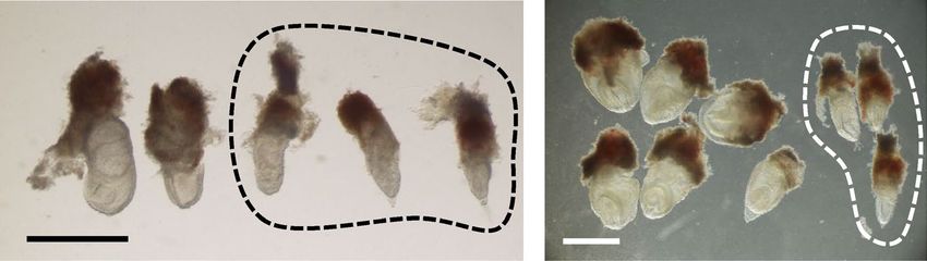
www.nature.com/scientificreports/
A Ets2 locus
Ets2-F Ets2-R 1 kbp
Exon 8
crRNA1 crRNA2 crRNA3
Genome editing
B Cas9 protein
tracrRNA gene mutated embryo
crRNA
Aggregation
chimeric embryo
Zygote
4n embryo
Electro fusion
C
564 bp
D
crRNA1 crRNA3
PAM PAM
WT ACCCAAGGACCACGACTCTCCAGAGʀʀʀGAGGAGCGACCCAGTCGAGCAAGGC
em1 ACCCAAG AGCAAGGC
Δ205 bp
crRNA1 crRNA2
PAM PAM
WT ACCCAAGGACCACGACTCTCCAGAGʀʀʀGGAACAGCCAGTCGTCCCTACTGGA
em2 ACCCAAG TACTGGA
Δ82 bp
Figure 1. Generation of new Ets2 mutant models using zygotes. (A) Design of crRNA targeting sites in exon
8 of the Ets2 gene and checking primer positions. (B) Strategy for obtaining Ets2 homozygous mutant l in F0
generation using the electroporation technique and the tetraploid method. (C) Genotyping of the two newly
generated Ets2 mutants in F0 generation. Both had a wildtype (WT: 564 bp) allele and a deletion allele (em1 or
em2), which were detected as shorter bands than WT. (D) DNA sequence of mutant alleles and crRNA targeted
sequences. The 205 bp deleted (Δ205 bp) allele was named em1, and the 82 bp deleted (Δ82 bp) allele was
named em2.
mutations were predicted to produce a transcriptional product that would undergo nonsense-mediated mRNA
decay (NMD) or, if translated, a protein lacking the ETS domain. The third one had the null mutant allele which
lacked all open reading frame regions. We found that some of the phenotypes exhibited by the frame-shift mutant
mice differed from the previous study, whose origin was investigated in this work.
Results
Generation of new Ets2 mutant mice by CRISPR/Cas9 system using zygotes. On the basis of
a previous study4, we designed three gRNA targeted to sites in exon 8 that would induce a frame-shift muta-
tion, leading to a deficiency in the ETS domain, encoded by exons 9 and 10 (Fig. 1A). The riboprotein complex,
which consisted of three designed crRNAs, tracrRNAs, and Cas9 protein, was electroporated into one-cell stage
Scientific Reports | (2021) 11:8297 | https://doi.org/10.1038/s41598-021-87751-5 2
Vol:.(1234567890)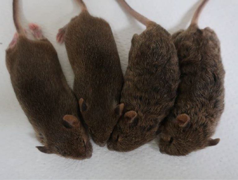
www.nature.com/scientificreports/
Parents genotype Genotype (*)
Female Male No. of offspring Average of litter size +/+ + /Δ Δ/Δ
Ets2+/em1 Ets2+/em2 16 5.3 10 6 0
Ets2+/em1 Ets2+/em1 136 6.5 ± 1.7 45 91 0
Table 1. Lethality of Ets2 mutant. (*) em1 or em2 allele shown as "Δ".
zygotes, which developed until the eight-cell stage. These genome-edited embryos were used for the tetraploid
complementation method in order to obtain homozygous mutant mice in the founder generation (Fig. 1B).
However, no homozygous mutant mice were born from the 29 transferred embryos.
Two out of three delivered pups had a heterozygous deletion mutation, which was determined by PCR
analysis. One mutation was a 205-bp deletion (hereafter referred to as em1), and the other was an 82 bp deletion
(hereafter referred to as em2) (Fig. 1C,D and full-length gel of Fig. 1C is presented in Supplemental Fig. S1).
Expectedly, both had frame-shift mutations.
Assessment of the development of the newly generated Ets2 mutant mice. Previous reports
indicated that Ets2db1/db1 mice exhibit an embryonic lethal phenotype due to a placental deficiency4. Sixteen pups
were obtained from three derivations, and as expected, none of the pups included the double mutant alleles
(em1/em2) (Table 1). Further, we analyzed the developmental ability of Ets2 mutant mice by performing a test
cross using Ets2+/em1 mice and assessed the genotypes of the offspring. No homozygous mutant pups (Ets2em1/em1)
were born (wild:hetero:homo = 45:91:0, Table 1).
A previous study reported that Ets2db1/db1 embryos were degenerated by the placental deficiency around E7.5
and disappeared after E8.5. To investigate whether the Ets2em1/em1 mutant phenocopies the Ets2db1/db1 mutant,
we crossed Ets2+/em1 animals and observed embryos at several stages. Ets2em1/em1 embryos at E7.5 were slightly
delayed in their developmental stage but clearly progressed in a comparable manner to embryos from Ets2db1/db1
animals. The Ets2em1/em1 embryos had survived at E8.5, but all of them were retarded. By E9.5 and E10.5, some
malformed Ets2em1/em1 embryos were present and developed before the turning of the embryo, which usually
occurred at approximately E8.5 (Fig. 2A; Table 2).
As the frame-shift mutation in Ets2em1/em1 is located in exon 8, the stop codon occurs before exon 9, and the
original stop codon is located in exon 10. For this reason, NMD might occur, such that the em1 mutant mRNA
may be degraded in Ets2em1/em1 embryos. To confirm this, we performed RT-PCR using E7.5 embryos. Embryos
were separated into the posterior trophectoderm (TE) and anterior epiblast (Epi). Both regions expressed em1
mutant mRNA (Fig. 2B, and full-length gels are presented in Supplemental Fig S2), and their sequences included
205 nt deletions that were predicted from the genomic sequence. This result showed that em1 mutant RNA had
escaped NMD. Furthermore, it is likely the case that the Ets2em1/em1 exhibited a distinct phenotype compared with
Ets2db1/db1 even if the em1 mRNA was translated into a protein product (Supplemental Fig. S7).
Establishment of Ets2 homozygous mutant ESC lines and phenotypic analysis after birth. By
rescuing placental function using the tetraploid complementation method, Ets2db1/db1 offspring were successfully
developed to term. Therefore, we attempted the same experiment to define whether the embryonic lethal pheno-
type of Ets2em1/em1 was dependent on the placental deficiency or not.
Before conducting tetraploid complementation, we established Ets2em1/em1 and Ets2em2/em2 ESC lines. In this
way, we improved the efficiency of obtaining homozygous mutant mice because the ratio of homozygous mutant
embryos was only one out of four when we used embryos from a heterozygous crossing. After crossing heterozy-
gotes, two-cell embryos were collected and developed until the blastocyst stage. ESC lines were established from
the collected blastocysts and analyzed by genotyping. The rate of homozygous mutant ESC line establishment
for both the Ets2em1/em1 and Ets2em2/em2 mutants followed Mendel’s law (Table 3).
Using the tetraploid complementation method, offspring were obtained from Ets2em1/em1 and Ets2em2/em2 ESC
lines (Fig. 3A–C; Supplemental Fig. S3, and Table 4). This result indicated that the embryonic lethality observed
for the Ets2em1/em1 and Ets2em2/em2 genotypes was due to a dysfunction of placental differentiation, the same as
that seen for the Ets2db1/db1 mutant. Unexpectedly, wavy hair and curly whisker phenotypes were not observed
in Ets2em1/em1 or Ets2em2/em2 mice (Fig. 3B,C; Supplemental Fig. S3).
To corroborate the relationship between Ets2 and the wavy hair phenotype, we established a null mutant
(hereafter referred to as Ets2null(em3/em3)) ES cell line, in which a region upstream of exon 2 through the 3′-UTR of
exon 10 was deleted, including all open reading frame (ORF) regions (Supplemental Fig. S4). Pups were then gen-
erated using the tetraploid complementation method (Fig. 3D–F; Table 4). Both the wavy hair and curly whisker
phenotypes were observed in Ets2null(em3/em3) mice from around 2-weeks of age, as was observed for Ets2db1/db1 mice.
Gene expression in newly established Ets2 mutant skins. In this study, newly established Ets2em1/em1
and Ets2em2/em2 mice exhibited an embryonic lethal phenotype due to placental dysfunction but did not exhibit
the wavy hair phenotype of Ets2db1/db1 and Ets2null(em3/em3) mice, despite having a frame-shift mutation. Therefore,
we next investigated the Ets2 gene expression from the Ets2 locus in their skins.
The expression of the mRNA was detected in the skin of wildtype, Ets2em1/em1 and Ets2em2/em2 mice, but not in
the skin of Ets2null(em3/em3) mice. Notably, two sizes of fragments were detected in Ets2em1/em1 and Ets2em2/em2 skin
samples (Fig. 4A, Supplemental Fig. S5A and full-length gels are presented in Supplemental Fig. S6).
Scientific Reports | (2021) 11:8297 | https://doi.org/10.1038/s41598-021-87751-5 3
Vol.:(0123456789)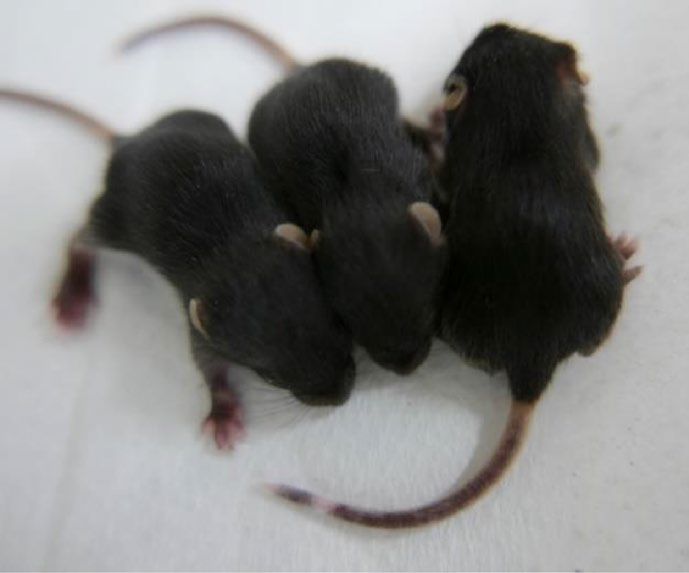
www.nature.com/scientificreports/
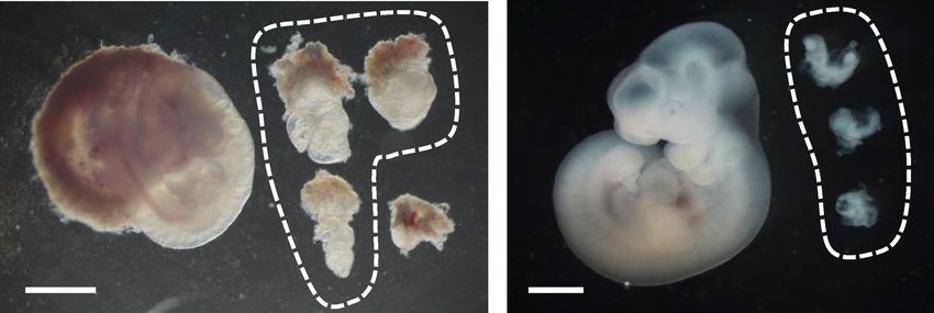
A
E7.5 E8.5
E9.5 E10.5
*
B Epi TE
Ets2
GAPDH
Figure 2. Development of Ets2em1/em1 embryos. (A) From E7.5 to E10.5, embryos were observed after crossing
Ets2+/em1 females and males. Ets2em1/em1 embryos are indicated as circles in each picture. The regions outside of
the circles correspond to Ets2+/+ or Ets2+/em1 embryos. The genotype of the embryo indicated by an asterisk at
E9.5 could not be determined. All scale bars indicate 1 mm. (B) Gene expressions of E7.5 WT and Ets2em1/em1
(em1) embryos. Embryos were separated into trophectodermal tissue (TE), including ectoplacental corn, and
epiblast (Epi), from which RNA and cDNA were prepared. Both em1 bands were shifted to be lower than the
WT bands. The DNA sequences of the em1 bands were the same as em1L in Fig. 4B.
Parents genotype Genotype
Female Male Age No. of embryos −/+ + /em1 em1/em1
E7.5 58 11 26 20
E8.5 51 14 28 9
Ets2+/em1 Ets2+/em1
E9.5 14 3 8 3
E10.5 16 5 8 3
Table 2. Developmental ability of Ets2 mutant embryos.
Scientific Reports | (2021) 11:8297 | https://doi.org/10.1038/s41598-021-87751-5 4
Vol:.(1234567890)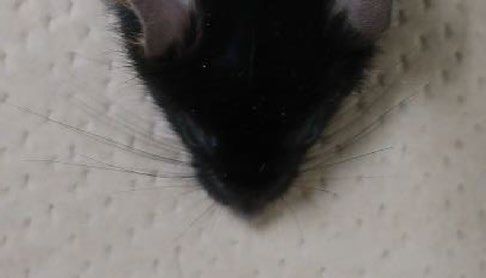
www.nature.com/scientificreports/
Parents genotype Genotype (*)
Female Male No. of blastocyst No. of established ESC lines +/+ + /Δ Δ/Δ
Ets2+/em1 Ets2+/em1 56 43 15 18 10
Ets2+/em2 Ets2+/em2 34 21 2 10 8
Table 3. Establishment of Ets2Δ/Δ ESC lines from blastocysts. (*) em1 or em2 allele shown as "Δ".
We hypothesized that the unexpected fragment size might be attributed to a splice variant and that this could
explain the differences in phenotype between new Ets2 mutants and Ets2db1/db1 animals. Therefore, we next ana-
lyzed the sequences of the potential splice variants. Although the large bands observed for skins and thymuses
of the Ets2em1/em1 and Ets2em2/em2 represented the expected frame-shifted sequences, the sequences of the smaller
bands showed skipping of exon 8, which was in-frame and consisted of 264 bp (Fig. 4B; Supplemental Fig. S5B).
Further, we examined the gene expression of MMP-3 and MMP-9 in Ets2em1/em1 skins, since a previous report
showed that expression of these genes was decreased in Ets2db1/db1 mice4. However, the expression levels of MMP-
3 and MMP-9 in 4-week-old Ets2em1/em1 skins were not reduced compared with the wildtype, even though they
were reduced in the Ets2null(em3/em3) (Fig. 4C).
Presence of Ets2 mutant protein in the skin and thymus. This Ets2 mutant protein, which skipped
exon 8 (Ets2 ΔEx8), was predicted to contain the ETS domain based on the SMART online database14 (Supple-
mental Fig. S7). To confirm the presence of Ets2 mutant protein in the skin and thymus, we performed western
blotting. Amino acid sequence sizes of Ets2, Ets2 em1, Ets2 em2, Ets2 ΔEx8 protein were of 468AA, 342AA,
298AA, and 380AA, respectively. Ets2 protein and Ets2 mutant proteins were distinguished by these molecular
weights and the lack of band in the null mutant tissues.
The Ets2 em1 protein was detected at its expected size in Ets2em1/em1 skin and thymus. The Ets2 em2 protein
was not found in both Ets2em2/em2 skin, even though Ets2 em2 mRNA (em2-L) was detected in the Ets2em2/em2 skin
(Supplemental Fig. S5A and Supplemental Fig. S6B). Ets2 ΔEx8 protein, which has ETS domain, was present
in the Ets2em1/em1 thymus, Ets2em2/em2 skin, but not clearly in the Ets2em1/em1 skin (Fig. 5, and full-length blots of
Fig. 5 are presented in Supplemental Fig. S8).
Discussion
In this work, we attempted to generate Ets2 homozygous mutant models at F0 generation by combination with
CRISPR/Cas9 system and tetraploid complementation. We could not obtain the Ets2 homozygous mutant model
at F0 generation using zygotes but generated those using ES cells. In the previous study, the theoretical birth rate
of Ets2db1/db1, which crossed with heterozygous parent and performed tetraploid complementation, was guessed
25% along with Mendelian law. However, it has been reported that the actual birth rate was 10%4. In this study,
the birth rate of offspring from Ets2null(em3/em3) ES cells by tetraploid complementation was comparable to that
from wild type ES cells as shown in Table 4. These results suggest that the mutant ES cells can be used to analyze
gene functions after birth if the mutations were deleterious to the trophectoderm lineages.
Here, we newly generated two Ets2 frame-shift mutant models, namely, em1 and em2. Since both have the
stop codon located before the ETS domain, we predicted that both mutant proteins would lack the ETS domain
if they were translated. Therefore, we expected that the phenotypes in these homozygous mutants would mimic
those described in previous reports for Ets2db1/db1 mice. Indeed, Ets2em1/em1 mice exhibited the same embryonic
lethal phenotype, but the wavy hair and curly whisker phenotypes observed in Ets2db1/db1 and Ets2null(em3/em3)
mice did not occur in Ets2em1/em1 and Ets2em2/em2 mice. Hair and whisker phenotypes of Ets2db1/db1 mice have been
caused due to the lack of ETS domain4. We found that the skin and thymus of Ets2em1/em1 and Ets2em2/em2mice
expressed a mutant mRNA lacking exon 8 that could potentially be translated into a protein, including the ETS
domain. We detected Ets2 ΔEx8 protein in Ets2em2/em2 skin and Ets2em1/em1 and Ets2em2/em2 thymuses. These data
demonstrated that Ets2 ΔEx8 mRNA was translated into the protein which contains the ETS domain. Therefore,
we hypothesize that this Ets2 ΔEx8 protein could rescue the Ets2 function in hair and whisker. This was strongly
suggested by the finding that these mice exhibited comparable levels of MMP-3 and MMP-9 mRNAs to wildtype,
and Ets2db1/db1 and Ets2null(em3/em3) mice exhibited decreased expression.
DNA mutations in a genetic locus frequently lead to exon skipping, and several human diseases are linked
to these types of exon skipping events. In addition, exon skipping can occur because of lack of exonic splicing
enhancer sequences or an exonic splicing silencer sequence inside of an e xon15,16. Some reports have also sug-
gested that unexpected exon skips can occur when using the CRISPR/Cas9 system, indicating that a frame-shifted
mutant exon induced by CRISPR/Cas9 is skipped but can be induced through alternative splicing or in-frame
exon skipping17–20. These studies indicated that mutations located on the three multiple exons could induce its
exon skipping. Due to our targeted exon 8 in Ets2 gene consists of 264 nts (88 multiples of three nts), exon skip-
ping may have occurred in the tissue specifically.
Since there was no report about the mutation within exon 8 of Ets2 in human by ClinVar database21, the new
Ets2 frame-shift mutant models could not applied to the human disease model. However, these new Ets2 frame-
shift mutant models had the following issue.
The exon skip observed was only identified in the skin and thymus but not detected during embryonic stages.
Thus, the induction of the exon skip was likely due to the changes in mRNA splicing that were dependent on the
Scientific Reports | (2021) 11:8297 | https://doi.org/10.1038/s41598-021-87751-5 5
Vol.:(0123456789)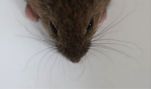
www.nature.com/scientificreports/

                                                                                                  Establishment of
                                                        Ets2Δ/Δ                                    Ets2Δ/Δ ESCs
                                            A

                                                          WT                4n embryo

                                            B                                                             C

                                                                                                                   em1/+

                                                     em1/em1                                                       em1/em1

                                                         Genome editing                       Establishment of
                                                                  ESC                        Ets2null(em3/em3) ESCs
                                            D

                                                        WT               4n embryo

                                            E                                                              F
                                                                                     #        #

                                                                                                                  WT

                                                                   WT        null (em3/em3)                        em3/em3

                                            Figure 3.  Generation of homozygous Ets2 mutant models using ESCs and assessment of hair and whisker
                                            phenotypes after birth. (A) Strategy for the generation of Ets2 homozygous mutant mouse after establishing
                                            the ESC from homozygous mutant blastocysts with tetraploid complementation. (B) 2-week-old Ets2em1/em1
                                            mice. (C) Faces of 4-week-old in Ets2+/em1 (left picture) and Ets2em1/em1 (right picture). Curly whiskers were not
                                            observed in Ets2em1/em1. (D) Strategy for the generation of Ets2 homozygous null mutant mouse after genome-
                                            editing to the ESC with tetraploid complementation. (E) The hair of the Ets2null(em3/em3) mutant mouse was wavy
                                            (indicated as #). (F) Some whiskers of the null mutant mouse were not straight (arrow in the lower picture).

          Scientific Reports |   (2021) 11:8297 |                https://doi.org/10.1038/s41598-021-87751-5                                                      6

Vol:.(1234567890)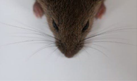
www.nature.com/scientificreports/

                                                                                                                                  Hair phenotype
                                   Genotype                  ESC lines   No. of transferred   No. of offspring      No. of wean   Normal   Wavy
                                                             1A1         40                   5                     2             2        0
                                   Ets2em1/em1               1A3         20                   3                     2             2        0
                                                             1B5         78                   8                     2             2        0
                                                             #11         20                   4                     2             2        0
                                   Ets2em2/em2
                                                             #22         20                   6                     3             3        0
                                   Ets2 null (em3/em3)       2-10G       56                   17                    17            0        17
                                   Ets2 WT                   mF1-05      57                   8                     8             8        0

                                  Table 4.  4n complementation of Ets2 homozygous mutant ESC lines.

                                                                                                                                        em1L
                                          A                                                                        B
                                                                   4wk skin

                                                                                                                                      205 bp del
                                       Ets2                                                   em1L
                                                                                              em1S                                     em1S

                                  GAPDH

                                                                                                                                      264 bp del

                                        C                    0.01                                                       0.0015
                                                                                   *
                                                                              **       **                                                          *
                                                                                                           Mmp9/Gapdh
                                               Mmp3/Gapdh

                                                                                                                         0.001

                                                            0.005

                                                                                                                        0.0005

                                                                   0                                                        0

                                  Figure 4.  Gene expression in the Ets2em1/em1 skin. (A) Gene expression from the Ets2 locus in skins of WT,
                                  Ets2null(em3/em3) mutant (null) and Ets2em1/em1 (em1) mice. From em1 skin, two types of mRNA were expressed,
                                  although both were shorter than WT. The larger mRNA was called em1L, and the smaller mRNA was called
                                  em1S. (B) Sequences of em1L and em1S. Em1L was the expected sequence, but em1S contained a deleted locus
                                  that matched exon 8, shown in Supplemental Fig. S5. (C) Gene expression of Mmp3 and Mmp9 in the Ets2em1/em1
                                  skin. There were significant differences in the expression level of Mmp3 between WT and null, em1 and null,
                                  and WT and em1. The expression level of Mmp9 in the null skin was slightly decreased compared with WT, but
                                  not significantly, and em1 showed a significant difference compared with null. *p < 0.05, **p < 0.01.

                                  cell type or developmental stage, despite having the same genomic mutation. This phenomenon has a possible
                                  effect on the appearance of phenotypes making it a novel finding of this study.
                                      Overall, our study suggested that all ORF-deletion models are adequate for analysis if we have to generate the
                                  gene knockouts. Furthermore, research related to indel mutation or point mutation in human disease models
                                  need to pay attention to the effects of alternative splicing.

Scientific Reports |   (2021) 11:8297 |                            https://doi.org/10.1038/s41598-021-87751-5                                               7

                                                                                                                                                       Vol.:(0123456789)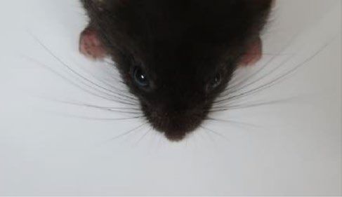
www.nature.com/scientificreports/
A 2wk skin 4wk skin
(KDa) (KDa)
70 70
Ets2 55 * 55 *
** **
***
***
35 35
##
25 25
GAPDH
thymus
B
(KDa)
70
Ets2 55 *
**
***
35
25
GAPDH
Figure 5. Presence of proteins in the Ets2 mutant skin and thymus. Western blotting with anti-Ets2 antibody
and anti-Gapdh antibody in the skin (A) and thymus (B). Native Ets2 protein (*) was detected only in WT
samples. Ets2 ΔEx8 (**) and Ets2 em1 (***) mutant proteins were not detected in WT and null mutant samples.
Ets2 em2 mutant protein (the predicted size indicates as ##) were not detected in Ets2em2/em2 (em2) skin.
Methods
Animals. All animal experiments were conducted in accordance with the guidelines of “Regulations and
By-Laws of Animal Experimentation at the Nara Institute for Science and Technology”, and were approved by
the Animal experimental Committee at the Nara Institute of Science and Technology (the approval no. 1639).
Study of the animal experiments were carried out in compliance with the ARRIVE guidelines22. B6D2F1 female
mice and ICR mice were purchased from SLC (Japan). C57BL/6J male mice were purchased from CLEA (Japan).
Collection of zygotes. Female mice were treated by PMSG and hCG for superovulation, then mated with
male mice. Pronuclear stage zygotes were collected from female oviducts after 20 h of hCG injection. After
removing cumulus cells using hyaluronidase, zygotes were incubated in KSOM at 37 °C under 5% CO2 in the air
until use. 2-cell stage zygotes were collected from female oviducts after 42–46 h of hCG injection by the flush-out
method. Collected 2-cell stage embryos were incubated until use the same as above.
Generation of Ets2 mutant zygote by CRISPR/Cas9 system using electroporation. Target sites
of guide RNA (gRNA) were designed using the web tool CRISPR direct23. Genome editing by the electroporation
was referred to as the previous study12.
CFB16-HB and LF501PT1-10 electrode (BEXCo.Ltd., Tokyo, Japan) were used for electroporation. 30–40
pronuclear stage zygotes were subjected to electroporation at one time. Zygotes were washed with Opti-MEM
I (Thermofisher) three times, subsequently placed in a line in the electrode gap filled with five μl the mixture of
120 ng/μl Cas9 protein (TaKaRa, Japan), 300 ng/μl tracrRNA, and 200 ng/μl crRNA (HPLC grade, Fasmac) in
Opti-MEM I. The electroporation condition was performed were 30 V (3 ms ON ± 97 ms OFF) four times. After
electroporation, zygotes were washed with KSOM three times then cultured until developing the eight-cell stage.
Eight-cell stage embryos were provided to the tetraploid complementation.
Establishment of Ets2 mutated ESC lines. To establish the Ets2 homozygous mutant model, collected
2-cell stage embryos from Ets2 heterozygous mutant parents were incubated until the blastocyst stage, remov-
ing the Zona pellucida (ZP) using Acidic Tyrode solution (Sigma T1788). Blastocyst embryos without the ZP
were seed on gelatin-coated 60-mm dishes and cultured on mouse embryonic fibroblast (MEF) with N2B27
medium supplemented with 3 μM CHIR99021(Axon1386), 1.5 μM CGP77675 (Sigma SML0314), and mouse
LIF (N2B27-a2i/L medium)24. After seven days, the outgrowth of blastocysts was disaggregated by 0.25% trypsin
Scientific Reports | (2021) 11:8297 | https://doi.org/10.1038/s41598-021-87751-5 8
Vol:.(1234567890)
www.nature.com/scientificreports/
in 1 mM EDTA in PBS (−). Half of the cells were seeded on MEF with the gelatin-coated dishes for expanding.
The others were seeded on the gelatin-coated dishes without MEF for genotyping by PCR. Ets2 homozygous
mutant ESC lines were provided for tetraploid complementation.
The Ets2 null mutant model was established using ESCs, referred to as the previous study25. mF1-05 ESC
line, which was newly established from 129X1and C57BL6/J F1 embryo, was seeded on MEF then transfected
with two designed pSpCas9(BB)-2A-Puro (pX459) V2.0 (Addgene #62988) plasmids using Lipofectamine 3000
(Thermofisher). Transfected cells were selected by transient treatment with 1 μg/ml puromycin, then ESC colonies
were subject to genotyping with PCR and sequencing. The Ets2 null mutant ESC line was provided for tetraploid
complementation.
Tetraploid complementation. Tetraploid embryos were prepared as described previously9,26. In brief,
ICR two-cell stage embryos were placed in the fusion buffer, and electrofusion was performed by applying 140 V
for 50 ms after aligning embryos between the electrodes. CFB16-HB and LF501PT1-10 electrode (BEXCo.Ltd.,
Tokyo, Japan) were used for cell fusion.
A wild-type tetraploid four-cell embryo and a genome-edited diploid eight-cell embryo were aggregated
after removing the Zonae pellucid for the aggregation method. For the injection method, Ets2 mutant ESCs
were injected into a wild-type tetraploid four-cell embryo or blastocyst. These embryos were cultured until the
blastocysts stage and transferred into the uterus of E2.5 pseudopregnant ICR mice. Offspring were recovered
by natural delivery or Caesarean section on E19.5. The mutation of offspring was detected by genotyping with
PCR and sequencing.
Genotyping. Genotyping primers for detecting Ets2-wild, em 1, and em 2 alleles were 5′-ctgagtttaagagt-
gctcggagg-3′ (Ets2_Fw) and 5′- gccctataggacttgtgtacagg-3′ (Ets2_Rev). Primers for Ets2 null (em3) mutant
allele(s) were 5′-tgtggagtctcacatcgaag-3′ (Ets2_Ex2_F) and 5′- gggcctgctcggtgccacgg-3′ (Ets2_EX10_R). DNA
fragments were amplified using GoTaq (Promega) for 40 cycles under the following conditions: 94 °C for 30 s,
60 °C for 30 s and 68 °C for 40 s for detecting wild, em1 or em2 allele, and 94 °C for 30 s, 60 °C for 30 s and 68 °C
for 20 s for detecting the null allele, respectively.
RNA expression analysis. Mouse cDNAs were prepared from 4-week old skin, adult skin, and adult thy-
mus using SuperScript III Reverse Transcriptase (Thermo Fisher Scientific) after purified RNA by Trizol reagent
(Thermo Fisher Scientific). RT-PCR was performed using 20 ng of cDNA with the following primers: 5′-CGT
GAATTTGCTCAACAACAATTCTG-3′ and 5′-GAGAGGCTATGCCGGT-3′ for Ets2, 5′-CCAGTATGACTC
CACTCACG-3′ and 5′-GACTCCACGACATACTCAGC-3 for Gapdh 27. cDNA fragments were amplified using
KOD Fx Neo (TOYOBO) or GoTaq (Promega) for 35 cycles under the following conditions: 94 °C for 30 s, 60 °C
for 30 s and 72 °C for 40 s for Ets2, and 94 °C for 30 s, 53 °C for 30 s and 72 °C for 30 s for Gapdh.
Quantitative Real-time PCR was performed using 20 ng of cDNA with following primers: 5′-TTAAAGACA
GGCACT T TTGG-3′ and 5′-CAGGGTGTGAATGCT T TTAG-3′for Mmp3, 5′-CGTCTGAGAATTGAATCA
GC-3′ and 5′-AGTAGGGGCAACTGAATACC-3′ for Mmp9 expression5. Gene expression level was normalized
by Gapdh, the same cDNA. The primer set for Gapdh was the same as above. Real-time PCR was performed by
LightCycler96 (Roche) using the Luna Universal qPCR Master Mix (NEB), and the data were analyzed by the
LightCycler96 software (Roche, version 1.1.0.1320).
Western blotting. Whole cell extracts of 2 weeks old skin or thymus were prepared by homogenization in
RIPA buffer (Nacalai). Equivalent amount of protein were separated by 4–12% Bolt Bis–Tris gel (Invitrogen) and
run under reducing conditions. After gel electrophoresis, they transferred to immobilon-P membrane (Merck).
Membranes were blocked in 5% skim milk powder in phosphate-buffered saline with Tween20. The membrane
was incubated with anti-Gapdh (WAKO, 010-25521) or anti-Ets2 (Genetex, GTX104527) followed by a second-
ary antibody conjugated with horseradish peroxidase (Cytiva, NA931, NA934). Peroxidase activity was detected
with Chemi-Lumi One (Nacalai) using the FUSION -Chemiluminescence Imaging System (Vilber-Lourmat).
Statistics analysis. The statistical difference was determined using the Student t-test. Differences were con-
sidered statistically significant if the P-value was less than 0.05.
Received: 2 October 2020; Accepted: 5 April 2021
References
1. Karim, F. D. et al. The ETS-domain: A new DNA-binding motif that recognizes a purine-rich core DNA sequence. Genes Dev. 4,
1451–1453. https://doi.org/10.1101/gad.4.9.1451 (1990).
2. Sharrocks, A. D. The ETS-domain transcription factor family. Nat. Rev. Mol. Cell Biol. 2, 827–837. https://doi.org/10.1038/35099
076 (2001).
3. Seidel, J. J. & Graves, B. J. An ERK2 docking site in the Pointed domain distinguishes a subset of ETS transcription factors. Genes
Dev. 16, 127–137. https://doi.org/10.1101/gad.950902 (2002).
4. Yamamoto, H. et al. Defective trophoblast function in mice with a targeted mutation of Ets2. Genes Dev. 12, 1315–1326. https://
doi.org/10.1101/gad.12.9.1315 (1998).
5. Man, A. K. et al. Ets2-dependent stromal regulation of mouse mammary tumors. Mol. Cell Biol. 23, 8614–8625. https://doi.org/
10.1128/mcb.23.23.8614-8625.2003 (2003).
Scientific Reports | (2021) 11:8297 | https://doi.org/10.1038/s41598-021-87751-5 9
Vol.:(0123456789)www.nature.com/scientificreports/
6. Wei, G. et al. Ets1 and Ets2 are required for endothelial cell survival during embryonic angiogenesis. Blood 114, 1123–1130. https://
doi.org/10.1182/blood-2009-03-211391 (2009).
7. Baran, C. P. et al. Transcription factor ets-2 plays an important role in the pathogenesis of pulmonary fibrosis. Am. J. Respir. Cell
Mol. Biol. 45, 999–1006. https://doi.org/10.1165/rcmb.2010-0490OC (2011).
8. Wei, G. et al. Activated Ets2 is required for persistent inflammatory responses in the motheaten viable model. J. Immunol. 173,
1374–1379. https://doi.org/10.4049/jimmunol.173.2.1374 (2004).
9. Okada, Y. et al. Complementation of placental defects and embryonic lethality by trophoblast-specific lentiviral gene transfer. Nat.
Biotechnol. 25, 233–237. https://doi.org/10.1038/nbt1280 (2007).
10. Cong, L. et al. Multiplex genome engineering using CRISPR/Cas systems. Science 339, 819–823. https://doi.org/10.1126/science.
1231143 (2013).
11. Mali, P. et al. RNA-guided human genome engineering via Cas9. Science 339, 823–826. https://doi.org/10.1126/science.1232033
(2013).
12. Hashimoto, M., Yamashita, Y. & Takemoto, T. Electroporation of Cas9 protein/sgRNA into early pronuclear zygotes generates
non-mosaic mutants in the mouse. Dev. Biol. 418, 1–9. https://doi.org/10.1016/j.ydbio.2016.07.017 (2016).
13. Nagy, A., Rossant, J., Nagy, R., Abramow-Newerly, W. & Roder, J. C. Derivation of completely cell culture-derived mice from
early-passage embryonic stem cells. Proc. Natl. Acad. Sci. U S A 90, 8424–8428, https://doi.org/10.1073/pnas.90.18.8424 (1993).
14. SMART. http://smart.embl-heidelberg.de/.
15. Cartegni, L., Chew, S. L. & Krainer, A. R. Listening to silence and understanding nonsense: Exonic mutations that affect splicing.
Nat. Rev. Genet. 3, 285–298. https://doi.org/10.1038/nrg775 (2002).
16. Baralle, F. E. & Giudice, J. Alternative splicing as a regulator of development and tissue identity. Nat. Rev. Mol. Cell Biol. 18, 437–451.
https://doi.org/10.1038/nrm.2017.27 (2017).
17. Mou, H. et al. CRISPR/Cas9-mediated genome editing induces exon skipping by alternative splicing or exon deletion. Genome
Biol. 18, 108. https://doi.org/10.1186/s13059-017-1237-8 (2017).
18. Chen, D. et al. CRISPR/Cas9-mediated genome editing induces exon skipping by complete or stochastic altering splicing in the
migratory locust. BMC Biotechnol. 18, 60. https://doi.org/10.1186/s12896-018-0465-7 (2018).
19. Sui, T. et al. CRISPR-induced exon skipping is dependent on premature termination codon mutations. Genome Biol. 19, 164.
https://doi.org/10.1186/s13059-018-1532-z (2018).
20. Tuladhar, R. et al. CRISPR-Cas9-based mutagenesis frequently provokes on-target mRNA misregulation. Nat. Commun. 10, 4056.
https://doi.org/10.1038/s41467-019-12028-5 (2019).
21. ClinVar. https://www.ncbi.nlm.nih.gov/clinvar/.
22. Kilkenny, C. et al. Improving bioscience research reporting: The ARRIVE guidelines for reporting animal research. PLoS Biol. 8,
e1000412. https://doi.org/10.1371/journal.pbio.1000412 (2010).
23. CRISPRdirect. http://crispr.dbcls.jp/.
24. Choi, J. et al. Prolonged Mek1/2 suppression impairs the developmental potential of embryonic stem cells. Nature 548, 219–223.
https://doi.org/10.1038/nature23274 (2017).
25. Oji, A. et al. CRISPR/Cas9 mediated genome editing in ES cells and its application for chimeric analysis in mice. Sci. Rep. 6, 31666.
https://doi.org/10.1038/srep31666 (2016).
26. Kokubu, C. et al. A transposon-based chromosomal engineering method to survey a large cis-regulatory landscape in mice. Nat.
Genet. 41, 946–952. https://doi.org/10.1038/ng.397 (2009).
27. Wen, F. et al. Ets2 is required for trophoblast stem cell self-renewal. Dev. Biol. 312, 284–299. https://doi.org/10.1016/j.ydbio.2007.
09.024 (2007).
Acknowledgements
This work was supported by JSPS KAKENHI Grant Number 16K07091, 18H04885, Start Up Fund for female
researchers in NAIST, and KAC 40th Anniversary Research Grant. The authors would like to thank Enago
(https://www.enago.jp) for the English language review.
Author contributions
A.I., Y.K., I.N., and W.H. performed most experiments, assisted by N.Y. who performed tetraploid complementa-
tion, and S.Y., performed qPCR. A.I., S.Y., and M.I. analyzed the data. A.I. wrote the manuscript and all authors
discussed the results and commented on the manuscript.
Competing interests
The authors declare no competing interests.
Additional information
Supplementary Information The online version contains supplementary material available at https://doi.org/
10.1038/s41598-021-87751-5.
Correspondence and requests for materials should be addressed to A.I.
Reprints and permissions information is available at www.nature.com/reprints.
Publisher’s note Springer Nature remains neutral with regard to jurisdictional claims in published maps and
institutional affiliations.
Open Access This article is licensed under a Creative Commons Attribution 4.0 International
License, which permits use, sharing, adaptation, distribution and reproduction in any medium or
format, as long as you give appropriate credit to the original author(s) and the source, provide a link to the
Creative Commons licence, and indicate if changes were made. The images or other third party material in this
article are included in the article’s Creative Commons licence, unless indicated otherwise in a credit line to the
material. If material is not included in the article’s Creative Commons licence and your intended use is not
permitted by statutory regulation or exceeds the permitted use, you will need to obtain permission directly from
the copyright holder. To view a copy of this licence, visit http://creativecommons.org/licenses/by/4.0/.
© The Author(s) 2021
Scientific Reports | (2021) 11:8297 | https://doi.org/10.1038/s41598-021-87751-5 10
Vol:.(1234567890)You can also read