Segmentation of Oral Leukoplakia (OL) and Proliferative Verrucous Leukoplakia (PVL) Using Artificial Intelligence Techniques
←
→
Page content transcription
If your browser does not render page correctly, please read the page content below

Hindawi
BioMed Research International
Volume 2022, Article ID 2363410, 11 pages
https://doi.org/10.1155/2022/2363410

Research Article
Segmentation of Oral Leukoplakia (OL) and Proliferative
Verrucous Leukoplakia (PVL) Using Artificial
Intelligence Techniques

          Samar Zuhair Alshawwa ,1 Asmaa Saleh,1 Malek Hasan ,2,3 and Mohd Asif Shah                                               4

          1
            Department of Pharmaceutical Sciences, College of Pharmacy, Princess Nourah bint Abdulrahman University, P.O. Box 84428,
            Riyadh 11671, Saudi Arabia
          2
            Department of Medical Instruments Engineering Techniques, Al-Farahidi University, Baghdad 10021, Iraq
          3
            The University of Mashreq, Research Center, Baghdad, Iraq
          4
            Kebri Dehar University, Ethiopia

          Correspondence should be addressed to Malek Hasan; malekb.has@yahoo.com

          Received 6 June 2022; Revised 27 June 2022; Accepted 30 June 2022; Published 21 July 2022

          Academic Editor: Dinesh Rokaya

          Copyright © 2022 Samar Zuhair Alshawwa et al. This is an open access article distributed under the Creative Commons
          Attribution License, which permits unrestricted use, distribution, and reproduction in any medium, provided the original work
          is properly cited.

          PVL (proliferative verrucous leukoplakia) has distinct clinical characteristics. They have a proclivity for multifocality, a high
          recurrence rate after treatment, and malignant transformation, and they can progress to verrucous or squamous cell
          carcinoma. AI can aid in the diagnosis and prognosis of cancers and other diseases. Computational algorithms can spot tissue
          changes that a pathologist might overlook. This method is only used in a few studies to diagnose LB and PVL. To see if their
          cellular nuclei differed and if this cellular compartment could classify them, researchers used a computational system and a
          polynomial classifier to compare OLs and PVLs. 161 OL and 3 PVL specimens in the lab were grown, photographed, and used
          for training and computation. Exam orders revealed patients’ sociodemographics and clinical pathologies. The nucleus was
          segmented using Mask R-CNN, and LB and PVL were classified using a polynomial classifier based on nucleus area, perimeter,
          eccentricity, orientation, solidity, entropies, and Moran Index (a measure of disorderliness). The majority of OL patients were
          male smokers; most PVL patients were female, with a third having malignant transformation. The neural network correctly
          identified cell nuclei 92.95% of the time. Except for solidity, 11 of the 13 nuclear characteristics compared between the PVL
          and the LB showed significant differences. The 97.6% under the curve of the polynomial classifier was used to classify the two
          lesions. These results demonstrate that computational methods can aid in diagnosing these two lesions.

1. Introduction                                                        Although each of them has specific histological aspects,
                                                                       some histological characteristics such as hyperkeratosis
According to the World Health Organization, cancer arises              (increase in keratin), hyperplasia (increase in the number
from transforming normal cells into tumor cells in a                   of cells), and even dysplasia (an architectural disorder of epi-
multistage process [1]. Among the various types of tumors,             thelial tissue accompanied by cytological atypia) may be
squamous cell carcinoma (SCC) of the oral cavity is usually            shared between them [3].
preceded by MPDs, which develop from etiological factors                   Premalignant cases are local lesions characterized by a
such as tobacco, alcohol, autoimmune diseases, and                     higher risk of malignant change than normal structures or
idiopathic or inherited genetic aberrations. We can mention            general conditions associated with an increased risk of can-
oral leukoplakia (OL), erythroplakia, oral submucosal fibro-            cer. Oral mucosa is an area where more than 5% of prema-
sis, palatine keratosis associated with inverted smoke, lichen         lignant cases turn into cancer [4, 5]. Therefore, the
planus, lupus erythematosus, and dyskeratosis congenita [2].           diagnosis of premalignant formations gains importance in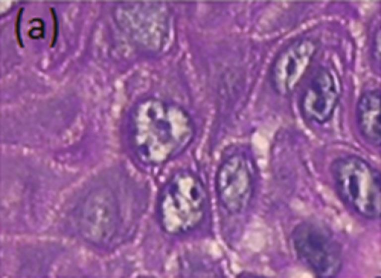
2                                                                                                   BioMed Research International

the early detection of possible malignant lesions. Problems          pathological data (lesion color, location, lesion type, dysplasia,
in evaluating premalignant cases mainly arise from two fac-          malignant transformation, and size) and sociodemographic
tors [5]. Although various clinical features are important in        (gender, age, and smoking status) of the patients were
determining the risk of malignant change in premalignant             obtained from the examination requests presented by the
cases, histological examinations are the most valid method           pathology laboratory. As a minimum inclusion criterion, only
today to determine these formations’ proper structure and            cases of OLs, PVLs, and SCCs had stained and well-preserved
malignant potential. Histologically, epithelial dysplasia and        slides, and their respective diagnoses were used. Otherwise, the
cellular atypia are prognostic indicators of premalignancy,          cases were excluded from the study.
which refers to impaired proliferation, maturation, and
organization of the epithelium. These changes are seen in            2.1. Computer Analysis. The slides selected from each case
three degrees mild, moderate, and severe. Although the               were photographed using a Leica DM500 optical microscope
relationship between epithelial dysplasia and future carci-          to study the nuclei. On average, ten fields/lesions were
noma is not certain, it is generally stated that the degree of       obtained at 400× magnification, and all images were saved
dysplasia and the transformation into cancer are directly            in JPEG and TIFF format with a resolution of 1600 ×
proportional [6].                                                    1200. From the capture of these fields, the regions of interest
    Technological advances in the last decade, especially            were extracted. Then, the steps were used sequentially: seg-
with the advent of high-resolution digitized images and AI,          mentation, postprocessing, feature extraction, and classifica-
have allowed pathology to adopt computational approaches,            tion, which are detailed below. All steps after extracting the
such as machine learning, to assess tissue aspects such as           ROIs were performed on a computer with the following con-
minimal or no human interference through algorithms capa-            figurations: AMD FX-8320 processor, 8GB of RAM, and
ble of predicting, for example, precancerous lesions’ risk of        NVIDIA GTX 1060 GPU with 6GB of VRAM. In the man-
malignant transformation [7]. Thus, several studies have             ual segmentation phase for network training, a Wacom
proposed using AI as a tool that adds to existing ones to            Monitor-Cintiq was used to bypass the cores; the GIMP soft-
understand the biology of these lesions better, but with the         ware was used both in this step and to obtain the ROIs
advantage of eliminating the pathologist’s subjectivity              obtained through the website https://www.gimp.org/. All
regarding the interpretation of histopathological findings [8].       software used in this work is available free of charge.

    Considering the potential of this tool, this study investi-      2.2. Segmentation. The first stage of segmentation is the
gated the nuclear aspects of OL and proliferative verrucous          training phase, in which the segmentation of the nuclei is
leukoplakia (PVL) cells with the main aim of detecting               performed manually from the ROIs extracted from the fields
nuclear alterations and creating a classification algorithm           obtained from each lesion, as explained above. For this step,
that can be used for differential diagnosis between the two           481 ROIs were used, distributed as follows: 74 ROIs from OL
lesions. We hypothesized that the nuclear aspects investi-           without dysplasia, 77 ROIs from OL with mild dysplasia, 114
gated by AI are different between the two lesions and that            ROIs from OL with moderate dysplasia, 43 ROIs from OL
this tool can be used for the purpose of differential diagnosis       with severe dysplasia, 59 PVL ROIs, and 114 SCC ROIs. A
between an OL and PVL, especially in the early stages of             total of 15,027 cores were manually segmented and used in
both lesions when the clinicopathological aspects overlap,           network training.
compromising the accuracy of the diagnosis both by the cli-              For training, the Mask R-CNN neural network was used
nician but mainly by the pathologist.                                together with the ResNet50 convolutional network to detect
                                                                     the cellular nuclei in the images. The ResNet50 convolu-
2. Material and Methods                                              tional network was first used in the learning stage of the
                                                                     characteristics of cell nuclei. The architecture of this network
Sixty-one cases of OL, three cases of PVL, and five cases of          is shown in Figure 1. The network model is composed of 50
SCC (all these cases were linked to malignant transformation         convolutional layers distributed over the input layer (E); four
of either OL or PVL) were collected from the Oral Pathology          blocks of convolutional layers called Block 1 (B1), Block 2
Laboratory of the Faculty of Dentistry of the University of          (B2), Block 3 (B3), and Block 4 (B4); and an output step.
Baghdad, Iraq, in the period from March 2021 to February             Briefly, layer E has 64 convolutional filters with a size of 7
2022 to be used in the study in the neural network training,          × 7 pixels that process the original image through a sliding
segmentation, and feature extraction phases. Initially, slides       window with an offset size of 2 pixels. Then, still in layer
stained with hematoxylin and eosin used in the clinical routine      E, a max-pooling filter, with a size of 2 × 2 pixels, is used,
were retrieved from their respective cases and reassessed to         with a displacement size equal to 2 pixels. This result is then
confirm the diagnosis of each of the lesions. In the case of          applied to layer C1 of B1, which has 64 convolutional filters
OLs and PVLs, the degrees of dysplasia were also checked.            of size 1 × 1 pixel, followed by C2, which has 64 filters of size
For this diagnostic confirmation step, the criteria established       3 × 3 pixels, and C3, which contains 256 filters of size 1 × 1
by the World Health Organization of 2017 were used [9].              pixel. B1 is repeated three times over the image processed
The fact of using routine slides stained with hematoxylin            in layer E, totaling nine convolutional layers in this block.
and eosin refers to the search in this work of the neural            The result of B1 is applied to B2, passing first through layer
network to learn from the conditions found in oral pathology         C1, with 128 filters of size 1 × 1 pixel, followed by C2, with
laboratories, ensuring better applicability of this tool. Clinico-   128 filters of size 3 × 3 pixels, and C3, with 512 filters of size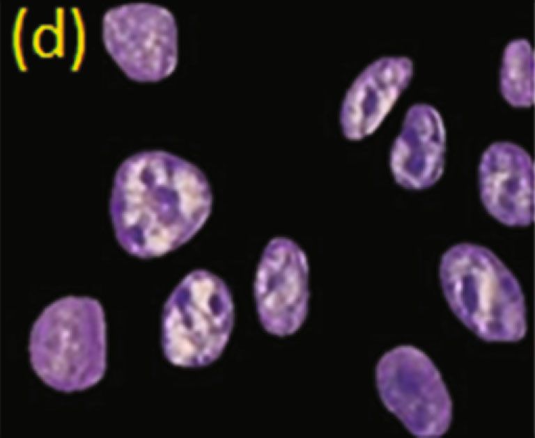
BioMed Research International                                                                                                         3

                                  2×2,                                                  B3
                                  Max                               B2                                   B4      Avg
                                                B1
                                  Pool                                                                           Pool
                          E                                                                                              Fc 1000

      Input limage Input layer           Block-1       Block-2                Block-3           Block-4
                   7×7, 64, Step 2)      C1;1×1,64     C1;1×1,128             C1;1×1,256        C1;1×1,512
                                         C2;3×3,64     C2;3×3,128             C2;3×3,256        C2;3×3,512
                                         C3;1×1,256    C3;1×1,512             C3;1×1,1024       C3;1×1,2048

        Figure 1: Architecture of the ResNet50 neural network used in the segmentation process for the learning of cell nuclei.

1 × 1 pixel. This block has four repetitions, resulting in 12            combined with the layers below, which are larger and have
convolutional layers. The result of B2 is then used in B3,               more detailed core features. As these results are passed
which has 256 filters of size 1 × 1 pixel in C1, 256 filters of            through these larger, more detailed layers, the identified
size 3 × 3 pixels in C2, and 1024 filters of size 1 × 1 pixel in          regions become more accurate and correct, while the bound-
C3. This block has six repetitions, totaling 18 layers. The              ing boxes are regressed. Finally, the R-CNN Mask through a
result of B3 is used in B4, which also has three layers: C1,             convolutional network used in conjunction with feature
which has 512 filters of size 1 × 1 pixel, C2, with 512 filters            maps produces a binary mask for each identified nucleus.
of size 3 × 3 pixels, and C3, with 2048 filters of size 1 × 1                 A multitasking loss rate is calculated for each region dur-
pixel. Block B4 is repeated three times, presenting nine                 ing the network training phase. This rate calculates the train-
layers in total. Finally, the generated data is transformed into         ing error percentage, that is, how much the training is
a vector by an average pooling filter, while the softmax func-            different from the gold standard (ROIs manually segmented
tion is used to classify objects between core or background              by the expert). Basically, it sums the rate of loss (error) of
region. For this step, a fully connected layer with 1000 neu-            classification and regression of boxes and masks, and its
rons is used. Between each block of convolutional layers (B1,            function is defined by
B2, B3, and B4), the feature matrix is reduced in size by a
proportionality of 2. For this, a sliding window with a dis-                                 L = Lcls + Lbox + Lmask :              ð1Þ
placement size of 2 pixels is used in the transition convolu-
tions between each block. For the network model used in
this work, the activation function adopted was the Rectified                  For the network model applied in this work, 60 ROIs
Linear Unit (ReLU) [10].                                                 were used for network training, 48 of them for training
    He and others [11] explained that the Mask R-CNN is a                and 12 for testing, with 40 epochs performed in total.
convolutional neural network that detects and separates can-                 416 ROIs were used to validate the model. Five of the
didate objects into distinct classes from regions. It consists of        total ROIs used were discarded during the process because
two stages; the first stage is called Region Proposal Network             they showed errors that prevented the program from
(RPN) and aims to generate sets of candidate objects in                  working.
bounding boxes (the smallest square that comprises an
                                                                         2.2.1. Postprocessing. After the network classifies the objects
object); the second stage aims to classify the objects con-
                                                                         between cores and the background region, some regions
tained in the bounding boxes and perform the regression
                                                                         may be incompletely filled or even present noise or artifacts
of these boxes. In parallel to the second stage, the network
                                                                         that can harm the segmentation process. To prevent this
also has a branch that provides a binary mask for each ROI.
                                                                         from happening, the postprocessing step is performed
    This network detects and segments nuclei through the
                                                                         through morphological operations to eliminate false-
core characteristics extracted by ResNet50. For this, Mask
                                                                         positive regions and refine the segmented nuclei.
R-CNN uses the feature maps generated by ResNet50 and
stacks them from the largest (most detailed) to smallest                 2.2.2. Classification. The classification was performed using
(least detailed) map forming a Feature Pyramid Network                   these vectors after the feature extraction process in which
(FPN). How ResNet50 has many layers that generate maps,                  the μ and σ of the extracted nuclear features were trans-
the use of all of them can generate a large pyramid of high              formed into a single vector for each ROI. The objective
execution complexity. To get around this problem, only                   was to separate the images in the two different classes of
the last layer of each block is used, that is, C3 of the last            lesions (OL and PVL) through the polynomial classifier
B1, C3 of the last B2, C3 of the last B3, and C3 of the last             (POL) [11]. This classifier employs polynomial expansion
B4. Then, the FPN layers are passed as input to the Region               over the extracted feature vector to define the coefficients
Proposal Network (RPN) to identify the cores in the top-                 used in the separation of classes. For this to occur, the fol-
down direction, from the smallest to the largest layer. With             lowing discriminant polynomial function is used:
this, we classify the regions between core and background
regions along with the bounding boxes in the first layer from
above (smaller and less detailed). These results are then                                       gðxÞ = aT pn ðxÞ:                   ð2Þ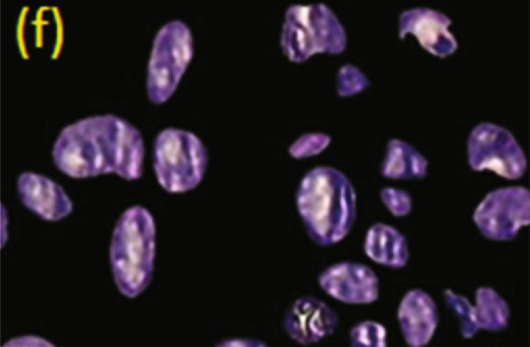
4                                                                                                BioMed Research International

2.3. Statistical Analysis. To assess the method’s effectiveness,   nuclear regions that were not identified in the gold standard
a comparison was performed by estimating the overlap              (manual segmentation) and therefore were called false-
between the segmented images and the gold standard. With          positive nuclear regions (Figure 2(c), red arrow). This may
this, measures of true positives, false positives, true nega-     be due to the similarity of coloration in the regions close to
tives, and false negatives are obtained, which were used in       the nuclear limits. Still in Figure 2(c), it can be seen that
segmentation and classification. The ROC curve is obtained         the neural network did not identify some nuclear regions,
from the graph plotted by sensitivity versus false-positive       as observed in the gold standard, indicating a false-negative
rate (TFP). In order to compare the morphological charac-         result (green arrow). In addition, some segmented images
teristics of the nuclei between OLs and PVLs, regardless of       presented noise that was wrongly classified as nuclei, which
the degree of dysplasia, the statistical Mann–Whitney tests       were considered false positives (Figure 2(e), red arrow). To
or the unpaired t-test were used, according to the data distri-   correct these and other irregularities, a postprocessing step
bution, as determined by the normality test, Shapiro-Wilk. It     was carried out.
was considered statistically significant when p < 0:05.
                                                                  3.1.2. Postprocessing. Postprocessing was carried out after
3. Results                                                        segmentation by the neural network to correct some segmen-
                                                                  tation irregularities. The result can be seen in Figure 3. In this
3.1. Clinicopathological Data. Most OL patients were male,        step, dilation (Figure 3(c)), hole filling (Figure 3(d)), and ero-
while all PVL cases were female. The mean age of the              sion (Figure 3(e)) operations were performed on an image. In
patients was 55 years, 54 in the OLs and 72 in the PVLs.          Figure 3(b), the region indicated by the green arrow indicates
Most patients with OL were smokers, while only one patient        the presence of a hole inside the core, where after the applica-
with PVL had information about smoking. The main loca-            tion of the expansion operation, the hole is reduced in size
tions of both lesions were the buccal mucosa, tongue, alveo-      and, subsequently, eliminated by the hole-filling operation.
lar ridge, and lip. For both OL and PVL, the most frequently      At the end, the erosion operation was applied so that the
found dysplastic alteration was mild dysplasia. Two cases of      nuclei returned to their respective original sizes. After the pro-
OL and one of PVL underwent a malignant transformation.           cess of identifying the cellular nuclei, the tissues started to be
Regarding the clinical characteristics of both lesions, most      classified based on the extraction of morphological character-
were white and had an average size of 1.5 cm.                     istics from the objects classified as nuclei. The morphological
                                                                  characteristics studied were area (A), eccentricity (E), perime-
3.1.1. Nuclear Segmentation and Neural Network. For the           ter (PE), orientation (OR), solidity (S), entropy (EN), and
analysis of nuclear segmentation of the lesions, photomicro-      Moran Index (IM). These selected characteristics are based
scopy of the lesions of OL, PVL, and SCC was obtained, as         on the work of which in turn was based on the studies [11, 12].
described in the methodology. Of the total, 568 images of               The area is calculated by the total value of the number of
OL, 45 images of PVL, and 58 images of SCC were taken.            pixels in an object; eccentricity concerns the difference in the
From these images, the following numbers of ROIs were             circumference of an object in relation to a circle, that is, the
captured for each image: 1,217 ROIs from OL, 119 ROIs             calculation of the elongation of objects; orientation calcu-
from PVL, and 133 ROIs from SCC. In this step, SCC sam-           lates the relationship between the major axis of an object
ples were used to increase the number of nuclei used for          about the x-axis; solidity analyzes the deformity of an object,
neural network training purposes and increase its accuracy        in the case of this work, the level of irregularity of the circu-
in the nuclear segmentation process. After obtaining the          lar shape by the number of invaginations present.
ROIs, the manual segmentation of the cellular nuclei belong-            The entropy measure analyses the intensity levels of a
ing to the 481 ROIs selected for learning the algorithm was       region and calculates the intensity variation present in the
performed, which represented a supervised training phase.         texture of a neighborhood of pixels. In this study, 7 neigh-
After that, the training of the Mask R-CNN network took           borhood sizes were used to extract entropy measurements,
place, where the network was fed with information about           as follows: 3 × 3, 5 × 5, 7 × 7, 9 × 9, 11 × 11, 13 × 13, and 15
the cores in the training and the segmentation of cores by         × 15 pixels based on in the work of Kleppe et al. (2018).
the network was performed in the separate ROIs for testing.       The Moran Index measures spatial autocorrelation by aver-
To evaluate Mask R-CNN’s performance in identifying and           aging the intensity of neighboring pixels and comparing
separating nuclei from background regions, both in testing        them with the central pixel.
and validation, the ROIs segmented by it were compared                  These 13 features were extracted for each core n present
to the gold standard.                                             in all ROIs. In this way, each ROI has a matrix of size 13 × n
    After training, Mask R-CNN was used to segment the            and, from these matrices, the mean (μ) and standard devia-
ROIs extracted from the dataset, and the results of the seg-      tion (σ) were extracted for each characteristic present in the
mentation process by Mask R-CNN can be seen in                    ROI, generating two vectors (one for average and another
Figures 2–4. Figure 2 shows the segmentation of an image          for standard deviation), each having the descriptors of these
with the nuclei being identified by the neural network. After      characteristics: MK = ½m1 , m2 ⋯ m13  and DK = ½d1 , d 2 ⋯
training, it was found that the network successfully sepa-        d 13 . In MK, there is a vector with the average of all 13 fea-
rated the nuclei from the background regions of the original      tures extracted from a given image, while in DK, there is a
image (Figure 2(a)). However, it was observed that the neu-       vector with the entire standard deviation of each feature
ral network detected perinuclear regions as belonging to the      found in the image in question. Finally, these vectors were
BioMed Research International                                                                                                          5

                                        (a)                                                  (b)

                                        (c)                                                  (d)

                                        (e)                                                  (f)

Figure 2: Segmentation of an OL image by the gold standard and the neural network. (a) Original image; (b) mask resulting from the gold
standard; (c) mask resulting from the Mask R-CNN neural network showing false-positive (red arrow) and false-negative (green arrow)
regions; (d) segmentation resulting from (c) and image of SCC showing a small noise that was classified as false positive (red arrow); (e)
mask resulting from Mask R-CNN; (f) segmentation resulting from (a).

concatenated (merged), forming a single vector that was               and the background regions in the histological images, pre-
used in the classification of each ROI.                                senting an average accuracy of 92.95%. Table 1 shows the
    The operation to eliminate small artifacts or noise classi-       means and standard deviations of sensitivity (SE), specificity
fied as false positives was also performed. Figure 4 shows the         (ES), accuracy (AC), correspondence rate (TC), and dice
result of the operation that aims to eliminate segmented              coefficient (DC) achieved by the Mask R-CNN network in
objects smaller than 30 pixels in an image, as identified by           segmenting the different tissue histopathological tests evalu-
the red arrow in Figure 4(a).                                         ated in this study. As can be seen, the values for each of the
    The segmentation performed by the neural network                  indices investigated in the neural network segmentation test
obtained a satisfactory performance in identifying the nuclei         were similar between the different lesions. In summary,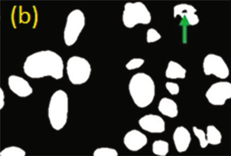
6                                                                                                     BioMed Research International

                                        (a)                                                  (b)

                                        (c)                                                  (d)

                                        (e)                                                  (f)

Figure 3: Postprocessing step in a SCC image: (a) original image, (b) mask after segmentation, (c) mask after the dilation operation, (d)
mask after applying the hole-filling operation, (e) mask after erosion operation, and (f) resulting segmentation.

these data indicate that the neural network presented similar         with p < 0:0001. Similarly, the mean nuclear perimeters for
sensitivity, specificity, and accuracy, indicating that the algo-      OL and PVL were 103.3 and 117.9, respectively, with p <
rithm performed well in identifying cell nuclei in all cases.         0:0001. As for solidity, an average of 0.9685 and 0.9621 for
                                                                      OL and PVL was observed, respectively, with p < 0:0001.
3.2. Morphological Features Extracted by the Neural                   Regarding the eccentricity and orientation characteristics,
Network. The nuclear features extracted by the neural net-            the means for OL and PVL were very similar and with p >
work were compared between OL and PVL. Altogether, the                0:05.
13 characteristics used in the classification were extracted
from the entire PVL ROI dataset and 1,196 of the 1,217                3.3. OL and PVL. For the separation between OL and PVL, a
OL ROIs. Regarding entropies, seven entropy measures were             polynomial classifier was used. For this, 119 ROIs from PVL
used, and the data for each entropy/lesion can be seen in             and 120 ROIs from OL randomly chosen were used by the
Table 2. The means for all entropies were always higher in            classifier to assess the degree of separability between them.
the PVLs when compared to the OLs, and the differences                 The classifier performed the cross-validation by dividing
were statistically significant (p < 0:0001).                           the ROIs into five groups (folds), where four of them were
    The Moran Index revealed a statistically significant dif-          used for training and one for testing, which were alternated
ference between OL and PVL, with means of 0.09702 and                 until all groups were trained and tested. As a result, the
0.1022, respectively (p = 0:0103). Regarding the area, the            mean AUC of the classifier was 97.06%, the mean sensitivity
averages for OL and PVL were 836.4 and 1,050, respectively,           was 95.83%, the mean specificity was 98.29%, and the mean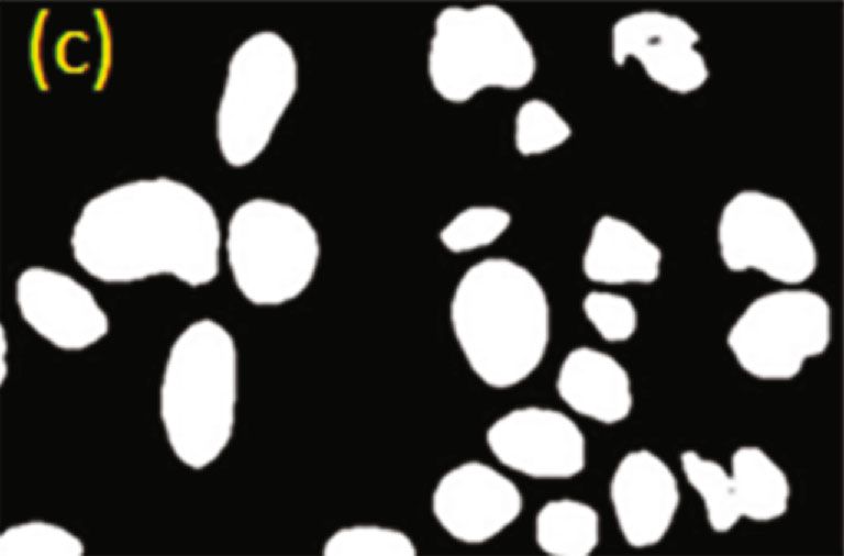
BioMed Research International                                                                                                            7

                                         (a)                                                   (b)

                                         (c)                                                  (d)

Figure 4: Operation to eliminate small artifacts in an SCC image: (a) resulting mask of the neural network; (b) segmentation resulting from
(a); (c) mask resulting from the process of eliminating objects smaller than 30 pixels; (d) segmentation resulting from (c).

         Table 1: Result of the segmentation of the Mask R-CNN in the different histopathological tissues of OL, PVL, and SCC.

Lesion                 SE (%)                     ES (%)                   AC (%)                      TC (%)                   DC (%)
OL                   81:89 ± 6:44              96:51 ± 2:28              93:15 ± 2:76                74:95 ± 8:21             83:80 ± 5:60
PVL                  80:79 ± 4:58              96:64 ± 1:61              92:93 ± 2:34                74:77 ± 5:71             83:82 ± 3:90
SCC                  80:24 ± 9:28              97:67 ± 1:83              94:63 ± 2:85                74:42 ± 8:81             83:47 ± 6:36

accuracy was 97.05%. These results show that the classifier             which can generate inconsistencies, difficulties, doubts, and
successfully distinguished the two lesions with a high degree          even diagnostic errors, according to pointed out by Al-
of sensitivity, specificity, and accuracy, indicating that such a       Rawi (2022) [13]. Since our results showed the presence of
tool has great potential for use in the differential diagnosis          detectable nuclear alterations among them, and that these
between them. From the obtained results, it is possible to             alterations in turn were able to differentiate them with high
observe the results obtained in each group and the mean                precision by the polynomial classifier, this study showed the
and standard deviation of the metrics used to evaluate the             importance of investigating the cell nucleus through
classifier’s performance.                                               machine learning in DBPMs for diagnostic purposes as well
                                                                       as in the elucidation of the characteristic nuclear properties
4. Discussion                                                          of each one of them. In light of advances in AI, this tool
                                                                       can be another foundation in the search for understanding
This study aimed to investigate the detection of nuclear               these lesions together with studies aimed at finding differen-
alterations in OLs and PVLs using a computer as well as                tial molecular biomarkers between them, which do not exist
the separation between them through the application of a               to date.
classifier based on morphological nuclear characteristics.                   Analyzing the clinicopathological data collected in this
The results of this study shed light on one of the problems            work, it can be seen that they are in line with the specific lit-
of oral pathology, which is to differentially diagnose a OL             erature. In general, the incidence of OL was higher in males
from a PVL, especially when the latter is at an early stage,           than in females; on the other hand, all cases of PVL were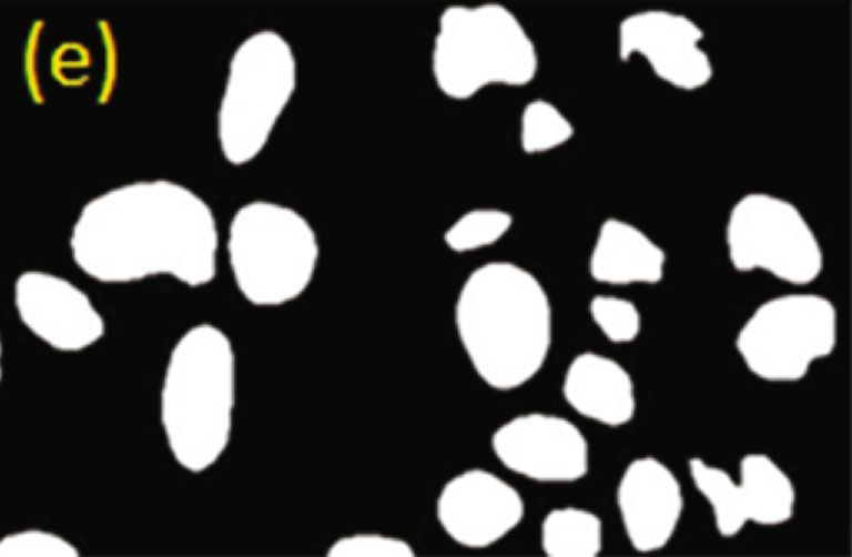
8                                                                                                     BioMed Research International

                          Table 2: Values of the seven levels of nuclear entropy evaluated between OL and PVL.

Variable                        Measures of central tendency and dispersion              LB                LVP                p-value
                                                 Average                                1968               2137
                                                 Median                                 1983               2149
Entropy 3 × 3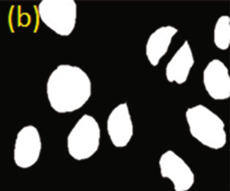
BioMed Research International                                                                                                        9

R-CNN proved to be the most effective in detecting nuclei in              Another characteristic evaluated in our study and related
studies that compared different methods of nuclear identifi-          to the nuclear texture is the Moran Index. There are no
cation. The study by Waal, Isaäc (2019) [16, 17], for exam-         works in the literature that use the Moran Index to assess
ple, segmented normal and abnormal nuclei of cervical               cell nuclei, except for Silva (2019). As in our study, in work
cells using the Mask R-CNN, obtaining an average accuracy           by Silva (2019), this feature proved useful in distinguishing
of 96%. They also compared this network with other seg-             the different lesions studied, with significant differences
mentation methods and found that these methods presented            between healthy tissue and tissue with dysplasia; in the pres-
lower results than the Mask R-CNN, such as the Multiscale           ent work, a significance was obtained between OL and PVL.
Watershed + Binary Classifier, which achieved an accuracy            In this sense, it can be concluded that both entropy and the
of 88%, the RGVF of 83%, and Patch-based FCM of 85%.                Moran Index can be used to detect changes in chromatin in
The work by Silva (2019) reached an average accuracy of             premalignant lesions, a fact reinforced by the study by
89.52% with the segmentation of nuclei of tongue epithelium         [20–22], who analyzed some nuclear characteristics and ver-
cells without and with mild, moderate, and severe dysplasias.       ified that the nuclear texture is an effective variable in differ-
Also in this study, other methods were tested in comparison         entiating the degrees of dysplasia in Barrett’s ssophagus, in
to his, and the results were inferior, such as the Otsu             addition to being efficient in predicting progression to can-
method, which presented an average accuracy of 60.78%,              cer, which, together with our results, further highlights the
K-means of 77.32%, and SegNet of 73.12%. These data reveal          importance of evaluating the nuclear textures in an attempt
that this neural network presents a good performance in             to elucidate the pathological conditions of premalignant
terms of nuclear segmentation, which was confirmed by                lesions.
our accuracy results in OLs, PVLs, and SCCs, corresponding               Regarding the area, our results point to a direct relation-
to 93.15%, 92.93%, and 94.63%, respectively. Future works           ship with some studies on DBPMs. The work of [23] showed
may further improve this network so that its use in the area        that the area of the cellular nuclei of the oral submucosal
of pathology becomes ubiquitous, as shown by Ananthara-             fibrosis lesion with dysplasia was greater than that of the
man et al. (2018), who developed a network based on the             normal nuclei, suggesting that this alteration could indicate
Mask R-CNN exclusively for the detection of nuclei called           the occurrence of tumorigenesis, reflecting the increase in
Nuclei R-CNN, with even better results than the original            the metabolic activity of these cells. Similar to the area,
Mask R-CNN.                                                         perimeter reflects the size of nuclei and is sometimes used
    As for the core features extracted from the entire OL and       together with area to infer changes in nuclear size. The work
PVL dataset after neural network training, including                by Krishnan et al. (2010) showed an increase in the perime-
entropy, Moran Index, area, perimeter, eccentricity, orienta-       ter of the nuclei between oral submucosal fibrosis with dys-
tion, and solidity, they were compared between the two              plasia and normal oral mucosa. Solidity is a descriptor used
lesions. Significant differences were found between OL and            in the assessment of nuclear deformity. In our results, a sig-
PVL in the seven entropy levels evaluated in our study.             nificant difference was found between the two injuries, also
Entropy is a measure that assesses the disorder of the nuclear      proving to be a useful variable in distinguishing between
texture, which, in turn, directly reflects the organization of       OL and PVL. Here, the mean solidity in the PVLs was
chromatin and, consequently, the genetic and epigenetic             slightly lower than in the OLs. Unlike our study, work by
changes that occur in the DNA molecule during the tumor-            Krishnan et al. [23] found no significant differences in the
igenesis process [18]. Thus, nuclear entropy has been studied       solidity of nuclei in an attempt to discriminate oral submu-
in several types of cancers, focusing on determining clinical       cosal fibrosis from normal oral mucosa. Similarly, the study
prognosis and tumor aggressiveness. In the study by, which          [19] also did not detect substantial differences in nuclear
investigated different types of cancers including the colon,         solidity between nonsmoking smokers and patients with
ovary, uterus, prostate, and endometrium, it was observed           PMBD. One hypothesis is that PVLs have a high power of
that patients who exhibited more heterogeneous texture pat-         malignant transformation compared to other MPDs,
terns, that is, higher entropy levels, had worse survival for all   increasing the chances of nuclear deformity and leaving the
tumors evaluated. In our study, it is interesting to note that      nuclei less convex and more irregular, a characteristic found
all mean entropy levels analyzed were higher in PVLs than           in cancer cells, as described. Solidity is a variable that is still
in OLs, suggesting a greater chromatin disorder in those            little investigated in studies involving the evaluation of
lesions to the detriment of these, which partly may explain         nuclear morphology by computational algorithms. There-
the greater potential of PVLs to progress to SCC.                   fore, it is expected that further research may include it in
    Similarly, [19] found a consecutive increase in entropy in      the evaluation of nuclei in different DBPMs as a possible
the epithelial cell nuclei of nonsmoking smokers and                descriptor that characterizes different lesions, as observed
patients with precancerous conditions. Therefore, these             in our study.
entropy results seem to be useful in distinguishing lesions              We found no significant differences in eccentricity and
with lower and higher malignant transformation potential,           orientation between OL and PVL. Interestingly, the study
in the case of LB and PVL, respectively, suggesting the             by [24–28] also found no differences in eccentricity between
occurrence of distinct genetic and epigenetic alterations           oral submucosal fibrosis nuclei and buccal mucosa. Thus,
between them. Further studies will assess whether higher            further studies should be conducted in order to determine
entropy indices in OLs indicate a greater risk of malignant         the value of these variables in DBPMs more clearly. Thus,
transformation, as seems to be the case for PVLs.                   it is possible to conclude from our investigation that both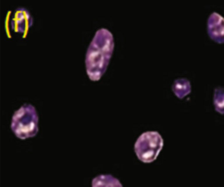
10                                                                                                BioMed Research International

lesions carry distinct nuclear alterations. Our findings also      time. In addition, the characteristics found in the nuclei of
support the importance of the nuclear study from computa-         the two lesions can provide important information in the
tional techniques evaluated by an AI, a promising area in         construction of a model to assess the risk of malignant trans-
medicine that, without a doubt, will shape the field of            formation of these disorders, being extremely important in
pathology soon, finding new ways to interpret the patholog-        making therapeutic decisions for each case. Added to this,
ical processes that occur. In various diseases. Based on the      the use of a classifier could also be used in the future as an
evidence found in our study and in the works mentioned            additional tool for cases of a difficult diagnosis. Finally, our
here, these descriptors may prove to be useful in predicting      investigations are added to the various works that show
the progression to SCC of PMBDs, such as OL and PVL.              machine learning as a new possibility for studies in pathol-
The investigated nuclear features were transformed into vec-      ogy, being an effective, low-cost method that will possibly
tors so that the classifier could dichotomize the samples          be used on a large scale in the near future in clinical routines,
between the two lesions. The classifier used all 119 PVL           adding speed, precision, and prediction in diagnoses.
ROIs and 120 OL ROIs to assess the degree of separability.
It is worth mentioning that for the classification to perform
well, it is important to have a balanced number among the         Data Availability
samples to be investigated. Therefore, only 120 OL ROIs           The data used to support the findings of this study are
were used by the classifier [29–33].                               included within the article.
     The polynomial classifier proved to be very effective in
the studies in which it was adopted, with extremely satisfac-
tory performance [15] in the classification between normal         Conflicts of Interest
and abnormal tissues of mammograms through texture
analysis, reaching an AUC of 98%, a performance superior          The authors declare that they have no conflicts of interest.
to the SVM, decision tree, and K-NN classifiers, which were
also compared in the study. Similarly, in work by Silva           Acknowledgments
(2019), the polynomial classifier achieved an average AUC
of 92% in the classification between healthy tissues and those     The authors extend their appreciation to Princess Nourah
with different degrees of dysplasia, that is, a superior result    bint Abdulrahman University Researchers Supporting Pro-
than the multilayer perceptron, decision tree, and random         ject number (PNURSP2022R141), Princess Nourah bint
classifiers. Forest was compared in their study. Similarly,        Abdulrahman University, Riyadh, Saudi Arabia.
our result was excellent, with an average UAC of 97.06%,
indicating that this classifier can be an additional tool for
pathologists in defining a histopathological diagnosis since       References
the diagnosis of PVLs is still a constant challenge in oral        [1] World Health Organization, WHO global report on trends in
pathology, and that can be easily confused with OLs, espe-             prevalence of tobacco smoking 2015, WHO, 2015, https://apps
cially in the initial cases. Thus, our study proposes the use          .who.int/iris/handle/10665/156262.
of AI as a tool that raises the criteria currently adopted in      [2] A. G. Zygogianni, G. Kyrgias, P. Karakitsos et al., “Oral squa-
the distinction between these two lesions, reducing the mar-           mous cell cancer: early detection and the role of alcohol and
gin of doubt and improving the accuracy of the diagnosis               smoking,” Head & Neck Oncology, vol. 3, no. 1, p. 2, 2011.
with less subjectivity. Further studies are needed to verify       [3] A. A. Hamad, M. L. Thivagar, M. B. Alazzam et al., “Dynamic
whether this method can be used in the early stages of PVLs,           systems enhanced by electronic circuits on 7D,” Advances in
when they are usually labeled as OLs without any evidence              Materials Science and Engineering, vol. 2021, Article ID
of evolution to more aggressive forms, which would be of               8148772, 2021.
great importance in determining an early diagnosis of PVLs,        [4] P. Speight, S. A. Khurram, and O. Kujan, “Oral potentially
increasing the chances of successful treatment.                        malignant disorders: risk of progression to malignancy,” Oral
                                                                       Surgery, Oral Medicine, Oral Pathology, Oral Radiology,
                                                                       vol. 125, no. 6, pp. 612–627, 2018.
5. Conclusion                                                      [5] E. Mustafa, S. Parmar, and P. Praveen, “Premalignant lesions
Based on the investigations carried out in this work, the              and conditions of the oral cavity,” in Oral and Maxillofacial
                                                                       Surgery for the Clinician, K. Bonanthaya, E. Panneerselvam,
present study showed that, despite being histologically simi-
                                                                       S. Manuel, V. V. Kumar, and A. Rai, Eds., Springer, Singapore,
lar, OLs and PVLs carry distinct nuclear properties that can
                                                                       2021.
be used for differential diagnosis between them, thus, help-
                                                                   [6] M. B. Alazzam, W. T. Mohammad, M. B. Younis et al., “Study-
ing to resolve one of the major challenges of oral pathology           ing the effects of cold plasma phosphorus using physiological
in the search for more effective and accurate ways to estab-            and digital image processing techniques,” Computational and
lish the differential diagnosis between OL and PVL. The fact            Mathematical Methods in Medicine, vol. 2022, 5 pages, 2022.
that the neural network has achieved an excellent perfor-          [7] L. A. Owki, E. Othieno, J. Wandabwa, and A. Okoth, “Preva-
mance in nuclear identification through the supervised                  lence of cancerous and pre-malignant lesions of cervical cancer
training performed reveals that this method can be a great             and their association with risk factors as seen among women in
ally in our later works involving histological studies, includ-        the regions of Uganda,” Journal of Clinical and Laboratory
ing the analysis of more cases of OLs and PVLs in the first             Medicine, vol. 2, 2019.
BioMed Research International 11
[8] P. Speight, “Update on oral epithelial dysplasia and progres- itoring system using interactive E-app,” Computational Intelli-
sion to cancer,” Head and Neck Pathology, vol. 1, no. 1, gence and Neuroscience, vol. 2021, 7 pages, 2021.
pp. 61–66, 2007. [24] I. Waal, “Oral leukoplakia: present views on diagnosis, man-
[9] H. Sawhney and C. Kumar, “Correlation of serum biomarkers agement, communication with patients, and research,” Cur-
(TSA & LSA) and epithelial dysplasia in early diagnosis of oral rent Oral Health Reports, vol. 6, no. 1, pp. 9–13, 2019.
precancer and oral cancer,” Cancer Biomarkers, vol. 10, [25] I. Y. A.-A. A. R. Saad, “Social intelligence and its relationship
pp. 43–49, 2011. to decision quality,” Scientific Journal Al-Imam University Col-
[10] Amjad Abbas Abdel Rahim Al-Baldawi, “The possibility of lege, vol. 1, pp. 1–22, 2022.
implementing industrial incubators and their role in the devel- [26] M. F. Jwaid and T. Baraskar, “An efficient technique for image
opment of small industry and medium in Iraq,” Scientific Jour- forgery detection using local binary pattern (Hessian and cen-
nal Al-Imam University College, vol. 1, pp. 1–22, 2022. ter symmetric) and transformation method,” Scientific Journal
[11] A. A. Hamad, M. L. Thivagar, M. B. Alazzam, F. Alassery, Al-Imam University College, vol. 1, pp. 1–11, 2022.
F. Hajjej, and A. A. Shihab, “Applying dynamic systems to [27] R. Anantharaman, M. Velazquez, and Y. Lee, “Utilizing Mask
social media by using controlling stability,” Computational R-CNN for detection and segmentation of oral diseases,” in
Intelligence and Neuroscience, vol. 2022, Article ID 4569879, 2018 IEEE International Conference on Bioinformatics and
2022. Biomedicine (BIBM), pp. 2197–2204, Madrid, Spain, 2018.
[12] B. Moxley-Wyles, R. Colling, and C. Verrill, “Artificial intelli- [28] H. Mahmood, M. Shaban, B. Indave, A. Santos-Silva,
gence in pathology: an overview,” Diagnostic Histopathology, N. Rajpoot, and S. A. Khurram, “Use of artificial intelligence
vol. 26, no. 11, pp. 513–520, 2020. in diagnosis of head and neck precancerous and cancerous
[13] N. Sinha, M. T. Nayak, S. Gupta, D. Dwivedi, N. Swarup, and lesions: a systematic review,” Oral Oncology, vol. 110, article
M. Agarwal, “Proliferative verrucous leukoplakia: a diagnostic 104885, 2020.
perplexity,” International Journal of Research and Reports in [29] S. Dey, R. Sarkar, K. Chatterjee, P. Datta, A. Barui, and S. P.
Dentistry, vol. 4, pp. 29–34, 2021. Maity, “Pre-cancer risk assessment in habitual smokers from
[14] M. García-Pola, E. Pons-Fuster, C. Suárez-Fernández, DIC images of oral exfoliative cells using active contour and
J. Seoane-Romero, A. Romero-Méndez, and P. López-Jornet, SVM analysis,” Tissue and Cell, vol. 49, no. 2, pp. 296–306,
“Role of artificial intelligence in the early diagnosis of oral 2017.
Cancer. A Scoping Review,” Cancers, vol. 13, no. 18, p. 4600, [30] M. M. R. Krishnan, C. Chakraborty, R. R. Paul, and A. K. Ray,
2021. “Hybrid segmentation, characterization and classification of
[15] K. He, G. Gkioxari, P. Dollár, and R. Girshick, Eds., “Mask R- basal cell nuclei from histopathological images of normal oral
CNN,” in 2017 IEEE International Conference on Computer mucosa and oral submucous fibrosis,” Expert Systems with
Vision (ICCV), pp. 2980–2988, Venice, Italy, 2017. Applications, vol. 39, no. 1, pp. 1062–1077, 2012.
[16] B. A. M. Muhammad, “The role of universities in developing [31] D. Zink, A. H. Fischer, and J. A. Nickerson, “Nuclear structure
societies by accreditation on scientific research,” Scientific in cancer cells,” Nature Reviews Cancer, vol. 4, no. 9, pp. 677–
Journal Al-Imam University College, vol. 1, pp. 1–19, 2022. 687, 2004.
[17] M. Z. Do Nascimento, A. S. Martins, L. A. Neves, R. P. Ramos, [32] A. Kleppe, F. Albregtsen, L. Vlatkovic et al., “Chromatin orga-
E. L. Flores, and G. A. Carrijo, “Classification of masses in nisation and cancer prognosis: a pan-cancer study,” The Lan-
mammographic image using wavelet domain features and cet Oncology, vol. 19, no. 3, pp. 356–369, 2018.
polynomial classifier,” Expert Systems with Applications, [33] E. Sabo, A. H. Beck, E. A. Montgomery et al., “Computerized
vol. 40, no. 15, pp. 6213–6221, 2013. morphometry as an aid in determining the grade of dysplasia
[18] L. Gongas, A. M. Moreno, and L. M. Bravo, “Automated diag- and progression to adenocarcinoma in Barrett's esophagus,”
nosis of breast cancer based on histological images,” in 2018 IX Laboratory Investigation, vol. 86, no. 12, pp. 1261–1271, 2006.
International Seminar of Biomedical Engineering (SIB), pp. 1–
6, Bogota, Colombia, 2018.
[19] A. B. Silva, Computational methods for analysis and classifica-
tion of dysplasias in oral cavity images, [M.S. thesis], Federal
University of Uberlândia, Uberlândia, 2019.
[20] N. Al-Rawi, A. Sultan, B. Rajai et al., “The effectiveness of arti-
ficial intelligence in detection of oral cancer,” International
Dental Journal, vol. 12, 2022.
[21] S. Khanagar, S. Naik, A. Kheraif et al., “Application and Per-
formance of Artificial intelligence technology in oral cancer
diagnosis and prediction of prognosis: a systematic review,”
Diagnostics, vol. 11, no. 6, p. 1004, 2021.
[22] R. Maghsoudi, A. Bagheri, and M. Maghsoudi, “Diagnosis pre-
diction of lichen planus, leukoplakia and oral squamous cell
carcinoma by using an intelligent system based on artificial
neural networks,” Journal of Dentomaxillofacial Radiology,
Pathology and Surgery, vol. 2, no. 2, pp. 1–8, 2013.
[23] M. B. Alazzam, H. Mansour, F. Alassery, and A. Almulihi,
“Machine learning implementation of a diabetic patient mon-You can also read