Role of detrusor PDGFRα+ cells in mouse model of cyclophosphamide induced detrusor overactivity - Nature
←
→
Page content transcription
If your browser does not render page correctly, please read the page content below

www.nature.com/scientificreports
OPEN Role of detrusor PDGFRα+
cells in mouse model
of cyclophosphamide‑induced
detrusor overactivity
Haeyeong Lee1*, Byoung H. Koh1, Lauren E. Peri1, Holly J. Woodward2, Brian A. Perrino1,
Kenton M. Sanders1 & Sang Don Koh1
Cyclophosphamide (CYP)-induced cystitis is a rodent model that shares many features common
to the cystitis occurring in patients, including detrusor overactivity (DO). Platelet-derived growth
factor receptor alpha positive (PDGFRα+) cells have been proposed to regulate muscle excitability in
murine bladders during filling. PDGFRα+ cells express small conductance Ca2+-activated K+ channels
(predominantly SK3) that provide stabilization of membrane potential during filling. We hypothesized
that down-regulation of the regulatory functions of PDGFRα+ cells and/or loss of PDGFRα+ cells
generates the DO in CYP-treated mice. After CYP treatment, transcripts of Pdgfrα and Kcnn3 and
PDGFRα and SK3 protein were reduced in detrusor muscle extracts. The distribution of PDGFRα+
cells was also reduced. Inflammatory markers were increased in CYP-treated detrusor muscles. An
SK channel agonist, CyPPA, increased outward current and hyperpolarization in PDGFRα+ cells.
This response was significantly depressed in PDGFRα+ cells from CYP-treated bladders. Contractile
experiments and ex vivo cystometry showed increased spontaneous contractions and transient
contractions, respectively in CYP-treated bladders with a reduction of apamin sensitivity, that could
be attributable to the reduction in the SK conductance expressed by PDGFRα+ cells. In summary,
PDGFRα+ cells were reduced and the SK3 conductance was downregulated in CYP-treated bladders.
These changes are consistent with the development of DO after CYP treatment.
Interstitial cystitis (IC) is characterized by suprapubic and/or bladder pain that is accompanied with an increase in
urinary urgency, frequency and nocturia1,2. The pathology of IC does not follow that of other diseases/syndromes
in the bladder, such as carcinoma, urinary tract infections or cystitis introduced by radiation or m edication3,4.
Although the pathogenesis of IC is still not fully understood, several factors have been suggested which include
neuronal, urothelium, and myogenic causes with likely associated i nflammation4.
Cyclophosphamide (CYP) causes cystitis in humans5,6. One of the commonly used models for cystitis in
rodents is CYP-induced cystitis, usually induced by injections7. This model shares many features with cystitis
occurring in human patients treated with CYP, as well as common features with bladder pain syndrome/inter-
stitial cystitis (BPS/IC)8–11. CYP causes functional and histological changes in humans and rodents12,13, such
as expression of receptors and signaling molecules in the urothelium/mucosa. CYP-induced changes include
up-regulation of nitric oxide s ynthase10,14,15, increased urothelial muscarinic M5 r eceptors16, and mucosal
permeability15. Furthermore, after CYP-injection, smaller micturition volumes have been noted17–19 that likely are
due to upregulated afferent and efferent neural effects16,20–22. CYP can also induce detrusor overactivity (DO)23.
CYP-treated mice showed upregulation of connexin 43 (GJA1) and gap junction blockers attenuated spontane-
ous contractions in CYP-treated strips, and decreased urinary frequency with increased total voided volume in
voiding behavior test of CYP-treated bladder24. However, the functional changes in the detrusor interstitial cells
responsible for DO have not been determined.
Recently, a unique population of interstitial cells were identified in the bladder. These cells are immunoposi-
tive for platelet-derived growth factor receptor alpha (PDGFRα), and therefore are referred to by this chemical
coding, i.e. PDGFRα+ cells. PDGFRα+ cells have been identified in murine, pig and human detrusor muscles25,26.
1
Department of Physiology and Cell Biology, School of Medicine, University of Nevada, Reno, NV 89557, USA. 2The
Roslin Institute, The University of Edinburgh, Easter Bush Campus, Midlothian EH25 9RG, UK. *email: hylee@
unr.edu
Scientific Reports | (2022) 12:5071 | https://doi.org/10.1038/s41598-022-09155-3 1
Vol.:(0123456789)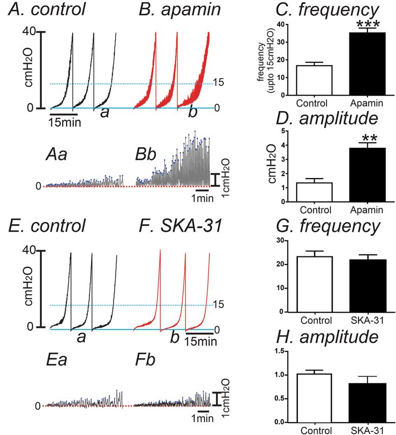
www.nature.com/scientificreports/
Figure 1. Quantitative analysis of transcripts in control and CYP-treated detrusor muscles. (A) Transcripts
for Pdgfra in control and CYP-treated detrusor muscle. (B) Transcripts for Kcnn in control and CYP-treated
detrusor muscle. (C) Transcripts for Tnf and Il6 in control and CYP-treated detrusor muscle. (D) Comparison
of transcriptional expression of Pdgfra and Kcnn3 from sorted PDGFRα+ cells in saline-injected (white bar)
and CYP-treated detrusors. (E) Comparison of transcriptional expression of Kcnma1and Cacna1c from
sorted smooth muscle cells (SMC) in saline-injected (white bar) and CYP-treated detrusors. Expression of all
transcripts was normalized to Gapdh. **Denotes P < 0.01 by unpaired t-test (n = 4 in all samples).
These cells are located on the edges of smooth muscle bundles and have multiple branches that may couple to and
form an electrical syncytium with smooth muscle cells (SMCs)25,27,28. The function of PDGFRα+ cells in detrusor
muscles has been investigated with molecular and electrophysiological techniques. PDGFRα+ cells displayed
higher current density of small conductance Ca2+-activated K+ (SK) channels26,29, as compared to detrusor SMCs.
Activation of SK channels in PDGFRα+ cells could provide a stabilizing influence on smooth muscle excitatiiby.
We hypothesized that loss-of-function in PDGFRα+ cells or SK channels could lead to DO in cystitis. This study
describes investigation into the molecular and functional changes that occur in detrusor PDGFRα+ cells and
how these changes relate to increased detrusor overactivity in CYP-injected bladder.
Results
Downregulation of PDGFRα and SK channel in murine bladders from CYP‑induced cysti‑
tis. We compared the transcriptional expression of Pdgfra and Kcnn1–4 in detrusor muscles from CYP-treated
and saline-treated mice. Pdgfra and Kcnn3 (SK3) were significantly downregulated (P < 0.01 by unpaired t-test),
but neither SK1–2 (Kcnn1 and Kcnn2) nor IK (Kcnn4) were changed significantly after CYP-treatment (n = 4,
Fig. 1A,B). Detrusor muscles from CYP-treated mice displayed a significant increase in Il6 and Tnf (inflamma-
tory markers) (n = 4, Fig. 1C) suggesting the onset of bladder inflammation after CYP treatment. We further
examined the transcriptional expression of Pdgfra and Kcnn3 in sorted PDGFRα+ cells from saline- and CYP-
treated PDGFRα/eGFP mice (see “Methods” section). Pdgfra and Kcnn3 were significantly decreased in CYP-
treated mice in comparison to mice treated with saline (n = 4, P < 0.01 in both genes, Fig. 1D). Transcriptional
changes were also evaluated in detrusor smooth muscle cells (SMC). SMCs were isolated from saline- and CYP-
treated smMHC/Cre/eGFP (see “Methods” section) mice, as previously described26. Main excitability-related
genes in detrusor SMC, Kcnma1 (BK channels) and Cacna1c (L-type Ca2+ channels) were unchanged in CYP-
treated SMC (n = 4, Fig. 1E).
Levels of transcripts do not necessarily translate linearly into protein expression. Therefore, we also employed
immunohistochemistry and Wes analysis to confirm parallel changes in protein expression. Immunohistochem-
istry revealed PDGFRα immune-positive cells were down-regulated in CYP-treated detrusor compared with
age-matched saline-injected controls (Fig. 2A,B). Figure 2C shows a negative control image in which the primary
antibody was omitted and the muscles were treated with only secondary antibody. Wes analysis showed in a
more quantitative way that PDGFRα and SK3 were significantly downregulated in CYP-treated detrusor mus-
cles, as compared to controls (n = 4, Fig. 3). These data are consistent with transcriptional data, and suggested
the possibility that down-regulation of Pdgfrα and/or Kcnn3 transcripts could be involved in generation of DO.
Scientific Reports | (2022) 12:5071 | https://doi.org/10.1038/s41598-022-09155-3 2
Vol:.(1234567890)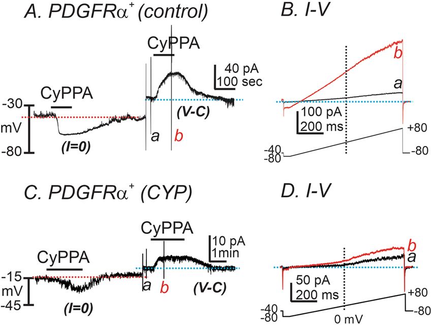
www.nature.com/scientificreports/
Figure 2. Immunohistochemistry of PDGFRα in murine detrusor from control and CYP-treated bladders. (A)
Whole mount detrusor with PDGFRα staining (green) from saline-treated control mouse. (B) Immunoreactivity
with PDGFRα (green) antibody was decreased in CYP-treated bladder detrusor compared with control (A). (C)
Secondary antibody only (no primary antibody control) produce little to no visual background. L and D denote
lamina propria and detrusor muscle, respectively.
Figure 3. Expression levels of PDGFRα and SK3 in control and CYP-treated detrusor muscles. (A,C)
Representative gels of PDGFRα (A) and SK3 (C) expression in control and CYP-treated detrusor muscle
[Original Wes image blots for PDGFRα and SK3 (Supplementary Material)]. (B,D) Quantification analysis of
expression level of PDGFRα (B) and SK3 (D) in control and CYP-treated detrusor muscle. **Denotes P < 0.005,
***denotes P < 0.001.
The effect of SK channel activator on the generation of outward currents and membrane
potentials in detrusor PDGFRα+ cells from CYP‑treated bladders. We tested the effect of a SK chan-
nel activator (CyPPA, 10 μM) on freshly dispersed detrusor PDGFRα+ cells. The cells were dialyzed with K+-rich
solution (see “Methods” section). CyPPA hyperpolarized control detrusor PDGFRα+ cells from − 31.4 ± 4.6
to − 61.6 ± 2.9 mV under current clamp (I = 0, red dot line, n = 5, Fig. 4A). In the same cells, CyPPA activated
outward current at a holding potential of − 40 mV in voltage-clamp (V-C) mode (Fig. 4A, blue dotted line).
CyPPA-activated current amplitude averaged 39.3 ± 7.1 pA. Figure 4B shows currents evoked by ramp depolari-
Scientific Reports | (2022) 12:5071 | https://doi.org/10.1038/s41598-022-09155-3 3
Vol.:(0123456789)www.nature.com/scientificreports/
Figure 4. The effect of SK channel activator on membrane currents and potential in PDGFRα+ cells from
control and CYP-treated mice. (A) In detrusor PDGFRα+ cells from control bladders, CyPPA (10 μM) induced
membrane hyperpolarization under current clamp (I = 0, red dot line). In the same cell, CyPPA activated
outward current (current above blue dotted line) at a holding potential of − 40 mV under voltage-clamp
mode (V-C). (B) Current responses to ramp-depolarizations from (A) before (a) and during (b) CyPPA. Inset
denotes voltage-protocol. (C) In detrusor PDGFRα+ cells from CYP-treated bladders, CyPPA (10 μM) induced
membrane hyperpolarization under current clamp (I = 0, red dot line). In the same cell, CyPPA activated
outward current (current above blue dotted line) at a holding potential of − 40 mV under voltage-clamp mode
(V-C). (D) Current responses to ramp-depolarizations from (C) before (a) and during (b) CyPPA. Inset denotes
voltage-protocol.
zation before (Fig. 4Ba, black trace) and in the presence of CyPPA (Fig. 4Bb, red trace). The resting membrane
potentials (RMP) of CYP-treated detrusor PDGFRα+ cells were depolarized to − 16 ± 1.4 mV (n = 5, P < 0.01, as
compared to untreated control PDGFRα+ cells). CyPPA induced less hyperpolarization in CYP-treated detru-
sor PDGFRα+ cells (∆mV 9.1 ± 2.0), as compared to untreated control PDGFRα+ cells (n = 5, P < 0.01, Fig. 4C)
under current-clamp mode (I = 0) and generated smaller outward currents under V-C mode (5.2 ± 0.9 pA; n = 5,
P < 0.01, Fig. 4D) at − 40 mV. These data suggest that SK current density was significantly decreased in PDGFRα+
cells isolated from CYP-treated bladders, as compared with current density in control PDGFRα+ cells isolated
from non-treated bladders.
The effect of SK channel blocker on detrusor muscle contractions in CYP‑treated blad‑
ders. We examined the effect of an SK channel blocker to compare changes in functional expression of SK
channels between saline-treated (control) and CYP-treated bladders using isometric force measurements. Det-
rusor muscle strips without submucosa exhibited spontaneous contractions. In saline-injected control, apamin
(300 nM, a selective blocker of SK channels) dramatically increased AUC from 62.9 ± 7.9 to 261.5 ± 22.5 mN s
during 5 min recordings (n = 8, P < 0.0001, Fig. 5A,C). These data are consistent with previous r eports30–32. In
CYP-treated detrusor muscle strips, spontaneous contractions were of high amplitude and irregular. AUC for
these contractions was calculated since averaging the frequency and amplitude of these irregular contractions
are not reliable measurements. AUC in CYP-treated muscles before and after apamin were 315.7 ± 69.8 mN s
and 341.3 ± 66.4 mN s (n = 6, Fig. 5B,D), respectively. Thus, apamin had no significant effect on spontaneous
contractile activity in CYP-treated muscles. We also calculated the apamin-sensitive contractions by normalized
the effect of apamin from control AUC (before apamin). The sensitivity to apamin was significantly decreased in
CYP-treated detrusor muscles (1.1 ± 0.1-fold) compared to saline-injected detrusor muscle strips (4.6 ± 0.6 fold,
P < 0.001, Fig. 5D).
The effect of SK channel blocker and agonist on CYP‑treated bladders using ex vivo prepara‑
tion. We also examined the pressure–volume relationships of excised bladders in ex vivo preparations to
investigate changes in functional expression of SK channel in control and CYP-treated bladders. Ex vivo pres-
sure–volume measurements exclude extrinsic neural regulation during filling, so this technique highlights regu-
lation via myogenic mechanisms. In in vivo cystometry, voiding contractions start around 20 c mH2O in murine
bladder33. Thus, we analyzed the pressure amplitude and frequency up to 15 c mH2O (see Fig. 6A,B) to associate
contractile activity (i.e. transient contractions or TCs) to the non-voiding contractions (NVCs) observed in
in vivo cystometry. The passive pressure underlying was normalized to 0 cmH2O (see Fig. 6Aa,Bb). In control
bladders, infusion of KRB solution (15 μl/min) induced a small increase in intravesical amplitude (1.3 ± 0.3
cmH2O) with the frequency of TCs equal to 17 ± 2 events at intravesical pressures up to 15 cmH2O. Addition of
apamin (300 nM) into the bath increased TC frequency to 35 ± 3 events (P < 0.001, Fig. 6A–C) and the amplitude
of TCs to 3.8 ± 0.4 c mH2O (n = 6, P < 0.01, Fig. 6A,B,D). We also tested the effect of SK chanel agonist, SKA-31
on control bladders. SKA-31( 10 µM) showed negligible effect on the frequency and amplitude of TCs (n = 7,
Scientific Reports | (2022) 12:5071 | https://doi.org/10.1038/s41598-022-09155-3 4
Vol:.(1234567890)www.nature.com/scientificreports/
Figure 5. The effect of SK channel blocker on contractions in saline- and CYP-treated detrusor muscle strips.
(A,B) Apamin increased contractility in saline-injected detrusor muscle (A), but did not show significant effect
in CYP-treated muscle strip (B). (C) Summarized the area under the curve for 5 min recordings before (control)
and after apamin in saline-injected (n = 8) and CYP-treated (n = 6) detrusor muscles. (D) Normalized apamin
sensitivity from (C) in saline and CYP treated detrusor muscles. ****Denotes P < 0.0001 by paired t-test in (C)
and ***denotes P < 0.001 by unpaired t-test in (D).
Fig. 6E–H) because SK channels may be already activated during filling. Thus, there is minmal activation by SK
channel opener. CYP-treated bladders displayed increased amplitude of TCs with high frequency before apamin
treatment (Fig. 7A–C). SKA-31 and apamin treatment did not significantly affect the amplitude or frequency of
TCs in CYP-treated bladders (n = 6, Fig. 7B,C).
Discussion
This study investigated the mechanisms of DO in CYP-induced cystitis. Gene transcripts and protein levels of
PDGFRα and SK3 were depressed in CYP-treated detrusor muscles and in PDGFRα+ cells sorted to purity by
FACS. The current density from SK channels evoked by a SK channel agonist, CyPPA, was also depressed in
PDGFRα+ cells isolated from CYP-treated detrusor muscles, suggesting that gene and protein expression levels
have functional consequences on bladder function. Contractile experiments supported the idea that apamin-
sensitive contractions were decreased in CYP-treated detrusor muscle strips. In ex vivo experiments, an SK
channel antagonist, apamin, increased the amplitude and frequency of TCs during filling in control bladders.
However, the effects of apamin were reduced significantly in CYP-treated bladders. Similarly the effects of an
SK agonist were also minimal in CYP-treated bladders. These data suggest that loss of PDGFRα+ cells and the
major functional conductance provided by SK channels in these cells results in increased TCs and induction of
DO in CYP-treated bladders.
Chemical cystitis is one of the adverse effects observed after administration of CYP chemotherapy in
humans34. CYP also induces cystitis in mice and r ats13,35. Therefore, CYP treatments of rodents has been used
widely as an experimental model of IC/BPS. Single CYP intraperitoneal injection leads to urinary bladder
inflammation36, visceral p ain8 and D
O23. However, the mechanisms of DO induced by CYP have not been
Scientific Reports | (2022) 12:5071 | https://doi.org/10.1038/s41598-022-09155-3 5
Vol.:(0123456789)www.nature.com/scientificreports/
Figure 6. The Effect of SK channel blocker on spontaneous activity using the ex vivo volume-pressure
relationship in control bladders. (A,B) Ex vivo compliance curves for control (A) and after apamin application
(B) from control bladders up to 15 cmH2O (see blue dotted line). Expanded time scales with adjustment
of baseline under control (Aa) and apamin (Bb) from panel (A,B) respectively. Blue dots were detected by
threshold (0.2cmH2O) and used for frequency analysis. (C,D) Summarized frequency (C) and amplitude (D)
under control and after apamin application (n = 6). ** and ***Denote P < 0.01 and P < 0.001, respectively. (E,F)
The effect of SK chanel agonist, SKA-31 (10 µM) on control bladders without (E) and with SKA-31 (F) in ex vivo
preparation. Expanded time scales with adjustment of baseline under control (Ea) and SKA-31 (Fb) from (E,F),
respectively. (G,H) Summarized frequency (G) and amplitude (H) under control and after SKA application
(n = 7).
clarified by previous studies. In the current study we tested the hypothesis that functional causes of DO after
CYP treatment could be related to loss-of-function of bladder regulation provided by PDGFRα+ cells.
Previous studies have shown that as the bladder fills, volume increases and the walls are stretched, but
intravesical pressure remains low during much of the period of fi lling37. This accommodation occurs even
though there is a natural tendency for detrusor SMCs to contract in response to stretch38,39. During bladder
filling, NVCs are detected in cystometric records from various s pecies40–43. NVCs appear to correspond to
localized contractions that are also observed in ex vivo bladder preparations and have been termed ‘spontane-
ous phasic contractions’, ‘micromotions’ or ‘TCs’44–48. TCs increase as bladder filling proceeds44. Activation of
non-selective cation channels expressed in detrusor SMCs38,39 generates TCs which initiate sensory inputs via
activation of afferent nerves44 that propagate to the central nervous system during bladder filling49–52. However,
under physiological condition, development of TCs can be restrained due to activation of SK channels in det-
rusor muscles26. For instance, SK channel activators reduce and SK channel blocker increase detrusor muscle
contractions30,32,53–55. Furthermore, Kcnn3 knockout mice show an increase in NVCs in in vivo cystometry and
TCs in ex vivo b ladders44,56. Previous reports demonstrate that Kcnn3 are highly expressed in detrusor PDGFRα+
cells26,29,57. The current density of SK channels is very low in SMCs compared to detrusor PDGFRα+ cells25.
Although there is no direct evidence of electrical coupling between PDGFRα+ cells and SMCs, it seems clear
that PDGFRα+ cells may serve an important stabilizing role in regulating contractile activity of SMCs during
Scientific Reports | (2022) 12:5071 | https://doi.org/10.1038/s41598-022-09155-3 6
Vol:.(1234567890)www.nature.com/scientificreports/
Figure 7. The Effect of SK channel activator and blocker on TCs in the ex vivo volume-preparation from CYP-
treated bladders. (A) Ex vivo compliance curves for control, SKA-31 and apamin application from CYP-treated
bladders. (B,C) Summarized frequency (B) and amplitude (C) under control, SKA31 and apamin application
(n = 6) from CYP-treated bladders.
bladder filling. Therefore, disruption of PDGFRα+ cells and/or downregulation of SK channels and intracellular
signaling pathways regulating SK channel activity could lead to DO.
In the present study we found that Kcnn3 and Pdgfra transcripts were depressed in CYP-treated detrusor
muscles and in detrusor PDGFRα+ cells from CYP-treated PDGFRα/eGP mice. In contrast, Kcnma1 and Cac-
na1C that encode important proteins that have been implicated in the regulation of bladder excitability were
unchanged from control SMCs in mice treated with CYP. Western analysis confirmed that both PDGFRα and
SK3 proteins were reduced in CYP-treated detrusor muscles.
Immunohistochemistry also showed a reduction in the density of PDGFRα+ cells in CYP-injected detrusor
muscle. At present we do not know what factors, activated by CYP, might be responsible for loss of PDGFRα+
cells and downregulation of SK channels in detrusor muscles, however the upregulation of inflammatory factors,
such as Tnfα and Il6, suggest that an inflammatory mechanism may be involved.
Functional studies to evaluate the state of the SK conductance in detrusor PDGFRα+ cells before and after
CYP treatment were performed using the patch clamp technique. Our recordings confirmed the presence of an
SK conductance in control (untreated) PDGFRα+ cells, and the availability of this conductance was decreased
in PDGFRα+ cells isolated from CYP-treated bladders. As above, CYP treatment led to reduced PDGFRα+ cell
density, and the patch clamp experiments showed that there was concomitant reduction in the SK conductance
normally prominent in these cells. Since SK channels provide stabilization of membrane potential and excit-
ability of SMCs during bladder filling, reducing the availability of the SK conductance would lead to increased
generation of TCs. This hypothesis was confirmed using isometric force measurments of detrusor muscle strips
and ex vivo bladder preparations of mice treated with CYP. We found an increase in TCs as the bladders were
filled and less sensitivity of apamin in comparison to the control mice.
In conclusion, we found that CYP treatments induced DO was caused by reduced detrusor PDGFRα+ cells
and reduction in the prominent SK conductance expressed by these cells that is utilized to regulate SMC excit-
ability during bladder filling. These experiments provide a novel understanding of detrusor PDGFRα+ cells and
how defects in these cells can contribute to the development of abnormal bladder activity.
Methods
Preparation of tissue. Male C57BL/6J, Pdgfratm11(EGFP)Sor/J (PDGFRα/eGFP) and smMHC/Cre/eGFP were
purchased from Jackson Laboratory, Bar Harbor, ME. The mice were maintained and experiments were carried
out in accordance with the National Institutes of Health Guide for the Care and Use of Laboratory Animals.
All methods are reported in accordance with ARRIVE guidelines. Animal protocols were approved by the Uni-
versity of Nevada, Reno Institutional Animal Care and Use Committee. Mice were housed in a pathogen-free
barrier facility on a 12-h light/dark cycle with free access to water and food (Prolab 5P76 Isopro 3000; 5.4% fat
by weight). All mice were males and used at 8–10 weeks of age for all experiments (purchased from Jackson
Laboratory, Bar Harbor, ME, USA). Mice were sacrificed with isoflurane inhalation (AErrane; Baxter, Deerfield,
Scientific Reports | (2022) 12:5071 | https://doi.org/10.1038/s41598-022-09155-3 7
Vol.:(0123456789)www.nature.com/scientificreports/
Gene name Primer sequences Accession number
F-GCCGATGCCCCCATGTTTGTGA
Gapdh NM_008084.3
R-GGGTGGCAGTGATGGCATGGAC
F-ATGACAGGAGGGAGGGCTTCAACG
Pdgfra NM_011058.2
R-CGGCACAGGTCACCACGATCGTTT
F-CTGAACTTCGGGGTGATCGG
Tnf NM_013693.3
R-GGCTTGTCACTCGAATTTTGAGA
F-TCCAGTTGCCTTCTTGGGAC
Il6 NM_031168.2
R-GTACTCCAGAAGACCAGAGG
F-GTAAGGATGAGTGAAGAAGCCGAGTAC
Cacna1c NM_009781
R-CAGAGCGAAGGAAACTCCTCTTTGG
F-GGTGATCTGTTCTGCAAAGCTCTG
Kcnma1 NM_001253358
R-GTTGGTACGAGCTCAAACTCGTAG
F-TGTGTTGTTGGTCTTCAGCG*
Kcnn1 NM_032397.2
R-ACACACCCTTCCCACAGTAG
F-TTCTAACAACCTGGCGCTCT
Kcnn2 NM_080465.2
R-CCAGCTTGTAGCCGATGTTC
F-CTGCTGGTGTTCAGCATCTCTCTG
Kcnn3 NM_080466.2
R-GTCCCCATAGCCAATGGAAAGGAAC
F-AAGATGCTGGCCGCCATCCACA
Kcnn4 NM_008433.4
R-TCTTCTCCAGGGCACGGTGCGA
Table 1. Primer sequences used for qPCR.
IL, USA) followed by cervical dislocation. The abdomens were opened and bladders were removed, then placed
in Krebs–Ringer Bicarbonate (KRB) buffer solution (see below). The bladders were opened and the urothelium
and detrusor layer were isolated by sharp dissection26.
Induction of CYP‑induced cystitis. Murine CYP-induced cystitis was established according to previ-
ously described p rotocols23,36,58. To induce acute cystitis, C57BL/6J, Pdgfratm11(EGFP)Sor/J (PDGFRα/eGFP) and
smMHC/Cre/eGFP were injected with 1.2 mg of CYP per 100 μl of saline solution and control mice were injected
only with saline solution. Both mice were sacrificed on day 7 after CYP treatment.
RNA isolation, reverse‑transcription PCR and quantitative PCR. For quantitative analysis of tran-
scripts, PDGFRα+ cells and SMCs purified by fluorescence‐activated cell sorting (FACS), and detrusor muscles
escribed26. Total RNA was isolated from the detrusor smooth mus-
were used for molecular tests as previously d
cle tissues and sorted cells using Direct-zol RNA miniPrep Kit (Zymo Research, Irvine, CA, USA), and first-
strand cDNA was synthesized using qScriptTM cDNA SuperMix (Quanta, Gaithersburg, MD, USA) according
to the manufacturer’s instructions. Endpoint PCR was performed with specific primers (Table 1) using Go-Taq
Green Master Mix (Promega Corp., Madison, WI, USA). Products of the end-point PCR were run on a 2% aga-
rose gel and visualized by ethidium bromide. Next, the standard curve method of Quantitative PCR (qPCR) was
performed as previously described in Bookout et al. 2005 with the same primers as PCR. The fast SYBR Green
Master Mix (Life Technologies, Grand Island, NY, USA) on the Quantstudio 3 Real Time PCR System (Applied
Biosystems) was also employed. In summary, mean values of duplicate samples for the diluted cDNA had regres-
sion analysis performed on them and this was used to generate standard curves. This resulted in transcriptional
quantification of each gene, log transformation of corresponding raw data was taken and transcription expres-
sion was given relative to the endogenous glyceraldehyde 3-phosphate dehydrogenase (Gapdh).
Immunohistochemistry. Whole bladder was fixed in paraformaldehyde [4% w/v in 0.1 m phosphate buffer
solution (PBS) for 60 min at 4 °C] and washed with PBS. Fixed tissue was passed through sucrose gradient (up to
30%). Tissues were bisected and snap frozen on liquid nitrogen in Tissue Tek OCT compound (Sakura Finetek,
USA). Ten micron cryosections were cut on a cryostat (Leica CM3050) and placed on to Vectabond (Vector
Labs, USA) coated slides. Tissues were washed 5 times and incubated in BSA (1%) for 1 h at room temperature
containing Triton X‐100 (0.3% in PBS) to reduce non‐specific antibody binding. Tissue sections were incubated
overnight in primary anti PDGFRα antibody (R&D Systems, 1:100 dilution) diluted in 0.5% Triton X-100 at
4 °C. Excess primary antibody was washed in PBS and were then incubated Alexa Fluor 488 (Invitrogen, Grand
Island, NY, USA) secondary antibody diluted 1:1000 in PBS for 1 h. Excess secondary antibody was washed in
PBS and mounted with Aqua mount mounting media (Lerner Laboratories, Pittsburgh, PA, USA). Secondary
antibody only was used for negative control to examine autofluorescence or non-specific staining. Sections on
glass slides and cover slipped were imaged with the Olympus FV1000 (Olympus America Inc., Center Valley,
PA, USA) and Carl Zeiss LSM 510 confocal microscope (Carl Zeiss Microimaging, LLC, Thornwood, NY, USA).
Adobe Photoshop CS5 (Adobe Systems Incorporated, San Jose, CA, USA) was used to arrange the images taken.
Wes Simple Western automated capillary electrophoresis and immunodetection. Tissue sam-
ples were prepared by homogenizing at 4 °C in 0.3 ml radioimmune precipitation assay (RIPA) buffer (1×/type)
Scientific Reports | (2022) 12:5071 | https://doi.org/10.1038/s41598-022-09155-3 8
Vol:.(1234567890)www.nature.com/scientificreports/
with added protease inhibitor tablet (Thermo-Fisher mini tablets EDTA free) with a Bullet Blender (5 min, speed
5, 1 stainless steel bead per detrusor muscle). The homogenate was centrifuged at 4 °C, 3000×g for 10 min, to
remove cell debris. The supernatant was aliquoted and stored at − 80 °C59. Other tissue samples were subjected to
differential centrifugation to obtain a plasma membrane-enriched fraction for PDGFRα and SK3 detection. In
this case, the tissues were homogenized in ice cold lysis buffer (mM; 50 Tris HCl pH 8.0, 60 beta-glycerophos-
phate, 100 NaF, 2 EGTA, 25 Na-pyrophosphate, 1 DTT, and protease inhibitor tablet). Each tissue was homog-
enized in 0.3 ml lysis buffer, centrifuged at 16,000×g at 4 °C for 10 min, and the supernatants centrifuged at
100,000×g for 1 h at 4 °C. The 100,000×g pellet was resuspended into 0.3 ml of lysis buffer, and stored at − 80 °C.
Protein concentrations of the supernatants were determined by the Bradford assay using bovine γ‐globulin as
the standard. Automated western blotting (Wes Simple Western, ProteinSimple, Santa Clara, CA) was utilized
to measure PDGFRα and SK3 protein levels. Simple Western analysis was performed according to the Pro-
teinSimple user manual. The primary antibodies and total protein lysate concentrations for each protein were
determined by initial titrations of lysate amounts and antibody dilutions. Final concentrations of bladder lysates
were 1.0 mg/ml and 0.3 mg/ml for SK3 and PDGFRα respectively. The antibody for SK3 (catalog #sc-28621,
Santa Cruz Biotechnologies, CA, USA) was used at a1:100 dilution. The antibody for PDGFRα (catalog #sc-338,
Santa Cruz Biotechnologies, CA, USA) was used at a 1:100 dilution. The boiled samples, biotinylated protein
ladder, blocking buffer, primary antibodies, secondary antibodies, chemiluminescent substrate, and wash buffer
were loaded into the plate (Wes 12–230 kDa Pre-filled Plates with Split Buffer, ProteinSimple). The plate was
then loaded onto the automatic size-based Simple Western system for protein separation, antibody incubation
and imaging using the Wes default parameters. Image reconstruction of the detected proteins was generated by
Compass software (ProteinSimple). The protein signals were quantified from the eletropherogram of the area
under the chemiluminescent intensity peak obtained by Compass software.
Electrophysiological recordings. Whole cell currents and membrane potentials were recorded using
whole cell voltage- and current-clamp techniques. Cells were placed in a 0.5 ml chamber mounted on an inverted
microscope (Nikon Eclipse Ti-E, Japan) equipped with fluorescence objective (40×, Nikon CFI Flour objective).
This microscope was equipped with Xenon arc illumination with a GFP filter set to visualize cells expressing
fluorescent reporters. PDGFRα+ cells, isolated from Pdgfratm11(EGFP)Sor/J mice, were identified by the fluorescence
of eGFP in nuclei under. Pipette tip resistances were: 4–6 MΩ for PDGFRα+ cells. An Axopatch 200B ampli-
fier with a CV-4 headstage (Molecular Devices, Sunnyvale, CA, USA) was used. All data were analysed using
pCLAMP software (Axon Instruments, USA) and Graphpad Prism (v. 3.0, Graphpad Software Inc., SanDiego,
CA, USA). All recordings were made at room temperature of ∼ 21 °C.
Isometric force measurements. Tension experiments were performed using standard organ bath tech-
niques to measure the changes in force. The bladders were cut from the neck to the base. The urothelium was
peeled off and the bladder was cut into 4 equal longitudinal strips of 1.5 × 5 mm. One end of the strip was
attached to a fix mount and the other to a force transducer (Grass FT03, Grass Instrument Co.). Muscles were
immersed in organ baths perfused with oxygenated (95% O2 and 5% CO2) KRB solution. The bath temperature
was maintained at 37.5 ± 0.5 °C. A resting force of 1 g was applied and the bladder strips were left to equilibrate
from 1 to 2 h. Mechanical responses were recorded on a computer running LabChart (ADInstruments, Colo-
rado Springs, CO, USA). The area under the curve (AUC) was calculated by Clampfit (version 10.1, Molecular
Devices, Sunnydale, CA, USA) after adjustment baseline to measure only active contractions.
Ex vivo preparation. This technique is used to establish the relationship between storage volume and pres-
sure. The bladders were extracted and the urethras ligated, close to the vesico-ureteric junctions. A catheter was
placed in the urethral opening to record pressure and filling. Intravesical pressure was recorded in reference
to atmospheric pressure. An amplifier was connected to a transducer that has been filled with water. A syringe
filled with KRB solution was connected to a pump. The infusion rate of the KRB solution was 15–25 µl/min by
automatic infusion and this was kept at 37 °C. The filling of the bladder with KRB solution was stopped when
the pressure reaches 45–50 cmH2O. This is to avoid distending the bladder which can cause permanent tissue
damage. Recordings obtained were analyzed by Clampfit (Molecular device) which had the baseline adjusted to
examine the amplitude and frequency of pressures during filling (see Fig. 5).
Solutions and chemicals. Whole-cell configuration was achieved in Ca2+-containing physiological saline
bath solution (mm): NaCl 135, KCl 5, M gCl2 1.2, C aCl2 2, glucose 10, Hepes 10, pH 7.4 with Tris-base. The
pipette solution contained (mm): KCl 135, C aCl2 0.012, MgATP 3, N a2GTP 0.1, creatine phosphate disodium
2.5, EGTA 0.1, glucose 10, Hepes 10, pH 7.2 with Tris-base. The KRB buffer solution contained (mM): NaCl 120,
KCl 5, CaCl2 2, MgCl2 1 NaHCO3 25, d-glucose 5.5. All drugs and reagents including apamin and CyPPA (N-
cyclohexyl-N-[2-(3,5-dimethyl-pyrazol-1-yl)-6-methyl-4-pyrimidinamine) were purchased from Sigma.
Statistical analyses. All data were expressed as means ± SEM. “n” denotes the number of animals used.
All statistical analyses were performed using Graphpad Prism. Student’s paired or non-paired t test were used
to compare groups of data and differences were considered to be significant at P < 0.05. Data analysis for the
westerns was performed using Compass software (ProteinSimple, San Jose, CA, USA), and expressed as intensity
area/ug of protein. Lane view images of the western blots were saved as JPEGs, opened with Adobe Photoshop,
converted to TIFFs; and saved after adjusting the image resolution with the Auto Res function.
Scientific Reports | (2022) 12:5071 | https://doi.org/10.1038/s41598-022-09155-3 9
Vol.:(0123456789)www.nature.com/scientificreports/
Data availability
The datasets used and/or analyzed during the current study available from the corresponding author on reason-
able request. All data generated or analyzed during this study are included in this published article.
Received: 21 January 2022; Accepted: 14 March 2022
References
1. Driscoll, A. & Teichman, J. M. How do patients with interstitial cystitis present? J. Urol. 166, 2118–2120 (2001).
2. Kim, A., Shin, D. M. & Choo, M. S. Stem cell therapy for interstitial cystitis/bladder pain syndrome. Curr. Urol. Rep. 17, 1. https://
doi.org/10.1007/s11934-015-0563-1 (2016).
3. Kuo, H. C. Potential urine and serum biomarkers for patients with bladder pain syndrome/interstitial cystitis. Int. J. Urol. 21(Suppl
1), 34–41. https://doi.org/10.1111/iju.12311 (2014).
4. Barr, S. Diagnosis and management of interstitial cystitis. Obstet. Gynecol. Clin. N. Am. 41, 397–407. https://doi.org/10.1016/j.ogc.
2014.04.001 (2014).
5. Emadi, A., Jones, R. J. & Brodsky, R. A. Cyclophosphamide and cancer: Golden anniversary. Nat. Rev. Clin. Oncol. 6, 638–647.
https://doi.org/10.1038/nrclinonc.2009.146 (2009).
6. Watson, N. A. & Notley, R. G. Urological complications of cyclophosphamide. Br. J. Urol. 45, 606–609 (1973).
7. Honda, M. et al. Effects of sensory neuron-specific receptor agonist on bladder function in a rat model of cystitis induced by
cyclophosphamide. Int. Urol. Nephrol. 46, 1953–1959. https://doi.org/10.1007/s11255-014-0734-x (2014).
8. Boucher, M. et al. Cyclophosphamide-induced cystitis in freely-moving conscious rats: Behavioral approach to a new model of
visceral pain. J. Urol. 164, 203–208 (2000).
9. Wantuch, C., Piesla, M. & Leventhal, L. Pharmacological validation of a model of cystitis pain in the mouse. Neurosci. Lett. 421,
250–252. https://doi.org/10.1016/j.neulet.2007.05.043 (2007).
10. Cho, K. H. et al. Expression of nitric oxide synthase and aquaporin-3 in cyclophosphamide treated rat bladder. Int. Neurourol. J.
14, 149–156. https://doi.org/10.5213/inj.2010.14.3.149 (2010).
11. Miki, T. et al. ONO-8130, a selective prostanoid EP1 receptor antagonist, relieves bladder pain in mice with cyclophosphamide-
induced cystitis. Pain 152, 1373–1381. https://doi.org/10.1016/j.pain.2011.02.019 (2011).
12. Stewart, F. A. Mechanism of bladder damage and repair after treatment with radiation and cytostatic drugs. Br. J. Cancer Suppl. 7,
280–291 (1986).
13. Hu, V. Y. et al. COX-2 and prostanoid expression in micturition pathways after cyclophosphamide-induced cystitis in the rat. Am.
J. Physiol. Regul. Integr. Comp. Physiol 284, R574–R585. https://doi.org/10.1152/ajpregu.00465.2002 (2003).
14. Matsuoka, Y., Masuda, H., Yokoyama, M. & Kihara, K. Protective effects of heme oxygenase-1 against cyclophosphamide-induced
haemorrhagic cystitis in rats. BJU Int. 100, 1402–1408. https://doi.org/10.1111/j.1464-410X.2007.07111.x (2007).
15. Vera, P. L., Wang, X. & Meyer-Siegler, K. L. Upregulation of macrophage migration inhibitory factor (MIF) and CD74, receptor
for MIF, in rat bladder during persistent cyclophosphamide-induced inflammation. Exp. Biol. Med. 233, 620–626. https://doi.org/
10.3181/0709-RM-240 (2008).
16. Giglio, D., Ryberg, A. T., To, K., Delbro, D. S. & Tobin, G. Altered muscarinic receptor subtype expression and functional responses
in cyclophosphamide induced cystitis in rats. Auton. Neurosci. Basic Clin. 122, 9–20. https://doi.org/10.1016/j.autneu.2005.07.005
(2005).
17. Wood, R., Eichel, L., Messing, E. M. & Schwarz, E. Automated noninvasive measurement of cyclophosphamide-induced changes
in murine micturition frequency and volume and demonstration of pharmacologic sensitivity. Urology 57, 115–116 (2001).
18. Ito, K., Iwami, A., Katsura, H. & Ikeda, M. Therapeutic effects of the putative P2X3/P2X2/3 antagonist A-317491 on cyclophospha-
mide-induced cystitis in rats. Naunyn Schmiedebergs Arch. Pharmacol. 377, 483–490. https://doi.org/10.1007/s00210-007-0197-z
(2008).
19. Andersson, M. et al. Pharmacological modulation of the micturition pattern in normal and cyclophosphamide pre-treated con-
scious rats. Auton. Neurosci. Basic Clin. 159, 77–83. https://doi.org/10.1016/j.autneu.2010.08.008 (2011).
20. Smith, C. P., Vemulakonda, V. M., Kiss, S., Boone, T. B. & Somogyi, G. T. Enhanced ATP release from rat bladder urothelium during
chronic bladder inflammation: Effect of botulinum toxin A. Neurochem. Int. 47, 291–297. https://doi.org/10.1016/j.neuint.2005.
04.021 (2005).
21. Nazif, O., Teichman, J. M. & Gebhart, G. F. Neural upregulation in interstitial cystitis. Urology 69, 24–33. https://doi.org/10.1016/j.
urology.2006.08.1108 (2007).
22. Dang, K., Lamb, K., Cohen, M., Bielefeldt, K. & Gebhart, G. F. Cyclophosphamide-induced bladder inflammation sensitizes and
enhances P2X receptor function in rat bladder sensory neurons. J. Neurophysiol. 99, 49–59. https://doi.org/10.1152/jn.00211.2007
(2008).
23. Juszczak, K., Krolczyk, G., Filipek, M., Dobrowolski, Z. F. & Thor, P. J. Animal models of overactive bladder: Cyclophosphamide
(CYP)-induced cystitis in rats. Folia Med. Cracov. 48, 113–123 (2007).
24. Okinami, T. et al. Altered detrusor gap junction communications induce storage symptoms in bladder inflammation: A mouse
cyclophosphamide-induced model of cystitis. PLoS ONE 9, e104216. https://doi.org/10.1371/journal.pone.0104216 (2014).
25. Monaghan, K. P., Johnston, L. & McCloskey, K. D. Identification of PDGFRalpha positive populations of interstitial cells in human
and guinea pig bladders. J. Urol. 188, 639–647. https://doi.org/10.1016/j.juro.2012.03.117 (2012).
26. Lee, H., Koh, B. H., Peri, L. E., Sanders, K. M. & Koh, S. D. Functional expression of SK channels in murine detrusor PDGFR+
cells. J. Physiol. 591, 503–513. https://doi.org/10.1113/jphysiol.2012.241505 (2013).
27. Kubota, Y. et al. Role of KIT-positive interstitial cells of cajal in the urinary bladder and possible therapeutic target for overactive
bladder. Adv. Urol. 2011, 816342. https://doi.org/10.1155/2011/816342 (2011).
28. Koh, B. H. et al. Platelet-derived growth factor receptor-alpha cells in mouse urinary bladder: A new class of interstitial cells. J.
Cell Mol. Med. 16, 691–700. https://doi.org/10.1111/j.1582-4934.2011.01506.x (2012).
29. Lee, H., Koh, B. H., Peri, L. E., Sanders, K. M. & Koh, S. D. Purinergic inhibitory regulation of murine detrusor muscles mediated
by PDGFRalpha+ interstitial cells. J. Physiol. 592, 1283–1293. https://doi.org/10.1113/jphysiol.2013.267989 (2014).
30. Herrera, G. M., Heppner, T. J. & Nelson, M. T. Regulation of urinary bladder smooth muscle contractions by ryanodine receptors
and BK and SK channels. Am. J. Physiol. Regul. Integr. Comp. Physiol. 279, R60–R68 (2000).
31. Herrera, G. M. & Nelson, M. T. Differential regulation of SK and BK channels by Ca(2+) signals from Ca(2+) channels and ryano-
dine receptors in guinea-pig urinary bladder myocytes. J. Physiol. 541, 483–492 (2002).
32. Parajuli, S. P., Soder, R. P., Hristov, K. L. & Petkov, G. V. Pharmacological activation of small conductance calcium-activated potas-
sium channels with naphtho[1,2-d]thiazol-2-ylamine decreases guinea pig detrusor smooth muscle excitability and contractility.
J. Pharmacol. Exp. Ther. 340, 114–123. https://doi.org/10.1124/jpet.111.186213 (2012).
33. Bjorling, D. E. et al. Evaluation of voiding assays in mice: Impact of genetic strains and sex. Am. J. Physiol. Renal Physiol. 308,
F1369-1378. https://doi.org/10.1152/ajprenal.00072.2015 (2015).
Scientific Reports | (2022) 12:5071 | https://doi.org/10.1038/s41598-022-09155-3 10
Vol:.(1234567890)www.nature.com/scientificreports/
34. Anderson, E. E., Cobb, O. E. & Glenn, J. F. Cyclophosphamide hemorrhagic cystitis. J. Urol. 97, 857–858. https://doi.org/10.1016/
s0022-5347(17)63134-3 (1967).
35. Boudes, M. et al. Functional characterization of a chronic cyclophosphamide-induced overactive bladder model in mice. Neurourol.
Urodyn. 30, 1659–1665. https://doi.org/10.1002/nau.21180 (2011).
36. Smaldone, M. C. et al. Multiplex analysis of urinary cytokine levels in rat model of cyclophosphamide-induced cystitis. Urology
73, 421–426. https://doi.org/10.1016/j.urology.2008.07.031 (2009).
37. Wein, A. J. 19–22 (2014).
38. Wellner, M. C. & Isenberg, G. Properties of stretch-activated channels in myocytes from the guinea-pig urinary bladder. J. Physiol.
466, 213–227 (1993).
39. Wellner, M. C. & Isenberg, G. Stretch effects on whole-cell currents of guinea-pig urinary bladder myocytes. J. Physiol. 480, 439–448
(1994).
40. Biallosterski, B. T., van Koeveringe, G. A., van Kerrebroeck, P. E., Gillespie, J. I. & de Wachter, S. G. Nonvoiding activity of the
guinea pig bladder. J. Urol. 186, 721–727. https://doi.org/10.1016/j.juro.2011.03.123 (2011).
41. Robertson, A. S. Behaviour of the human bladder during natural filling: The Newcastle experience of ambulatory monitoring and
conventional artificial filling cystometry. Scand. J. Urol. Nephrol. Suppl. 201, 19–24. https://doi.org/10.1080/003655999750042105
(1999).
42. Streng, T., Hedlund, P., Talo, A., Andersson, K. E. & Gillespie, J. I. Phasic non-micturition contractions in the bladder of the
anaesthetized and awake rat. BJU Int. 97, 1094–1101. https://doi.org/10.1111/j.1464-410X.2006.06137.x (2006).
43. Zvara, P. et al. A non-anesthetized mouse model for recording sensory urinary bladder activity. Front. Neurol. 1, 127. https://doi.
org/10.3389/fneur.2010.00127 (2010).
44. Heppner, T. J., Tykocki, N. R., Hill-Eubanks, D. & Nelson, M. T. Transient contractions of urinary bladder smooth muscle are
drivers of afferent nerve activity during filling. J. Gen. Physiol. 147, 323–335. https://doi.org/10.1085/jgp.201511550 (2016).
45. Drake, M. J. et al. Partial outlet obstruction enhances modular autonomous activity in the isolated rat bladder. J. Urol. 170, 276–279.
https://doi.org/10.1097/01.ju.0000069722.35137.e0 (2003).
46. Gillespie, J. I. Phosphodiesterase-linked inhibition of nonmicturition activity in the isolated bladder. BJU Int. 93, 1325–1332.
https://doi.org/10.1111/j.1464-410X.2004.04840.x (2004).
47. Parsons, B. A., Drake, M. J., Gammie, A., Fry, C. H. & Vahabi, B. The validation of a functional, isolated pig bladder model for
physiological experimentation. Front. Pharmacol. 3, 52. https://doi.org/10.3389/fphar.2012.00052 (2012).
48. Vahabi, B. & Drake, M. J. Physiological and pathophysiological implications of micromotion activity in urinary bladder function.
Acta Physiol. (Oxf.) 213, 360–370. https://doi.org/10.1111/apha.12373 (2015).
49. Iijima, K., Igawa, Y., Wyndaele, J. J. & De Wachter, S. Mechanosensitive primary bladder afferent activity in rats with and without
spinal cord transection. J. Urol. 182, 2504–2510. https://doi.org/10.1016/j.juro.2009.07.012 (2009).
50. Kanai, A. & Andersson, K. E. Bladder afferent signaling: Recent findings. J. Urol. 183, 1288–1295. https://doi.org/10.1016/j.juro.
2009.12.060 (2010).
51. Satchell, P. & Vaughan, C. Efferent pelvic nerve activity, ganglionic filtering, and the feline bladder. Am. J. Physiol. 256, R1269–R1273
(1989).
52. Yu, Y. & de Groat, W. C. Sensitization of pelvic afferent nerves in the in vitro rat urinary bladder-pelvic nerve preparation by
purinergic agonists and cyclophosphamide pretreatment. Am. J. Physiol. Renal Physiol. 294, F1146–F1156. https://doi.org/10.
1152/ajprenal.00592.2007 (2008).
53. Afeli, S. A., Rovner, E. S. & Petkov, G. V. SK but not IK channels regulate human detrusor smooth muscle spontaneous and nerve-
evoked contractions. Am. J. Physiol. Renal Physiol. 303, F559–F568. https://doi.org/10.1152/ajprenal.00615.2011 (2012).
54. Hashitani, H. & Brading, A. F. Electrical properties of detrusor smooth muscles from the pig and human urinary bladder. Br. J.
Pharmacol. 140, 146–158. https://doi.org/10.1038/sj.bjp.0705319 (2003).
55. Thorneloe, K. S. et al. Small-conductance, Ca(2+)-activated K+ channel 2 is the key functional component of SK channels in
mouse urinary bladder. Am. J. Physiol. Regul. Integr. Comp. Physiol. 294, R1737–R1743. https://doi.org/10.1152/ajpregu.00840.
2006 (2008).
56. Herrera, G. M. et al. Urinary bladder instability induced by selective suppression of the murine small conductance calcium-activated
potassium (SK3) channel. J. Physiol. 551, 893–903. https://doi.org/10.1113/jphysiol.2003.045914 (2003).
57. Lee, H. et al. Premature contractions of the bladder are suppressed by interactions between TRPV4 and SK3 channels in murine
detrusor PDGFRalpha+ cells. Sci. Rep. 7, 12245. https://doi.org/10.1038/s41598-017-12561-7 (2017).
58. Auge, C., Game, X., Vergnolle, N., Lluel, P. & Chabot, S. Characterization and validation of a chronic model of cyclophosphamide-
induced interstitial cystitis/bladder pain syndrome in rats. Front. Pharmacol. 11, 1305. https://doi.org/10.3389/fphar.2020.01305
(2020).
59. Li, W., Sasse, K. C., Bayguinov, Y., Ward, S. M. & Perrino, B. A. Contractile protein expression and phosphorylation and contractility
of gastric smooth muscles from obese patients and patients with obesity and diabetes. J. Diabetes Res. 2018, 8743874. https://doi.
org/10.1155/2018/8743874 (2018).
Author contributions
H.L., K.M.S. and S.D.K. designed research. H.L., B.H.K., L.E.P., H.J.W. and B.A.P. performed patch clamp, immu-
nohistochemistry, molecular, ex vivo and Wes experiments and analysis, respectively. H.L., S.D.K. and K.M.S.
edited, reviewed the M.S. All authors approved the final version of the manuscript.
Funding
This research was supported by the Interstitial Cystite Association, AUA for HL and NIH/NIDDK R01 DK098388
to SDK and DK119491 to KMS and SDK.
Competing interests
The authors declare no competing interests.
Additional information
Supplementary Information The online version contains supplementary material available at https://doi.org/
10.1038/s41598-022-09155-3.
Correspondence and requests for materials should be addressed to H.L.
Reprints and permissions information is available at www.nature.com/reprints.
Scientific Reports | (2022) 12:5071 | https://doi.org/10.1038/s41598-022-09155-3 11
Vol.:(0123456789)www.nature.com/scientificreports/
Publisher’s note Springer Nature remains neutral with regard to jurisdictional claims in published maps and
institutional affiliations.
Open Access This article is licensed under a Creative Commons Attribution 4.0 International
License, which permits use, sharing, adaptation, distribution and reproduction in any medium or
format, as long as you give appropriate credit to the original author(s) and the source, provide a link to the
Creative Commons licence, and indicate if changes were made. The images or other third party material in this
article are included in the article’s Creative Commons licence, unless indicated otherwise in a credit line to the
material. If material is not included in the article’s Creative Commons licence and your intended use is not
permitted by statutory regulation or exceeds the permitted use, you will need to obtain permission directly from
the copyright holder. To view a copy of this licence, visit http://creativecommons.org/licenses/by/4.0/.
© The Author(s) 2022
Scientific Reports | (2022) 12:5071 | https://doi.org/10.1038/s41598-022-09155-3 12
Vol:.(1234567890)You can also read