Rickettsiae in Red Fox (Vulpes Vulpes), Marbled Polecat (Vormela Peregusna) and Their Ticks in Northwestern China - Research Square
←
→
Page content transcription
If your browser does not render page correctly, please read the page content below
Rickettsiae in Red Fox (Vulpes Vulpes), Marbled
Polecat (Vormela Peregusna) and Their Ticks in
Northwestern China
Gang Liu
Shihezi University
Shanshan Zhao
Shihezi University
Wenbo Tan
Shihezi University
Sándor Hornok
University of Veterinary Medicine
Wumei Yuan
Shihezi University
Ligu Mi
Shihezi University
Suwen Wang
Shihezi University
Zhiqiang Liu
Xinjiang Academy of Animal Science
Yanyan Zhang
Xinjiang Academy of Agricultural and Reclamation Science
Wurelihazi Hazihan
Shihezi University
Xinli Gu
Shihezi University
Yuanzhi Wang ( wangyuanzhi621@126.com )
Shihezi University
Short report
Keywords: Rickettsia, red fox, marbled polecat, ticks, northwestern China
Posted Date: December 16th, 2020
DOI: https://doi.org/10.21203/rs.3.rs-127289/v1
Page 1/12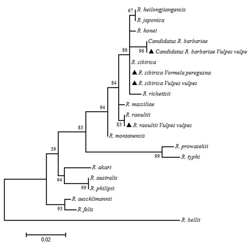
License: This work is licensed under a Creative Commons Attribution 4.0 International License.
Read Full License
Version of Record: A version of this preprint was published at Parasites & Vectors on April 19th, 2021. See
the published version at https://doi.org/10.1186/s13071-021-04718-1.
Page 2/12Abstract
Background
Previously, twelve Rickettsia species were found in ticks, fleas, sheep keds (Melophagus ovinus), bats
(common pipistrelle: Pipistrellus pipistrellus) and a tick-bitten patient in Xinjiang Uygur Autonomous
Region (XUAR), northwestern China. Here we aimed to molecularly detect rickettsial agents in red fox
(Vulpes vulpes), marbled polecat (Vormela peregusna) and their ticks.
Methods
During 2018-2019, 12 red foxes, 1 marbled polecat and their ticks were sampled in two counties and a
city of Xinjiang Uygur Autonomous Region (northwestern China). The heart, liver, spleen, lung and kidney
of these 13 carnivores were dissected, followed by DNA extraction. Hard ticks were identified both
morphologically and molecularly. All samples were examined for the presence of rickettsiae by
amplifying four genetic markers.
Results
A total of 26 adult ticks and 28 nymphs (38 Ixodes canisuga, nine Ixodes kaiseri, six Haemaphysalis
erinacei and one Dermacentor marginatus) were collected from red foxes, and four H. erinacei ticks were
removed from a marbled polecat. Analysis of cytochrome c oxidase subunit I (COI) gene sequences
indicated that 2-32 nucleotides differed between I. canisuga, I. kaiseri and H. erinacei from northwestern
China and Europe. Rickettsia raoultii was detected in three red foxes, Candidatus Rickettsia barbariae in a
red fox, Rickettsia sibirica in a red fox and a marbled polecat, and R. raoultii in two tick species (I.
canisuga and D. marginatus).
Conclusions
To the best of our knowledge, I. canisuga and I. kaiseri have not been previously reported from red foxes
in China. The DNA of R. sibirica and R. raoultii was detected for the first time in organs of red foxes, and
R. sibirica in organs of marbled polecat. This is also the first molecular evidence for the presence of R.
raoultii in I. canisuga. Our findings add to the range of tick-borne pathogens in wildlife species and
associated ticks in China.
Background
The red fox (Vulpes vulpes) is widely distributed throughout Europe, Asia, North Africa, and North America
[1]. Its habitats highly overlap with those of other wildlife species, domestic animals and even humans
[2]. Previously, red foxes were reported to harbor several vector-borne pathogens of veterinary-medical
importance, such as tick-borne encephalitis virus [3], Borrelia burgdorferi [4], Ehrlichia canis [5],
Leishmania infantum [6], Hepatozoon canis [7] and Babesia vulpes [8, 9]. Serological investigation of red
foxes indicated that 50.3% had antigens of SFG rickettsiae, including Rickettsia massiliae and Rickettsia
Page 3/12conorii in Spain [10]. In addition, immuno-fluorescence assay showed that 1.9% of red foxes had
antibodies to Rickettsia typhi, and 6.7% of them to Rickettsiaslovaca in Spain [4].
The geographical range of marbled polecat (Vormela peregusna) covers Central Asia, northwestern China
and Europe [2]. Considering studies on its epidemiological role, seroconversion was detected in a marbled
polecat to plague F1 antigen in Xinjiang Uygur Autonomous Region (XUAR), northwestern China. Borrelia
burgdorferi sensu lato and Babesia sp. were molecularly identified in a marbled polecat in Romania and
China, respectively [11, 12]. Furthermore, Rickettsia raoultii and Candidatus Rickettsia barbariae were
molecularly identified in marbled polecats in XUAR [12].
In the temperate climate zone, hard ticks (Acari: Ixodidae) are regarded as the most important vectors of
pathogens [13]. Among them, Ixodes persulcatus, Ixodes ricinus, Ixodes hexagonus, Ixodes kaiseri,
Ixodescanisuga, Dermacentor reticulatus, Dermacentor marginatus, Haemaphysalis punctata and
Rhipicephalus sanguineus were reported from red foxes [10, 14-16]. In Spain, Rickettsia massiliae,
Rickettsia aeschlimannii and Rickettsia slovaca were detected in red fox ticks [10]. In addition,
Haemaphysalis erinacei from marbled polecats contained the DNA of Rickettsia raoultii in China [17].
The aim of the present study was to investigate rickettsial agents in 12 red foxes, a marbled polecat and
their ticks in China.
Methods
Sample collection and species identification
A total of 12 illegally hunted or road-killed red foxes and one naturally died marbled polecat were
sampled in two counties and a city of XUAR during 2018-2019 (data shown in Additional file 1). The red
foxes and the marbled polecat were morphologically identified by an experienced zoologist. The heart,
liver, spleen, lung and kidney of all 13 carcasses were removed. Simultaneously, the entire body surface
of each individual was checked for ticks, all of which were removed. The ticks were morphologically
identified to the species level according to the standard taxonomic keys as previously described [18]. This
was also confirmed by molecular and phylogenic analyses based on two mitochondrial markers, the 16S
rDNA and the cytochrome c oxidase subunit I (COI) genes [16].
Detection, sequencing and phylogenetic analysis of rickettsiae
Genomic DNA was extracted from organs (heart, liver, spleen, lung and kidney) of wild carnivores, as well
as from their ticks using the TIANamp Genomic DNA Kit (TIANGEN, Beijing, China). To investigate the
presence of rickettsiae in ticks, four genetic markers were targeted, including the 17 kDa antigen (17-kDa),
the citrate synthase (gltA), the outer membrane protein A (ompA) and the surface cell antigen 1 (sca1)
genes. Two (the gltA and ompA) genes were used to detect rickettsiae in organs of wild carnivores [19].
The primers and PCR cycling conditions of this study are shown in Additional file 2. Each PCR assay
included a negative control (distilled water instead of DNA template) and a positive control (containing
Page 4/12sequence-verified DNA of R. massiliae from Rhipicephalus turanicus ticks collected in XUAR) [20].
Purification and sequencing of the PCR products were performed as described before [21, 22]. Sequences
were manually edited, aligned and compared to reference GenBank sequences by nucleotide BLASTn
program (https://blast.ncbi.nlm.nih.gov). A phylogenetic tree was constructed using the Maximum-
Likelihood method in MEGA 7.0 software [17].
Results
Tick identification
A total of 26 adult ticks and 28 nymphs (38 I. canisuga, nine I. kaiseri, six H. erinacei and one D.
marginatus) were collected from 12 red foxes, and four H. erinacei ticks were found on the marbled
polecat. Morphological characteristics are shown in Supplementary Figure 1.
Molecular and phylogenetic analyses
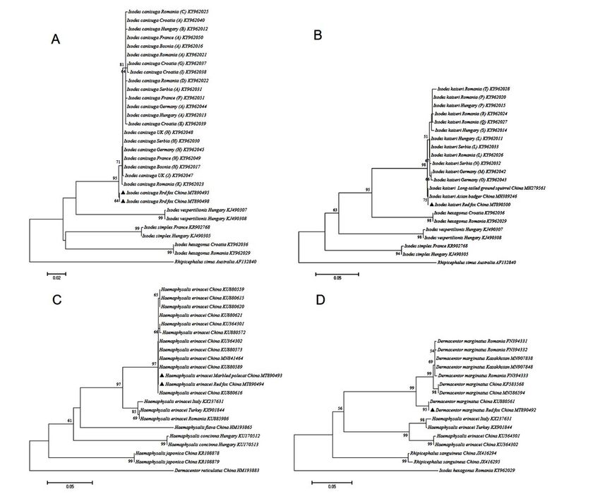
Analysis of COI sequences revealed 2-32 nucleotide differences in case of I. canisuga (3-6 bp), I. kaiseri
(2-7 bp) and H. erinacei (30-32 bp) between Europe and China. Phylogenetic analysis showed that i) I.
canisuga in XUAR was in a basal position to eleven European haplotypes (“A to K”) [18] (Fig. 1A); ii) I.
kaiseri from red foxes in XUAR was also in basal position to nine European haplotypes (“L to T”), and had
identical sequence with conspecific ticks from long-tailed ground squirrels and Asian badgers [16, 19, 23]
(Fig. 1B); iii) H. erinacei from red foxes and marbled polecat had identical sequences, and formed a
distinct clade from those reported in Turkey, Italy and Romania (Fig. 1C); and iv) D. marginatus from red
fox #2 had identical sequence with the off-host tick collected formerly in Altaw City, XUAR.
Red fox #3, #5 and #11 were positive for R. raoultii, and red fox #8 was positive for Candidatus R.
barbariae. At the same time, red fox #12 and the marbled polecat were positive for Rickettsia sibirica. In
addition, R. raoultii was detected in I. canisuga from red fox #11 (Manas County) and D. marginatus from
red fox #2 (Nilka County). Nucleotide sequences of rickettsial agents were deposited in the GenBank
database (MT890502-MT890525). Phylogenetic analyses are shown in Fig. 2 and Additional Figure 2.
Discussion
Molecular studies on I. canisuga and I. kaiseri were mostly reported from Europe, where these tick species
typically infest Eurasian badgers, red foxes, steppe polecats, raccoon dogs, common hedgehogs and
domestic dogs. Among them, dogs and red foxes can also be co-infested with I. canisuga and I. kaiseri
[16, 24]. In this study, I. canisuga and I. kaiseri were found on red foxes, as also confirmed by 16S rDNA
gene sequences (GenBank: MT889694-MT889698 and MT889701-MT889705). To the best of our
knowledge, I. canisuga and I. kaiseri have been discovered here for the first time on red foxes in China.
Phylogenetic analysis of the COI gene showed that i) I. canisuga specimens collected from the same host
species (red fox) are genetically different between China (MT890495-MT890498) and Germany
(KY962044-KY962045), Croatia (KY962037-KY962040), Bosnia-Herzegovina (KY962016-KY962017),
Page 5/12Serbia (KY962030-KY962031), Romania (KY962025, KY962021-KY962023); ii) I. kaiseri from red foxes
and long-tailed ground squirrels in XUAR had 100% identity, but clustered in a separate phylogenic
position compared to European ticks; and iii) H. erinacei infesting red fox and marble polecat in XUAR
shared identical COI gene sequences (MT890493 and MT890494), but differed from those collected in
Italy, Turkey and Romania (Fig. 1C). These findings support that the genetic diversity of I. kaiseri, I.
canisuga and H. erinacei might reflect geographical distribution rather than host associations,
consistently with Klompen et al [25].
Considering previous reports, Rickettsia helvetica was detected in the blood sample of a red fox in
Switzerland [26]. In addition, R. raoultii and Candidatus Rickettsia barbariae were reported in a marbled
polecat in Altaw City, XUAR [12]. Here, R. raoultii was detected for the first time in organs of red foxes, and
R. sibirica in the lung and kidney of a marbled polecat and in the liver of a red fox. Moreover, the DNA of
R. raoultii was shown to be present in I. canisuga. In our previous work, R. raoultii and R. sibirica were
identified in several tick species [12, 17, 19, 27, 28], and R. raoultii was even found in organs/tissues of
bats (Pipistrellus pipistrellus) and a tick-bitten patient [19, 29]. Based on the above findings, the
epidemiological role of red foxes, marble polecats and their ticks in transmitting rickettisiae can be
postulated. However, further studies (including transmission experiments) are necessary to eventually
verify this. In addition, the scope of this work should be extended to more wildlife species from China and
Central Asia.
Conclusions
To our knowledge, I. canisuga and I. kaiseri have not been previously identified from red foxes in China.
The genetic diversity of I. kaiseri, I. canisuga and H. erinacei might be more related to geographical
distribution than parasitized hosts. Rickettsia raoultii in I. canisuga and organs of red foxes, and R.
sibirica in organs of a red fox and a marbled polecat are reported here for the first time. Our findings add
to the range of tick-borne pathogens in wildlife species and associated ticks.
Abbreviations
COI: cytochrome c oxidase subunit I; ompA: outer membrane protein A; gltA: citrate synthase; 17-kDa: 17-
kDa antigen; sca1: cell surface antigen 1; XUAR: Xinjiang Uygur Autonomous Region
Declarations
Acknowledgements
The authors thank the contributions by the staff at the School of Medicine and School of Animal Science
and Technology, Shihezi University.
Funding
Page 6/12This work was supported by the National Key Research & Development Program of China
(2018ZX10101002-007), National Natural Science Foundation of China (81960379 and 31960709),
International Scientific and Technological Cooperation in Bingtuan (2020BC008) and International
Cooperation Projects of and Xinjiang Uygur Autonomous Region (2020E01008)..
Authors’ contributions
GL, SZ, WT and YW conceived and designed the study, and wrote the manuscript. WY, LM, SW, YZ, ZL, WH
and XG performed the experiments, analyzed the data. SH contributed to study design and edited the
manuscript. All authors read and approved the final manuscript.
Ethical approval and consent to participate
This study was approved by the Animal Ethics Committee of Shihezi University (Approval No.
AECSU2015-11).
Consent for publication
Not applicable.
Competing interests
The authors declare that they have no competing interests.
Author details
1
Department of Basic Medicine, School of Medicine, Shihezi University, Shihezi, Xinjiang, Uygur
Autonomous Region,China. 2Department of Parasitology and Zoology, University of Veterinary Medicine,
Budapest, Hungary. 3Institute of Veterinary Medicine, Xinjiang Academy of Animal Science, Urumqi,
Xinjiang, Uygur Autonomous Region, China. 4State Key Laboratory of Sheep Genetic Improvement and
Healthy Production, Institute of Animal Husbandry and Veterinary, Xinjiang Academy of Agricultural and
Reclamation Science, Shihezi, Xinjiang, Uygur Autonomous Region, China. 5Department of Veterinary
Medicine, College of Animal & Science, Shihezi University, Shihezi, Xinjiang, Uygur Autonomous Region,
China.
References
1. Wilson DE, Reeder DM. Mammal species of the world. A taxonomic and geographic reference. 3rd ed.
Baltimore: Johns Hopkins University Press; 2005.
2. Hunter L, Barrett P. A field guide to the carnivores of the world. Bloomsbury Publishing; 2018.
3. Wurm R, Dobler G, Peters M, Kiessig ST. Serological investigations of red foxes (Vulpes vulpes L.) for
determination of the spread of tick-borne encephalitis in Northrhine-Westphalia. J Vet Med B Infect
Dis Vet Public Health. 2000;47:503-9.
Page 7/124. Lledó L, Serrano JL, Isabel Gegúndez M, Giménez-Pardo C, Saz JV. Antibodies to Rickettsia spp. and
Borrelia burgdorferi in Spanish Wild Red Foxes (Vulpes vulpes). J Wildl Dis. 2016;52:122-5.
5. Fishman Z, Gonen L, Harrus S, Strauss-Ayali D, King R, Baneth G. A serosurvey of Hepatozoon canis
and Ehrlichia canis antibodies in wild red foxes (Vulpes vulpes) from Israel. Vet Parasitol.
2004;119:21-6.
6. Davoust B, Mary C, Marié JL. Detection of Leishmania in red foxes (Vulpes vulpes) from
southeastern France using real-time quantitative PCR. J Wildl Dis. 2014;50:130-2.
7. Gabrielli S, Kumlien S, Calderini P, Brozzi A, Iori A, Cancrini G. The first report of Hepatozoon canis
identified in Vulpes vulpes and ticks from Italy. Vector Borne Zoonotic Dis. 2010;10:855-9.
8. Checa R, López-Beceiro AM, Montoya A, Barrera JP, Ortega N, Gálvez R, et al. Babesia microti-like
piroplasm (syn. Babesia vulpes) infection in red foxes (Vulpes vulpes) in NW Spain (Galicia) and its
relationship with Ixodes hexagonus. Vet Parasitol. 2018;252:22-28.
9. Dezdek D, Vojta L, Curković S, Lipej Z, Mihaljević Z, Cvetnić Z, et al. Molecular detection of Theileria
annae and Hepatozoon canis in foxes (Vulpes vulpes) in Croatia. Vet Parasitol. 2010;172:333-6.
10. Ortuño A, Sanfeliu I, Nogueras M, Pons I, López-Claessens S, Castellà J, et al. Detection of Rickettsia
massiliae/Bar29 and Rickettsia conorii in red foxes (Vulpes vulpes) and their Rhipicephalus
sanguineus complex ticks. Ticks Tick Borne Dis. 2018;2;629
11. Gherman CM, Sándor AD, Kalmár Z, Marinov M, Mihalca AD. First report of Borrelia burgdorferi sensu
lato in two threatened carnivores: the marbled polecat, Vormela peregusna and the European mink,
Mustela lutreola (Mammalia: Mustelidae). BMC Vet Res. 2012;8:137.
12. Liu XF, Yang MH, Liu GY, Zhao SS, Yuan WM, Xiao RH, et al. Molecular evidence of Rickettsia raoultii,
"Candidatus Rickettsia barbariae" and a novel Babesia genotype in marbled polecats (Vormela
peregusna) at the China-Kazakhstan border. Parasit Vectors. 2018;11:450.
13. Jongejan F, Uilenberg G. The global importance of ticks, 2004. Parasitology. 2004;129:3-14.
14. Isogai E, Isogai H, Kawabata H, Masuzawa T, Yanagihara Y, Kimura K, et al. Lyme disease spirochetes
in a wild fox (Vulpes vulpes schrencki) and in ticks. J Wildl Dis. 1994;30:439-44.
15. Jemeršić L, Dežđek D, Brnić D, Prpić J, Janicki Z, Keros T, et al. Detection and genetic
characterization of tick-borne encephalitis virus (TBEV) derived from ticks removed from red foxes
(Vulpes vulpes) and isolated from spleen samples of red deer (Cervus elaphus) in Croatia. Ticks Tick
Borne Dis. 2014;5:7-13.
16. Hornok S, Sándor AD, Beck R, Farkas R, Beati L, Kontschán J, et al. Contributions to the phylogeny of
Ixodes (Pholeoixodes) canisuga, (Ph.) kaiseri, I. (Ph.) hexagonus, and a simple pictorial key for the
identification of their females. Parasit Vectors. 2017;10:545.
17. Guo LP, Mu LM, Xu J, Jiang SH, Wang AD, Chen CF, et al. Rickettsia raoultii in Haemaphysalis erinacei
from marbled polecats, China-Kazakhstan border. Parasit Vectors. 2015;8:461.
18. Estrada-Peña A, Mihalca AD, Petney TN: Ticks of Europe and North Africa: A Guide to Species
Identification. Springer Int; 2017.
Page 8/1219. Zhao S, Yang MH, Jiang MM, Yan B, Zhao SS, Yuan WM, et al. Rickettsia raoultii and Rickettsia
sibirica in ticks from the long-tailed ground squirrel near the China-Kazakhstan border. Exp Appl
Acarol. 2019;77:425-433.
20. Wei QQ, Guo LP, Wang AD, Mu LM, Zhang K, Chen CF, et al. The first detection of Rickettsia
aeschlimannii and Rickettsia massiliae in Rhipicephalus turanicus ticks, in northwest China. Parasit
Vectors. 2015;8:631.
21. Anstead CA, Chilton NB. A novel Rickettsia species detected in Vole Ticks (Ixodes angustus) from
Western Canada. Appl Environ Microbiol. 2013;79:7583-9.
22. Anstead CA, Chilton NB. Detection of a novel Rickettsia (Alphaproteobacteria: Rickettsiales) in rotund
ticks (Ixodes kingi) from Saskatchewan, Canada. Ticks Tick Borne Dis. 2013;4:202-6.
23. Sheng JL, Jiang MM, Yang MH, Bo XW, Zhao SS, Zhang YY, et al. Tick distribution in border regions
of Northwestern China. Ticks Tick Borne Dis. 2019;10:665-9.
24. ZipcodeDev Team (2018). http://zipco dezoo .com/
25. Klompen JS, Black WC, Keirans JE, Oliver JH. Evolution of ticks. Annu Rev Entomol. 1996;41:141-61.
26. Hofmann-Lehmann R, Wagmann N, Meli ML, Riond B, Novacco M, Joekel D, et al. Detection of
“Candidatus Neoehrlichia mikurensis” and other Anaplasmataceae and Rickettsiaceae in Canidae in
Switzerland and Mediterranean countries. Schweiz Arch Tierheilkd. 2016;158:691-700.
27. Tian ZC, Liu GY, Shen H, Xie JR, Luo J, Tian MY. First report on the occurrence of Rickettsia slovaca
and Rickettsia raoultii in Dermacentor silvarum in China. Parasit Vectors. 2012;5:19.
28. Spitalská E, Stefanidesová K, Kocianová E, Boldiš V. Rickettsia slovaca and Rickettsia raoultii in
Dermacentor marginatus and Dermacentor reticulatus ticks from Slovak Republic. Exp Appl Acarol.
2012;57:189-97.
29. Dong ZH, Yang YC, Wang Q, Xie SS, Zhao SS, Tan WB, et al. A case with neurological abnormalities
caused by Rickettsia raoultii in northwestern China. BMC Infect Dis. 2019; 19:796.
Figures
Page 9/12Figure 1
Phylogenic tree based on COI sequences of ticks collected from 12 red foxes and a marble polecat in
northwestern China. The evolutionary history was inferred using the Maximum Likelihood method
(bootstrap replicates: 1000) with MEGA 7.0. New sequences obtained in this study are indicated by black
triangles. A. Ixodes canisuga, B. Ixodes kaiseri, C. Haemaphysalis erinacei, and D. Dermacentor
marginatus.
Page 10/12Figure 2
Phylogenetic tree of the ompA-gltA concatenated sequences of rickettsial agents in 12 red foxes and a
marble polecat.
Supplementary Files
This is a list of supplementary files associated with this preprint. Click to download.
Page 11/12Additionalfile1SH.docx
Additionalfile2SH.doc
GraphicalAbstract.tif
SupplementaryFigure1SH.docx
SupplementaryFigure2SH.docx
Page 12/12You can also read