Reversal of G-Quadruplexes' Role in Translation Control When Present in the Context of an IRES
←
→
Page content transcription
If your browser does not render page correctly, please read the page content below

biomolecules

Review
Reversal of G-Quadruplexes’ Role in Translation Control When
Present in the Context of an IRES
Mohammed Enamul Hoque, Thulasi Mahendran                              and Soumitra Basu *

                                          Department of Chemistry and Biochemistry, Kent State University, Kent, OH 44242, USA;
                                          mhoque1@kent.edu (M.E.H.); tmahend1@kent.edu (T.M.)
                                          * Correspondence: sbasu@kent.edu; Tel.: +1-330-9908107

                                          Abstract: G-quadruplexes (GQs) are secondary nucleic acid structures that play regulatory roles in
                                          various cellular processes. G-quadruplex-forming sequences present within the 50 UTR of mRNAs
                                          can function not only as repressors of translation but also as elements required for optimum function.
                                          Based upon previous reports, the majority of the 50 UTR GQ structures inhibit translation, presumably
                                          by blocking the ribosome scanning process that is essential for detection of the initiation codon.
                                          However, there are certain mRNAs containing GQs that have been identified as positive regulators of
                                          translation, as they are needed for translation initiation. While most cellular mRNAs utilize the 50 cap
                                          structure to undergo cap-dependent translation initiation, many rely on cap-independent translation
                                          under certain conditions in which the cap-dependent initiation mechanism is not viable or slowed
                                          down, for example, during development, under stress and in many diseases. Cap-independent
                                          translation mainly occurs via Internal Ribosomal Entry Sites (IRESs) that are located in the 50 UTR of
                                          mRNAs and are equipped with structural features that can recruit the ribosome or other factors to
                                initiate translation without the need for a 50 cap. In this review, we will focus only on the role of RNA
                                   GQs present in the 50 UTR of mRNAs, where they play a critical role in translation initiation, and
Citation: Hoque, M.E.;                    discuss the potential mechanism of this phenomenon, which is yet to be fully delineated.
Mahendran, T.; Basu, S. Reversal of
G-Quadruplexes’ Role in Translation       Keywords: G-quadruplex; IRES; context dependent; cap-independent translation; cellular mRNAs;
Control When Present in the Context       ribosome recruitment mechanism
of an IRES. Biomolecules 2022, 12, 314.
https://doi.org/10.3390/
biom12020314

Academic Editors: Pavel Ivanov,           1. Introduction
Prakash Kharel, Anna Varizhuk and               A G-quadruplex (GQ) is a secondary structure adopted by both DNA and RNA, and
Valentina Pirota                          is formed by the stacking of G-tetrad units. G-tetrads are assembled by the combination of
Received: 29 December 2021
                                          four guanines via the Hoogsteen hydrogen bonds, and the GQ structures are stabilized in the
Accepted: 14 February 2022
                                          presence of metal cations bound in a plane or in between two tetrads (Figure 1) [1–4]. It has been
Published: 16 February 2022
                                          found that DNA GQs play regulatory roles in replication, transcription, cell proliferation,
                                          genome recombination and telomere maintenance [5–10]. On the other hand RNA GQs
Publisher’s Note: MDPI stays neutral
                                          were found to be involved in translational regulation [11–24], 30 -end processing [25,26],
with regard to jurisdictional claims in
                                          transcription termination [27], alternative splicing [28–32], mRNA localization [33–35],
published maps and institutional affil-
                                          protein binding [36–38] and telomeric RNA biology [39–43]. Compared with their DNA
iations.
                                          counterparts, RNA GQs are easier to form, since they do not have to compete with a comple-
                                          mentary strand. RNA GQs have been found to be more stable in the folded form, presumably
                                          because of the 20 -OH group’s ability to participate in hydrogen bonding [18,44–46].
Copyright: © 2022 by the authors.
                                                Bioinformatic analysis has revealed more than 4000 GQ motifs in the 50 UTR
                                            0
                                          (5 untranslated region) of human mRNAs [47–50]. The functional consequences of GQ
Licensee MDPI, Basel, Switzerland.
This article is an open access article    have been experimentally validated in more than 30 50 UTR sequences from human mR-
distributed under the terms and           NAs [20,24]. It has been demonstrated that GQ-forming sequences present within the
conditions of the Creative Commons        50 UTR can function both as vital elements and as repressors of in vitro and in vivo trans-
Attribution (CC BY) license (https://     lation [14,18,21,22,51,52]. More interestingly, the role of GQ structures in the modulation
creativecommons.org/licenses/by/          of translation depends on the context in which the GQ structure exists [53,54]. It is well
4.0/).                                    known that GQ structures modulate translation in several clinically important mRNAs.

Biomolecules 2022, 12, 314. https://doi.org/10.3390/biom12020314                                      https://www.mdpi.com/journal/biomolecules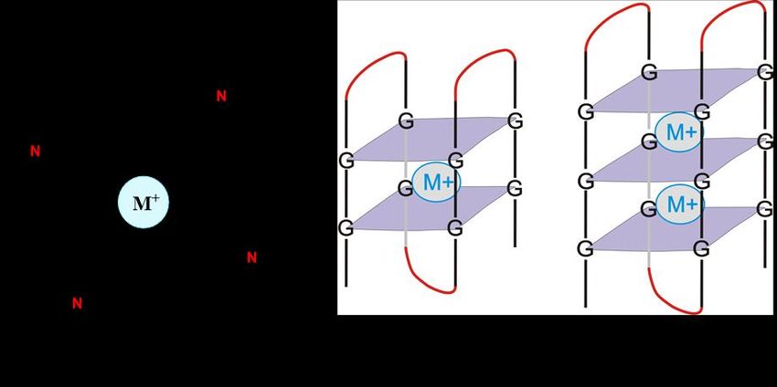
Biomolecules 2022, 12, 314                                                                                          2 of 14

                             Detailed studies of GQ-forming sequences in the 50 UTR region of various mRNAs, includ-
                             ing NRAS [18], ZIC-1 [11], MT3-MMP [22] and many others [13,15,16,19,23–25,36,37,55–64],
                             have shown that GQ structures usually serve as repressors of translation and, in most
                             cases, they drive mRNA translation through the cap-dependent mechanism [20,65,66].
                             The inhibitory effect is often a function of the stability and location of the GQ within the
                             50 UTR. The most commonly accepted mechanism of inhibition is GQ-mediated hindrance
                             of ribosomal scanning as it marches toward finding the initiation codon. Other mecha-
                             nisms of inhibition have also been reported. For example, in case of P1-HNF4A, the two
                             long side chains within the 50 UTR GQ recruit RNA binding proteins that stabilize the
                             GQ and result in strong repression of translation [67]. In another case, the NRAS GQ
                             was able to inhibit in vitro translation only when it was located in close proximity to the
                             50 cap [54], indicating that position matters in the case of the inhibitory effects of the GQ
                             when present within the 50 UTR of mRNAs. In contrast to the widely reported inhibitory
                             role of the RNA GQs in translation, other reports have demonstrated that the RNA GQs in
                             the 50 UTR of FGF2 [14], VEGF [21], ARPC2 [38,68], TGFβ2 [51], α-synuclein (SNCA) [69],
                             NRF2 [70,71], BAG-1 [72] and mTOR [73] mRNAs are essential for optimal translation.
                             Interestingly, several of these mRNAs harbor Internal Ribosomal Entry Sites (IRESs) that
                             are known to initiate translation in a cap-independent manner. IRES elements are reported
                             to be important during development, stress and many diseases [74–78]. Because IRESs are
                             important in cap-independent translation initiation, GQs embedded in such sequences may
                             play a role in such a process. In fact, in the case of VEGF IRES-A, the 40S ribosomal subunit
                             has been shown to interact with the GQ-containing domain [79]. However, in most cases,
                             the function of GQ structures in the IRES-driven cap-independent translation initiation
                             remains to be uncovered Although initially observed in viral mRNAs, IRES-mediated
                             translation initiation has also been identified in many cellular mRNAs [80–82]. Generally,
                             cellular IRESs contain fewer RNA structures than viral IRESs and share little sequence
                             conservation among them, making it difficult to classify and predict novel endogenous
                             IRESs in eukaryotic mRNAs. A recent screen using an in vivo translation reporter assay
                             has demonstrated that about 10% of mammalian mRNAs contain certain elements that are
                             involved in IRES function [83]. However, compared with the viral IRESs, the mechanistic as-
                             pects of cellular IRES function are poorly understood [66,84–86]. Most studies have shown
                             that cap-independent initiation mechanism utilizes IRES elements in case of impaired m7G
                             cap structure recognition at the 50 -end of mRNAs or non-canonical scanning models of
                             translation initiation in eukaryotes [80,85]. However, cap-independent translation initiation
                             can also occur in the absence of an IRES. Some cellular mRNAs use a separate mechanism,
                             known as ‘cap-independent translation enhancer (CITE)-mediated translation’ under apop-
                             totic conditions, which relies on ribosomal scanning of the 50 UTR [78,87]. Recently, it has
                             also been mentioned that mRNAs containing N6-methyladenosine (m6A) in their 50 UTR
                             may also be translated cap-independently [88–90]. In this mini review, we will primarily
                             concentrate on the role of GQ structures in the context of IRES-mediated cap-independent
                             translation initiation, along with the experimental systems used to investigate them. Re-
                             lated topics such as the regulatory roles of 50 UTR mRNAs in translation can be found
                             in many recent reviews [20,24,54,91,92], and the biological functions of IRES-mediated
                             cap-independent translation and regulation have also been reviewed recently [65,66,93,94].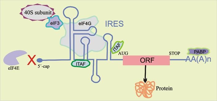
Biomolecules 2022, 12, x FOR PEER REVIEW 3 of 15
Biomolecules 2022,12,
Biomolecules2022, 12,314
x FOR PEER REVIEW 32ofof14
15
sequence conservation among them, making it difficult to classify and predict novel en-
dogenous IRESs in eukaryotic mRNAs. A recent screen using an in vivo translation re-
porter assay has demonstrated that about 10% of mammalian mRNAs contain certain el-
ements that are involved in IRES function [83]. However, compared with the viral IRESs,
the mechanistic aspects of cellular IRES function are poorly understood [66,84–86]. Most
studies have shown that cap-independent initiation mechanism utilizes IRES elements in
case of impaired m7G cap structure recognition at the 5′-end of mRNAs or non-canonical
scanning models of translation initiation in eukaryotes [80,85]. However, cap-independ-
ent translation initiation can also occur in the absence of an IRES. Some cellular mRNAs
use a separate mechanism, known as ‘cap-independent translation enhancer (CITE)-me-
diated translation’ under apoptotic conditions, which relies on ribosomal scanning of the
5′ UTR [78,87]. Recently, it has also been mentioned that mRNAs containing N6-methyl-
adenosine (m6A) in their 5′ UTR may also be translated cap-independently [88–90]. In this
mini review, we will primarily concentrate on the role of GQ structures in the context of
IRES-mediated cap-independent translation initiation, along with the experimental sys-
tems used to investigate them. Related topics such as the regulatory roles of 5′ UTR
mRNAs in translation can be found in many recent reviews [20,24,54,91,92], and the bio-
logical functions of IRES-mediated cap-independent translation and regulation have also
Figure1.1. The
Figure The chemical
chemical structure
structureofofa aguanine
guaninetetrad featuring
tetrad a central
featuring metal
a central cation
metal (G-tetrad),
cation two-
(G-tetrad),
been reviewed recently [65,66,93,94].
tier (bi-tetrad) and three-tier (tri-tetrad) G-quadruplexes.
two-tier (bi-tetrad) and three-tier (tri-tetrad) G-quadruplexes.
2. The Role of RNA G-Quadruplexes on IRES-Driven Cap-Independent Translation
2. TheBioinformatic
Role of RNAanalysis has revealed
G-Quadruplexes more than 4000
on IRES-Driven GQ motifs in theTranslation
Cap-Independent 5′ UTR (5′ un-
The
translated traditional
region) of translation
human mRNAs initiation mechanism
[47–50]. relied
The functional on eIF4E binding
consequences to the 5′ cap
The traditional translation initiation mechanism relied on eIF4E binding toofthe GQ 50have
cap
of mRNAs
been for
experimentally cap-dependent
validated translation
in more until
than 301988,
5′ when
UTR Pelletier
sequences and
from Sonenberg
human [95]
mRNAs
of mRNAs for cap-dependent translation until 1988, when Pelletier and Sonenberg [95]
showed that
[20,24]. that
It hassome mRNAs
beenmRNAs have a mechanism
demonstrated that GQ-forming for circumventing the needwithin
sequences present for eIF4E
the bind-
5′bind-
UTR
showed some have a mechanism for circumventing the need for eIF4E
ing,
can which
function was termed
both as IRES-mediated
vital elements and translation
as repressorsinitiation.
of in This
vitro mode
and in of initiation
vivo of
translation
ing, which was termed IRES-mediated translation initiation. This mode of initiation of
translation is usually
[14,18,21,22,51,52]. independent of the identification of 5′ cap structure but may include
translation is usuallyMore interestingly,
independent the role of GQ
of the identification of 5structures
0 cap structurein thebutmodulation
may include of
scanning
translation in a search for the AUG start codon or recruiting the 40S ribosomal subunit di-
scanning in adepends
search for onthe
theAUGcontextstart incodon
whichorthe GQ structure
recruiting the 40Sexists [53,54].
ribosomal It is well
subunit di-
rectly
known inthat
the GQ vicinity of the modulate
structures start codon. Recruitment
translation in of the 40S subunit may takemRNAs. place
rectly in the vicinity of the start codon. Recruitment of several clinically
the 40S subunit important
may take place either
either in the complete absence of any other protein factors or with the assistance of specific
Detailed
in studies
the complete of GQ-forming
absence of any other sequences in the or
protein factors 5′ with
UTRthe region of various
assistance mRNAs,
of specific combi-in-
combinations of canonical initiation factors (such as eIF4G and eIF3) and IRES trans-acting
cluding NRAS [18], ZIC-1 [11], MT3-MMP [22] and many
nations of canonical initiation factors (such as eIF4G and eIF3) and IRES trans-acting factors others [13,15,16,19,23–
factors (ITAFs) (Figure 2) [96,97]. ITAFs are known to help recruit the 40S ribosomal sub-
25,36,37,55–64],
(ITAFs) (Figure 2)have shown
[96,97]. ITAFsthatareGQ structures
known to helpusually
recruit serve
the 40S asribosomal
repressorssubunitof translation
on the
unit on the mRNA by different interactions or by stabilizing specific active IRES confor-
and, inby
mRNA most cases,interactions
different they drive or mRNA translation
by stabilizing through
specific activethe cap-dependent
IRES conformationsmechanism[81,98,99].
mations [81,98,99]. Recently, several studies demonstrated the effect of RNA GQ struc-
[20,65,66].
Recently, The inhibitory
several effect is often
studies demonstrated thea function
effect of RNAof theGQ stability and on
structures location of the GQ
IRES-mediated
tures on IRES-mediated cap-independent translation when these structures were located
within the 5′ UTR.
cap-independent The mostwhen
translation commonly accepted mechanism
these structures were located ofeither
inhibitionin theisvicinity
GQ-mediatedof the
either in the vicinity of the IRES elements or as a part of them [14,21,68,72,73]. Below, we
hindrance
IRES elements of ribosomal
or as a partscanning
of them as it marches toward
[14,21,68,72,73]. Below,finding
we review the initiation
the known codon.
role ofOther
GQ
review the known role of GQ structures on IRES activity during cap-independent transla-
mechanisms
structures of inhibition
on IRES have also
activity during been reported.translation
cap-independent For example, in casemechanisms.
initiation of P1-HNF4A, the
tion initiation mechanisms.
two long side chains within the 5′ UTR GQ recruit RNA binding proteins that stabilize the
GQ and result in strong repression of translation [67]. In another case, the NRAS GQ was
able to inhibit in vitro translation only when it was located in close proximity to the 5′ cap
[54], indicating that position matters in the case of the inhibitory effects of the GQ when
present within the 5′ UTR of mRNAs. In contrast to the widely reported inhibitory role of
the RNA GQs in translation, other reports have demonstrated that the RNA GQs in the 5′
UTR of FGF2 [14], VEGF [21], ARPC2 [38,68], TGFβ2 [51], α-synuclein (SNCA) [69], NRF2
[70,71], BAG-1 [72] and mTOR [73] mRNAs are essential for optimal translation. Interest-
ingly, several of these mRNAs harbor Internal Ribosomal Entry Sites (IRESs) that are
known to initiate translation in a cap-independent manner. IRES elements are reported to
be important during development, stress and many diseases [74–78]. Because IRESs are
important in cap-independent translation initiation, GQs embedded in such sequences
may play a role in such a process. In fact, in the case of VEGF IRES-A, the 40S ribosomal
subunit has been shown to interact with the GQ-containing domain [79]. However, in
most cases, the function of GQ structures in the IRES-driven cap-independent translation
Figure 2. Schematic
initiation representation
remainsrepresentation
to be uncovered of cap-independent
Although initially translation.
observed Stem–loop
in viral secondary
mRNAs,structures
IRES-me-
Figure 2. Schematic of cap-independent translation. Stem–loop secondary structures
within
diated the IRES
translationin 5’
0 UTR may
initiation recruit
has the
also 40S
beenribosomal
identifiedsubunit
in directly
many to the
cellular start codon (AUG)
mRNAs [80–82].
within the IRES in 5 UTR may recruit the 40S ribosomal subunit directly to the start codon (AUG) of
the open reading frame (ORF) or its vicinity through direct or indirect interactions, requiring thelittle
Generally, cellular IRESs contain fewer RNA structures than viral IRESs and share aid
of certain canonical initiation factors (eIFs) and/or IRES trans-acting factors (ITAFs).Biomolecules 2022, 12, 314 4 of 14
The role of RNA GQ in the 50 UTR of mRNAs is mostly described as a repressor of
translation, leading to the suggestion that RNA GQ is an inhibitory element for gene expres-
sion [17]. However, RNA GQ formation has been shown to promote translation initiation in
several cases in a cap-independent manner. Several IRES elements in the 50 UTRs of cellular
mRNAs have been reported to contain GQ motifs. In 2003, Bonnal et al. [14] reported an
IRES element within the 50 UTR of the human fibroblast growth factor 2 (FGF2) mRNA
from an analysis of the cis-acting elements. They demonstrated that IRES harbors a GQ
motif, which is responsible for IRES activity in the translation initiation. They identified the
five G-quartets’ RNA GQ structure in the 50 UTR of FGF2 mRNA by chemical probing and
enzymatic footprinting experiments. Moreover, the mutation and deletion studies revealed
that a single 176-nucleotide-long IRES contains two RNA stem–loops and a GQ motif,
and the contribution of these structures on IRES activity was able to control translation
initiation at four downstream initiation codons within the FGF2 mRNA. In a recent study,
IRES-mediated translation control was demonstrated for the differential expression of
FGF9 protein in response to the metastasis of colon cancer cells [100]. Reports indicated
that FGF9 protein synthesis is normally low due to upstream open reading frame (uORF)-
mediated translational repression; however, it is upregulated in response to hypoxia via an
IRES-dependent translational control.
Morris et al. [21] reported that the RNA GQ structure is essential for IRES-mediated
translation initiation in the human vascular endothelial growth factor (hVEGF) mRNA.
The hVEGF mRNA possesses a relatively long (1038 nt) 50 UTR and harbors two separate
IRESs, which are independently capable of initiating cap-independent translation [52,101].
RNase T1 and DMS footprinting assays revealed that a 17-nt (GGAGGAGGGGGAGGAGG)
guanine-rich sequence of VEGF IRES-A (293 nt) has a GQ structure. To investigate the roles
of GQ on IRESs, we performed a dual-luciferase reporter assay in HeLa cells by introducing
mutations in the IRES-A. It was found that the mutation of one of the minimal GQ forming
segments had no effect on IRES activity, whereas the quadruple mutant (lacking sufficient
Gs to adopt an intramolecular G-quadruplex structure) had lost its activity. We proposed
that the double mutant that retained the activity has accommodated an alternate GQ
structure by which it can maintain an activity similar to the wild-type. Furthermore, it
was proven that the IRES-A forms a switchable, two-tier GQ structure, which is essential
for its functional role. The different double mutants analyzed in this study were able to
utilize different G-stretches and form alternate quadruplex structures (possible by using
different combinations of the five G-stretches) with varying levels of activity. We proposed
that the structural flexibility and malleability of the VEGF IRES-A GQ is crucial for its
unique function that helps it bind to the factors necessary for translation initiation in a
cap-independent manner.
In another study, a subsequent mechanistic analysis by using in vitro RNA footprinting
showed that the GQ located in the IRES element of the VEGF mRNA interacts directly with the
40S ribosomal subunit in the absence of other protein factors [79]. In 2015, Cammas et al. [102]
also analyzed the effect of GQs in VEGF mRNA translation. They increased GQ stability by
inserting or replacing the sequence to form a 3-G-quartet quadruplex structure and further
stabilized it by utilizing GQ ligands. These led to an inhibitory effect on translation, similar to
the previously characterized GQs in the 50 UTR of several mRNAs.
In a recent study, Al-Zeer et al. [68] provided structural and functional evidence that a
GQ structure within the 50 UTR of the actin-related protein 2/3 complex subunit 2 (ARPC2)
mRNA is essential for IRES-mediated cap-independent translation. The 50 UTR of ARPC2
mRNA exists in two variants; the longer variant adopts IRES, which harbors a GQ motif in
its central stem-loop element. They investigated the cellular function of the IRES element
by measuring the ARPC2 expression levels as a function of increased cell density-related
stress. They observed that the relative ARPC2 protein level increased with cell density,
thus suggesting the expected role of IRES elements in the expression of certain genes that
represses cap-dependent translation under stressful conditions [76]. Furthermore, they
proposed a model for IRES element folding in the 50 UTR of the ARPC2 mRNA based onBiomolecules 2022, 12, 314 5 of 14
structural probing experiments and bioinformatic prediction programs. They demonstrated
that RNA folds into three hairpins, the middle of which includes an exposed GQ motif on
top of the stem–loop structure. Disruption of the GQ structures by site-directed mutagenesis
led to an approximately 30% decrease in translational efficiency. The decreased translation
efficiency was less pronounced than the value reported previously for the GQ-containing
IRES element in the 50 UTR of VEGF; nevertheless, it showed the functional effect of GQ
in IRES-mediated translation initiation [21]. These results indicated that the GQ in the
ARPC2 IRES is at least partly required for its full functionality. In contrast, another study
demonstrated that the GQ motif located in the 50 UTR region of ARPC2 mRNA plays an
inhibitory role in translation. They utilized biophysical characterization for GQ structure
confirmation and luciferase reporter assays for translational inhibitory activity, but the exact
mechanism of GQ’s roles in the 50 UTR of ARPC2 mRNA was not demonstrated [38,103].
Koukouraki and Doxakis [69] reported another IRES element that harbors a GQ motif
in the 50 UTR of the α-synuclein (SNCA) mRNA. SNCA is a neuronal protein which is
likely to be involved in the modulation of synaptic neurotransmission. Several approaches
revealed that the 50 UTR of SNCA mRNA has an internal ribosome entry site (IRES) element,
which encompasses most of the 50 UTR. They observed that different cellular conditions,
such as depolarization of the plasma membrane, serum malnutrition and oxidative stress,
stimulated the translation of α-synuclein protein through its IRES activity. The increased
IRES activity not only enhanced expression of a luciferase reporter but also showed a
significant increase in endogenous α-synuclein expression [69]. It was shown that the
50 UTR initiates SNCA mRNA translation in a cap-independent process when cap-dependent
translation was diminished by rapamycin treatment. The human SNCA mRNA has a mod-
erately long 50 UTR of 264 nt with 66% of GC content that, as expected, has stable stem–loop
structures and also includes a GQ motif. To investigate the role of GQ structures in SNCA
translation or IRES activity, the authors used mutagenized reporter constructs and per-
formed a dual luciferase assay. Functional studies using dual luciferase and real time
RT-PCR assays suggested that the GQ in the 50 UTR of α-synuclein mRNA was not abso-
lutely required for translational activity or IRES functionality, but certainly increased its
efficiency. This study serves as another example of GQs’ role in optimal IRES activity.
Recently, Jodoin et al. [72] showed that an RNA GQ located towards the 50 end of the
BAG-1 50 UTR influences both cap-dependent as well as cap-independent mRNA transla-
tion, making it the first report of the involvement of a quadruplex in both mechanisms of
translation initiation. In a previous study, the same group identified that the GQ structure
within 6 and 35 nt of the BAG-1 50 UTR repressed the translation of the luciferase gene [63].
They confirmed that this particular quadruplex represses the cap-dependent translation of
the main BAG-1 isoform, which agrees with the role of the several previously identified
50 UTR GQs. Furthermore, they proved that a mutation in the GQ-forming sequence led
to the inhibition of cap-independent translation as well, even though it was not present
within the IRES. In order to elucidate this phenomenon, they analyzed the effect of deleting
the GQ structure in other secondary structural elements in the 50 UTR, including its IRES,
using the selective 20 -hydroxyl acylation analyzed by primer extension (SHAPE) technique.
They found that disruption of the GQ structure created base pairing differences mainly in
the IRES region of the 50 UTR, thereby affecting the global folded secondary structure of the
50 UTR, which, in turn, created the more stable minimal IRES subdomain in the quadruplex
mutant compared with the wild-type. On the basis of this observation, they proposed that
the structural stability altered because the quadruplex disruption made it more difficult for
the IRES to bind to the ITAFs that are essential for 40S subunit recruitment. In conclusion,
the study provided an example of a GQ that is essential for maintaining a specific IRES
secondary structure to aid cap-independent translation.
Based on a bioinformatics analysis, it has been revealed that transforming growth factor
β2 (TGFβ2) mRNA has a 23-nt putative GQ-forming sequence in its 50 UTR. Agarwala et al.
performed spectroscopic studies and a luciferase reporter assay to characterize the thermo-
dynamic stability of the GQ and its role in modulating gene expression at the translationalBiomolecules 2022, 12, 314 6 of 14
level [51]. They found that a construct with the GQ forming sequence together with the
flanking sequences, as in the context of the entire 50 UTR of TGFβ2, significantly increased
the luciferase expression levels compared with its corresponding mutant. This observation
indicated that the GQ within the 50 UTR of the TGFβ2 mRNA has a context-dependent
effect on translation in which the GQ structure acts as an enhancer of gene expression [51].
The enhancing effect of the GQ structure could be attributed to its location in the 50 UTR,
i.e., it lies further away from both the 50 end of the UTR and from the translation start
site. Unlike the GQ structure in VEGF 50 UTR, which has been previously referred to as a
switchable quadruplex that positively regulates cap-independent translation initiation, the
TGFβ2 quadruplex is a stable quadruplex within an unusually long 50 UTR that positively
regulates translation in cap-dependent translation. Although there is an IRES element
present in the 50 UTR of TGFβ2, there is no correlation between the role of GQ and IRES
functionality. Further study is needed to delineate the precise role of GQ in the context of
IRES activity in TGFβ2 translation initiation.
Human nuclear factor erythroid 2–related factor 2 (NRF2) mRNA contains a 555-nt
sequence of the 50 UTR with 70% GC content, which has a 31-nt putative GQ-forming
sequence. In 2017, Lee et al. [70] demonstrated the presence of a GQ structure in the 50 UTR
of NRF2 mRNA by utilizing biophysical and biochemical methods, and found that the GQ
structure is important for 50 UTR activity. To identify the exact role of the GQ structure
in NRF2 protein translation under oxidative stress, they used proteomics to reveal that
elongation factor 1 alpha (EF1a) binds to the GQ sequence. Furthermore, they measured
the binding interaction of the EF1a protein with the GQ-forming sequence of 50 UTR by
electrophoretic mobility shift assays (EMSAs) along with RNA–protein interaction assays
for cells treated with H2 O2 . In addition, their use of small interfering RNA (siRNA) to
knock down EF1a by using a reporter assay suggested that the presence of the GQ is
important for cellular-level activation of NRF2 50 UTR under oxidative stress. In 2010,
Li et al. [104] studied cap-independent translation initiation in NRF2 mRNA via an IRES-
mediated mechanism under oxidative stress. They discovered that translation initiation was
induced by the binding of 18S rRNA of the ribosome to a highly conserved RNA binding
site located within the IRES. According to these two studies, there are two mechanisms
through which cap-independent translation is mediated in NRF2 mRNA, via either the
IRES or the GQ. Therefore, it would be interesting to study collectively whether there is
any influence of the GQ on IRES-mediated translation initiation, since they both are located
in close proximity to each other.
3. Mechanism of the Role of G-Quadruplexes in IRES-Mediated Translation Initiation
The initiation of protein synthesis in eukaryotes needs the recruitment of the 40S
ribosomal subunit to eventually recognize the start codon (AUG) for translation of the
mRNA [105–107]. Canonical eukaryotic cap-dependent translation initiation incorporates
a complex mechanism for the recruitment and positioning of ribosomes at the start sites,
during which, many factors interact with the ribosome [106,108,109]. Alternatively, several
viruses and some eukaryotic mRNAs use a cap-independent pathway through highly struc-
tured IRES sequences present in the 50 UTR of mRNAs, which drives pre-initiation complex
formation by either positioning the ribosome on or just upstream of the translation start
site [66,88,110] (Figure 2). Several studies have shown that GQ structures reside in proxim-
ity to the IRES element in 50 UTR of mRNAs and modulate IRES activity [21,70,71,73,79].
For example, the presence of a switchable GQ structure in the IRES-A of human vascular
growth factor (hVEGF) mRNA was deemed to be essential for optimum cap-independent
translation initiation [21]. To identify the exact role of GQ in IRES-A-mediated translation
initiation of hVEGF, we utilized structure mapping analyses [79]. Our findings indicated
that a 17-nucleotide independently folding RNA GQ domain within the 294-nucleotide
IRES-A interacted directly with the 40S ribosomal subunit without the aid of other protein
factors (Figure 3). In addition, we showed that the GQ-forming domain particularly dictates
the function and binding affinity of IRES-A towards the 40S subunit compared with the bac-cap-independent translation initiation [21]. To identify the exact role of GQ in IRES-A-
mediated translation initiation of hVEGF, we utilized structure mapping analyses [79].
Our findings indicated that a 17-nucleotide independently folding RNA GQ domain
within the 294-nucleotide IRES-A interacted directly with the 40S ribosomal subunit with-
Biomolecules 2022, 12, 314 7 of 14
out the aid of other protein factors (Figure 3). In addition, we showed that the GQ-forming
domain particularly dictates the function and binding affinity of IRES-A towards the 40S
subunit compared with the bacterial 30S subunit. Further, we proved that GQ domain
terial 30S subunit.
deletion hinderedFurther,
the 40Swe proved
subunit thatbinding
from GQ domain deletion
to the hindered
IRES and the 40Scap-inde-
deteriorated subunit
from binding to the IRES and deteriorated cap-independent translation initiation,
pendent translation initiation, indicating the necessity of the GQ structure in IRES indicat-
func-
ing
tion. Similarly, Marques-Ramos et al. [73] also demonstrated the direct binding al.
the necessity of the GQ structure in IRES function. Similarly, Marques-Ramos et [73]
interac-
also demonstrated
tions the direct binding interactions of GC-rich secondary structures
of GC-rich secondary structures of mTOR 5′ UTR with the 40S ribosomal subunit of mTOR
0 UTR with the 40S ribosomal subunit without the involvement of any initiation factors.
5without the involvement of any initiation factors. They revealed that the mTOR 5′ UTR
0 UTR binds to the ribosomal subunit with a similar binding
They revealed that the mTOR 5
binds to the ribosomal subunit with a similar binding affinity to the CFSV viral IRES by
affinity to the CFSV viral IRES by using a filter binding assay.
using a filter binding assay.
Figure 3. Schematic representation of direct recruitment of the 40S ribosomal subunit by G-quadru-
Figure 3. Schematic representation of direct recruitment of the 40S ribosomal subunit by G-
plex structures to drive cap-independent translation initiation. Adapted with permission from
quadruplex structures to drive cap-independent translation initiation. Adapted with permission from
Bhattacharyya et al., ACS Biochemistry [79].
Bhattacharyya et al., ACS Biochemistry [79].
InInanother
anotherstudy,
study,indirect
indirectevidence
evidenceofofGQ’s GQ’srole
roleininthe
thebinding
bindingofofIRESIREStotothethe40S
40S
ribosomalsubunit
ribosomal subunithas hasbeen
beeninvestigated
investigated[72]. [72]. The
The authors
authors analyzed
analyzed the theeffects
effectsofofGQGQ
structure disruption on other secondary structural elements in
structure disruption on other secondary structural elements in the 5 UTR, including its the 5′
0 UTR, including its
IRES using SHAPE and found that GQ disruption made it more difficult for the IRES to bindto
IRES using SHAPE and found that GQ disruption made it more difficult for the IRES
tobind to ITAFs,
ITAFs, which which is necessary
is necessary for 40Sfor 40S subunit
subunit recruitment.
recruitment. In 2016, InLee
2016, Lee[70]
et al. et al. [70] re-
reported
ported that a GQ-forming sequence was important 0 for the 5′ UTR
that a GQ-forming sequence was important for the 5 UTR activity of NRF2 mRNA through activity of NRF2 mRNA
through structural
structural mapping mapping
experiments experiments
[111]. To[111]. To investigate
investigate the exactthe exact
role role structure
of GQ of GQ struc-
in
ture in NRF2 protein translation under oxidative stress, they
NRF2 protein translation under oxidative stress, they utilized proteomics and EMSA, and utilized proteomics and
EMSA, and
revealed the revealed the bindingof
binding interaction interaction of the elongation
the elongation factor 1 alpha factor 1 alpha
(EF1a) (EF1a)
protein protein
with the
GQ-forming sequence within the 5 UTR. Their siRNA-mediated knockdown of EF1α byof
with the GQ-forming sequence within
0 the 5′ UTR. Their siRNA-mediated knockdown
EF1αaby
using using assay
reporter a reporter assaythat
suggested suggested that the
the presence presence
of the of the GQfor
GQ is important is NRF2
important
50 UTRfor
NRF2 5′ under
activation UTR activation underAlthough
oxidative stress. oxidativesome stress. Although
studies some studies the
have demonstrated have demon-
direct and
strated role
indirect the direct
of GQsand indirect
in IRES role in
activity ofthe
GQs in IRES activitytranslation
cap-independent in the cap-independent
of various mRNAs, trans-
lation
the exactofmechanism
various mRNAs, the exact
and potential mechanism
variations in GQs’androle
potential
in ITAFvariations
binding inin GQs’
IRES role in
function
ITAF binding in IRES function
still need to be adequately deciphered. still need to be adequately deciphered.
4. Future Perspectives of G-Quadruplexes’ Effects on IRES-Mediated Translation
G-quadruplexes present within the 50 UTR modulate translation in both a cap-dependent
and cap-independent fashion. IRESs are segments located in 50 UTR of mRNAs that are
capable of recruiting the ribosome and initiating translation independently of the well-
understood 50 cap-dependent initiation mechanism [105,107]. IRESs generally function
when 50 cap-dependent translation initiation has been repressed due to an impaired m7 GBiomolecules 2022, 12, 314 8 of 14
cap structure recognition during development, stress and in many diseases [73,74,79],
thereby mediating translation initiation in a cap-independent pathway. There is evidence
that the cap-independent mechanism is regulated through RNA secondary structures such
as the GQs present within IRESs. However, a limited number of experimentally verified
IRES elements have been reported due to the absence of structural and sequence similarities,
which make it harder to predict new IRESs in human mRNAs. In 2016, Kwok et al. [112]
reported that out of the 3383 RNA GQs observed in the mRNAs, 540 were found in the
50 UTR. They also identified that in the presence of an rG4 constraint, most of the RNA
secondary structures differed extensively, proposing that it not only yields different RNA
conformations but can also influence the folding of distal elements. On the basis of these
facts and the examples that have been discussed above, we propose that the GQs present
within or in the vicinity of an IRES have the potential to either influence IRESs’ functionality
or folding [21,51,70,72,104].
Now the question arises as how to predict the putative IRES structures using the
mRNA 50 UTR sequence amid the lack of structural and sequence similarities among the
IRESs. Very recently, Tzu-Hsien et al. [113] retrieved 659 human IRESs with experimental
evidence based on analyses of databases including IRESite [114,115] and IRESbase [116]
together with published literature. Though the number is 659, many of them are from the tran-
script variants of a particular gene. Combining all the previously identified databases (for ex-
ample, IRESsite and IRESbase) and available tools, including IRESPred [117], IRESfinder [118]
and IRESpy [119], together with the recent reports published by Tzu-Hsien et al. [113] and
Bohálová et al. [120], we can find out the experimentally validated IRES elements as well the
mRNAs that have the putative IRES-forming sequence in the 50 UTR. Given the sequences
of these selected mRNAs, we can easily predict the presence of GQ sequences within their
IRES-forming motifs or in the 50 UTR in close proximity to the IRES. Currently, GQ detection
software such as QGRS Mapper [50], G4 Hunter [121] and QuadBase [122] are available.
We analyzed the 50 UTR sequences of 659 previously identified human IRESs to check
individually whether any of them contained potential GQ-forming sequences. Our analysis
using QGRS Mapper revealed that there are 51 50 UTR sequences corresponding to the
mRNAs of 51 different genes that have potential GQ-forming regions with a G score of
20 and above. Among the 51, there are 14 sequences with G scores of above 30 which can
form either a three-tiered or a four-tiered GQ, including PTCH1, which had a very high
score of 60 (Table 1).
The targets identified using QGRS Mapper were also analyzed using the G4 RNA
screener tool in order to find other scores such the G4 Hunter score (G4H), the cGcC score
and the neural network score (G4NN). G4 Hunter (G4H) [123] predicts a G quadruplex
propensity score based on the G richness and G skewness in a given sequence. G4H uses an
optimal threshold score of 0.9 to classify an RNA sequence as a G4 RNA. Bedrat et al. [121]
proposed that a G4H score above the threshold of 1.2 would be a good measure for the
identification of G4-forming potential in a given sequence, though there will be some false
positives and false negatives. All the targets given in Table 1 except two have a G4H score
above 1.2. In the case of G4NN, the optimal threshold for G4RNA is 0.5, as it assigns
scores between 1 and 0 for G4 and non-G4 RNAs, respectively [124]. Most of our predicted
targets have a score that is very close to 1, except for insulin receptor (INSR). The cGcC
score is used to evaluate the competition between the formation of GQ and Watson–Crick
rule-based structures, as the G stretches involved in the formation of the GQ can base-pair
with the nearby C stretches and inhibit GQ formation [125]. The given threshold for the
cGcC score in G4 Screener is 4.5. We evaluated the GQ-forming sequences given in Table 1,
accompanied by an additional 30 nucleotides upstream and downstream to predict the
cGcC score. Beaudoin et al. [125], based on their analysis using 12 potential GQ-forming
motifs, suggested that C runs that are present as far as 20 to 50 nucleotides from the GQ
motif can influence its folding. They also predicted that candidates above the threshold
score of 2.05 can fold into a GQ structure [125]. Among the targets that we analyzed in
Table 1, all except FBXW7 and RUNX1 satisfied this particular threshold score.Biomolecules 2022, 12, 314 9 of 14
Table 1. List of mRNAs comprising high G scores (≥30) at the 50 UTR according to QGRS Mapper.
Predicted Highest G Score
Name of the mRNAs cGcC G4H G4NN
(QGRS Mapper)
Leucine zipper protein 6 (LUZP6) 40 6.7778 2.1176 0.9985
Patched 1 (PTCH1) 60 6.0323 2.8000 0.9991
Baculoviral IAP repeat containing 2 (BIRC2) 42 2.6731 1.6333 0.9395
Nuclear factor erythroid 2 like 2 (NFE2L2) 35 1.5571 2.2963 0.9918
MYCN proto-oncogene, bHLH transcription factor (MYCN) 35 4.1154 1.7083 0.9746
Lymphoid enhancer binding factor 1 (LEF1) 36 2.6429 1.4286 0.9449
F-box and WD repeat domain containing 7 (FBXW7) 42 1.1585 1.8889 0.9857
Fibroblast growth factor 2 (FGF2) 41 3.2069 2.3070 0.9962
APC regulator of WNT signaling pathway (APC) 34 3.1556 1.2500 0.9211
Serine hydroxymethyltransferase 1 (SHMT1) 40 3.3871 1.6400 0.9595
MAX network transcriptional repressor (MNT) 38 3.7647 1.5000 0.8408
Insulin receptor (INSR) 34 2.0533 1.0000 0.6700
RUNX family transcription factor 1 (RUNX1) 40 1.1207 1.8333 0.9901
SNF2 histone linker PHD RING helicase (SHPRH) 42 7.3125 2.4000 0.9983
The GQs that were computationally predicted using the available software can be further validated using
biophysical and biochemical approaches [126,127] in order to determine further details of the regulatory roles of
the GQ structures in IRES-mediated translation initiation.
5. Conclusions
The essential role of GQs within a set of 50 UTR mRNA in upregulating cap-independent
translation is still not fully delineated. Unlike the inhibitory effect of GQ within the 50 UTR,
the ones that are involved in the positive regulation are few and far between. G-quadruplexes
present in VEGF, alpha synuclein, ARPC2 and BAG-1 were found to be involved in cap-
independent translation that occurs via 50 UTR IRESs. Although, the abovementioned GQs
positively regulate IRES-mediated cap-independent translation initiation, only the GQs of
VEGF, alpha synuclein and ARPC2 are present within the IRES, whereas BAG-1 is located
upstream of the IRES. G-quadruplexes in the 50 UTR of TGFβ2 and NRF2 can also positively
influence translation, but their role in the context of IRES is yet to be reported. According
to our analysis, the highly structured nature of the IRES is important in modulating cap-
independent translation, especially in binding to ITAFs and recruiting the 40S ribosomal
subunit. The GQs, when present within or in the vicinity of IRESs, may help display such
structural features in order to recruit trans-acting factors. We believe that, considering the
context in which GQs are located and their interactions with translation initiation factors as
well as ribosomal subunits, experimental analysis of more mRNAs would help us to better
understand the role of GQs in IRES-mediated cap-independent translation initiation. Such
efforts are poised to be more successful as more IRES databases and GQ prediction tools
become available and their findings are coupled with experimental approaches.
Author Contributions: Conceptualization, S.B., M.E.H. and T.M.; writing—original draft preparation,
M.E.H. and T.M.; writing—review and editing, S.B., M.E.H. and T.M.; funding acquisition, S.B. All
authors have read and agreed to the published version of the manuscript.
Funding: The research was supported by funding from Kent State University to S.B.
Institutional Review Board Statement: Not applicable.
Informed Consent Statement: Not applicable.
Conflicts of Interest: The authors declare no conflict of interest.
References
1. Bhattacharyya, D.; Mirihana Arachchilage, G.; Basu, S. Metal Cations in G-Quadruplex Folding and Stability. Front. Chem. 2016, 4, 38.
[CrossRef] [PubMed]
2. Gilbert, D.E.; Feigon, J. Multistranded DNA Structures. Curr. Opin. Struct. Biol. 1999, 9, 305–314. [CrossRef]Biomolecules 2022, 12, 314 10 of 14
3. Henderson, E.; Hardin, C.C.; Walk, S.K.; Tinoco, I.; Blackburn, E.H. Telomeric DNA Oligonucleotides Form Novel Intramolecular
Structures Containing Guanine-Guanine Base Pairs. Cell 1987, 51, 899–908. [CrossRef]
4. Kim, J.; Cheong, C.; Moore, P.B. Tetramerization of an RNA Oligonucleotide Containing a GGGG Sequence. Nature 1991, 351,
331–332. [CrossRef]
5. Balasubramanian, S.; Hurley, L.H.; Neidle, S. Targeting G-Quadruplexes in Gene Promoters: A Novel Anticancer Strategy? Nat.
Rev. Drug Discov. 2011, 10, 261–275. [CrossRef] [PubMed]
6. Brooks, T.A.; Kendrick, S.; Hurley, L. Making Sense of G-Quadruplex and i-Motif Functions in Oncogene Promoters. FEBS J. 2010,
277, 3459–3469. [CrossRef] [PubMed]
7. Du, Z.; Zhao, Y.; Li, N. Genome-Wide Analysis Reveals Regulatory Role of G4 DNA in Gene Transcription. Genome Res. 2008, 18,
233–241. [CrossRef]
8. Moore, M.J.B.; Schultes, C.M.; Cuesta, J.; Cuenca, F.; Gunaratnam, M.; Tanious, F.A.; Wilson, W.D.; Neidle, S. Trisubstituted
Acridines as G-Quadruplex Telomere Targeting Agents. Effects of Extensions of the 3,6- and 9-Side Chains on Quadruplex
Binding, Telomerase Activity, and Cell Proliferation. J. Med. Chem. 2006, 49, 582–599. [CrossRef]
9. Rizzo, A.; Salvati, E.; Porru, M.; D’Angelo, C.; Stevens, M.F.; D’Incalci, M.; Leonetti, C.; Gilson, E.; Zupi, G.; Biroccio, A.
Stabilization of Quadruplex DNA Perturbs Telomere Replication Leading to the Activation of an ATR-Dependent ATM Signaling
Pathway. Nucleic Acids Res. 2009, 37, 5353–5364. [CrossRef]
10. Siddiqui-Jain, A.; Grand, C.L.; Bearss, D.J.; Hurley, L.H. Direct Evidence for a G-Quadruplex in a Promoter Region and Its
Targeting with a Small Molecule to Repress c-MYC Transcription. Proc. Natl. Acad. Sci. USA 2002, 99, 11593–11598. [CrossRef]
11. Arora, A.; Dutkiewicz, M.; Scaria, V.; Hariharan, M.; Maiti, S.; Kurreck, J. Inhibition of Translation in Living Eukaryotic Cells by
an RNA G-Quadruplex Motif. RNA 2008, 14, 1290–1296. [CrossRef] [PubMed]
12. Arora, A.; Suess, B. An RNA G-Quadruplex in the 3’ UTR of the Proto-Oncogene PIM1 Represses Translation. RNA Biol. 2011, 8,
802–805. [CrossRef]
13. Balkwill, G.D.; Derecka, K.; Garner, T.P.; Hodgman, C.; Flint, A.P.F.; Searle, M.S. Repression of Translation of Human Estrogen
Receptor Alpha by G-Quadruplex Formation. Biochemistry 2009, 48, 11487–11495. [CrossRef] [PubMed]
14. Bonnal, S.; Schaeffer, C.; Créancier, L.; Clamens, S.; Moine, H.; Prats, A.-C.; Vagner, S. A Single Internal Ribosome Entry Site
Containing a G Quartet RNA Structure Drives Fibroblast Growth Factor 2 Gene Expression at Four Alternative Translation
Initiation Codons. J. Biol. Chem. 2003, 278, 39330–39336. [CrossRef] [PubMed]
15. Derecka, K.; Balkwill, G.D.; Garner, T.P.; Hodgman, C.; Flint, A.P.F.; Searle, M.S. Occurrence of a Quadruplex Motif in a Unique
Insert within Exon C of the Bovine Estrogen Receptor α Gene (ESR1). Biochemistry 2010, 49, 7625–7633. [CrossRef]
16. Gomez, D.; Guédin, A.; Mergny, J.-L.; Salles, B.; Riou, J.-F.; Teulade-Fichou, M.-P.; Calsou, P. A G-Quadruplex Structure within the
50 -UTR of TRF2 mRNA Represses Translation in Human Cells. Nucleic Acids Res. 2010, 38, 7187–7198. [CrossRef]
17. Halder, K.; Wieland, M.; Hartig, J.S. Predictable Suppression of Gene Expression by 50 -UTR-Based RNA Quadruplexes. Nucleic
Acids Res. 2009, 37, 6811–6817. [CrossRef]
18. Kumari, S.; Bugaut, A.; Huppert, J.L.; Balasubramanian, S. An RNA G-Quadruplex in the 50 UTR of the NRAS Proto-Oncogene
Modulates Translation. Nat. Chem. Biol. 2007, 3, 218–221. [CrossRef]
19. Lammich, S.; Kamp, F.; Wagner, J.; Nuscher, B.; Zilow, S.; Ludwig, A.-K.; Willem, M.; Haass, C. Translational Repression of the
Disintegrin and Metalloprotease ADAM10 by a Stable G-Quadruplex Secondary Structure in Its 50 -Untranslated Region. J. Biol.
Chem. 2011, 286, 45063–45072. [CrossRef]
20. Leppek, K.; Das, R.; Barna, M. Functional 50 UTR mRNA Structures in Eukaryotic Translation Regulation and How to Find Them.
Nat. Rev. Mol. Cell Biol. 2018, 19, 158–174. [CrossRef]
21. Morris, M.J.; Negishi, Y.; Pazsint, C.; Schonhoft, J.D.; Basu, S. An RNA G-Quadruplex Is Essential for Cap-Independent Translation
Initiation in Human VEGF IRES. J. Am. Chem. Soc. 2010, 132, 17831–17839. [CrossRef]
22. Morris, M.J.; Basu, S. An Unusually Stable G-Quadruplex within the 50 -UTR of the MT3 Matrix Metalloproteinase mRNA
Represses Translation in Eukaryotic Cells. Biochemistry 2009, 48, 5313–5319. [CrossRef]
23. Shahid, R.; Bugaut, A.; Balasubramanian, S. The BCL-2 50 Untranslated Region Contains an RNA G-Quadruplex-Forming Motif
That Modulates Protein Expression. Biochemistry 2010, 49, 8300–8306. [CrossRef]
24. Song, J.; Perreault, J.-P.; Topisirovic, I.; Richard, S. RNA G-Quadruplexes and Their Potential Regulatory Roles in Translation.
Translation 2016, 4, e1244031. [CrossRef]
25. Christiansen, J.; Kofod, M.; Nielsen, F.C. A Guanosine Quadruplex and Two Stable Hairpins Flank a Major Cleavage Site in
Insulin-like Growth Factor II mRNA. Nucleic Acids Res. 1994, 22, 5709–5716. [CrossRef] [PubMed]
26. Decorsière, A.; Cayrel, A.; Vagner, S.; Millevoi, S. Essential Role for the Interaction between HnRNP H/F and a G Quadruplex in
Maintaining P53 Pre-mRNA 3’-End Processing and Function during DNA Damage. Genes Dev. 2011, 25, 220–225. [CrossRef]
27. Wanrooij, P.H.; Uhler, J.P.; Simonsson, T.; Falkenberg, M.; Gustafsson, C.M. G-Quadruplex Structures in RNA Stimulate
Mitochondrial Transcription Termination and Primer Formation. Proc. Natl. Acad. Sci. USA 2010, 107, 16072–16077. [CrossRef]
28. Didiot, M.-C.; Tian, Z.; Schaeffer, C.; Subramanian, M.; Mandel, J.-L.; Moine, H. The G-Quartet Containing FMRP Binding Site in
FMR1 mRNA Is a Potent Exonic Splicing Enhancer. Nucleic Acids Res. 2008, 36, 4902–4912. [CrossRef]
29. Gomez, D.; Lemarteleur, T.; Lacroix, L.; Mailliet, P.; Mergny, J.-L.; Riou, J.-F. Telomerase Downregulation Induced by the G-
Quadruplex Ligand 12459 in A549 Cells Is Mediated by HTERT RNA Alternative Splicing. Nucleic Acids Res. 2004, 32, 371–379.
[CrossRef]Biomolecules 2022, 12, 314 11 of 14
30. Huang, H.; Zhang, J.; Harvey, S.E.; Hu, X.; Cheng, C. RNA G-Quadruplex Secondary Structure Promotes Alternative Splicing via
the RNA-Binding Protein HnRNPF. Genes Dev. 2017, 31, 2296–2309. [CrossRef]
31. Marcel, V.; Tran, P.L.T.; Sagne, C.; Martel-Planche, G.; Vaslin, L.; Teulade-Fichou, M.-P.; Hall, J.; Mergny, J.-L.; Hainaut, P.; Van
Dyck, E. G-Quadruplex Structures in TP53 Intron 3: Role in Alternative Splicing and in Production of P53 MRNA Isoforms.
Carcinogenesis 2011, 32, 271–278. [CrossRef]
32. Verma, S.P.; Das, P. Novel Splicing in IGFN1 Intron 15 and Role of Stable G-Quadruplex in the Regulation of Splicing in Renal
Cell Carcinoma. PLoS ONE 2018, 13, e0205660. [CrossRef]
33. Imperatore, J.A.; McAninch, D.S.; Valdez-Sinon, A.N.; Bassell, G.J.; Mihailescu, M.R. FUS Recognizes G Quadruplex Structures
Within Neuronal mRNAs. Front. Mol. Biosci. 2020, 7, 6. [CrossRef]
34. Maltby, C.J.; Schofield, J.P.R.; Houghton, S.D.; O’Kelly, I.; Vargas-Caballero, M.; Deinhardt, K.; Coldwell, M.J. A 50 UTR GGN
Repeat Controls Localisation and Translation of a Potassium Leak Channel mRNA through G-Quadruplex Formation. Nucleic
Acids Res. 2020, 48, 9822–9839. [CrossRef]
35. Subramanian, M.; Rage, F.; Tabet, R.; Flatter, E.; Mandel, J.-L.; Moine, H. G-Quadruplex RNA Structure as a Signal for Neurite
MRNA Targeting. EMBO Rep. 2011, 12, 697–704. [CrossRef]
36. Darnell, J.C.; Jensen, K.B.; Jin, P.; Brown, V.; Warren, S.T.; Darnell, R.B. Fragile X Mental Retardation Protein Targets G Quartet
mRNAs Important for Neuronal Function. Cell 2001, 107, 489–499. [CrossRef]
37. Schaeffer, C.; Bardoni, B.; Mandel, J.L.; Ehresmann, B.; Ehresmann, C.; Moine, H. The Fragile X Mental Retardation Protein Binds
Specifically to Its mRNA via a Purine Quartet Motif. EMBO J. 2001, 20, 4803–4813. [CrossRef]
38. Serikawa, T.; Spanos, C.; von Hacht, A.; Budisa, N.; Rappsilber, J.; Kurreck, J. Comprehensive Identification of Proteins Binding to
RNA G-Quadruplex Motifs in the 50 UTR of Tumor-Associated mRNAs. Biochimie 2018, 144, 169–184. [CrossRef]
39. Deng, Z.; Norseen, J.; Wiedmer, A.; Riethman, H.; Lieberman, P.M. TERRA RNA Binding to TRF2 Facilitates Heterochromatin
Formation and ORC Recruitment at Telomeres. Mol. Cell 2009, 35, 403–413. [CrossRef]
40. Huppert, J.L.; Bugaut, A.; Kumari, S.; Balasubramanian, S. G-Quadruplexes: The Beginning and End of UTRs. Nucleic Acids Res.
2008, 36, 6260–6268. [CrossRef]
41. López de Silanes, I.; Stagno d’Alcontres, M.; Blasco, M.A. TERRA Transcripts Are Bound by a Complex Array of RNA-Binding
Proteins. Nat. Commun. 2010, 1, 33. [CrossRef]
42. Patel, D.J.; Phan, A.T.; Kuryavyi, V. Human Telomere, Oncogenic Promoter and 50 -UTR G-Quadruplexes: Diverse Higher Order
DNA and RNA Targets for Cancer Therapeutics. Nucleic Acids Res. 2007, 35, 7429–7455. [CrossRef]
43. Xu, Y.; Suzuki, Y.; Ito, K.; Komiyama, M. Telomeric Repeat-Containing RNA Structure in Living Cells. Proc. Natl. Acad. Sci. USA
2010, 107, 14579–14584. [CrossRef]
44. Cheong, C.; Moore, P.B. Solution Structure of an Unusually Stable RNA Tetraplex Containing G- and U-Quartet Structures.
Biochemistry 1992, 31, 8406–8414. [CrossRef]
45. Liu, H.; Matsugami, A.; Katahira, M.; Uesugi, S. A Dimeric RNA Quadruplex Architecture Comprised of Two G:G(:A):G:G(:A)
Hexads, G:G:G:G Tetrads and UUUU Loops. J. Mol. Biol. 2002, 322, 955–970. [CrossRef]
46. Saccà, B.; Lacroix, L.; Mergny, J.-L. The Effect of Chemical Modifications on the Thermal Stability of Different G-Quadruplex-
Forming Oligonucleotides. Nucleic Acids Res. 2005, 33, 1182–1192. [CrossRef]
47. Cobbold, L.C.; Spriggs, K.A.; Haines, S.J.; Dobbyn, H.C.; Hayes, C.; de Moor, C.H.; Lilley, K.S.; Bushell, M.; Willis, A.E.
Identification of Internal Ribosome Entry Segment (IRES)-Trans-Acting Factors for the Myc Family of IRESs. Mol. Cell. Biol. 2008,
28, 40–49. [CrossRef]
48. Garant, J.-M.; Perreault, J.-P.; Scott, M.S. G4RNA Screener Web Server: User Focused Interface for RNA G-Quadruplex Prediction.
Biochimie 2018, 151, 115–118. [CrossRef]
49. Garst, A.D.; Batey, R.T. A Switch in Time: Detailing the Life of a Riboswitch. Biochim. Biophys. Acta 2009, 1789, 584–591. [CrossRef]
50. Kikin, O.; D’Antonio, L.; Bagga, P.S. QGRS Mapper: A Web-Based Server for Predicting G-Quadruplexes in Nucleotide Sequences.
Nucleic Acids Res. 2006, 34, W676–W682. [CrossRef]
51. Agarwala, P.; Pandey, S.; Mapa, K.; Maiti, S. The G-Quadruplex Augments Translation in the 50 Untranslated Region of
Transforming Growth Factor B2. Biochemistry 2013, 52, 1528–1538. [CrossRef] [PubMed]
52. Bornes, S.; Boulard, M.; Hieblot, C.; Zanibellato, C.; Iacovoni, J.S.; Prats, H.; Touriol, C. Control of the Vascular Endothelial
Growth Factor Internal Ribosome Entry Site (IRES) Activity and Translation Initiation by Alternatively Spliced Coding Sequences.
J. Biol. Chem. 2004, 279, 18717–18726. [CrossRef]
53. Bhattacharyya, D.; Morris, M.J.; Kharel, P.; Mirihana Arachchilage, G.; Fedeli, K.M.; Basu, S. Engineered Domain Swapping
Indicates Context Dependent Functional Role of RNA G-Quadruplexes. Biochimie 2017, 137, 147–150. [CrossRef] [PubMed]
54. Bugaut, A.; Balasubramanian, S. 50 -UTR RNA G-Quadruplexes: Translation Regulation and Targeting. Nucleic Acids Res. 2012, 40,
4727–4741. [CrossRef]
55. Beaudoin, J.-D.; Perreault, J.-P. 50 -UTR G-Quadruplex Structures Acting as Translational Repressors. Nucleic Acids Res. 2010, 38,
7022–7036. [CrossRef] [PubMed]
56. Serikawa, T.; Eberle, J.; Kurreck, J. Effects of Genomic Disruption of a Guanine Quadruplex in the 50 UTR of the Bcl-2 MRNA in
Melanoma Cells. FEBS Lett. 2017, 591, 3649–3659. [CrossRef]
57. Rouleau, S.G.; Beaudoin, J.-D.; Bisaillon, M.; Perreault, J.-P. Small Antisense Oligonucleotides against G-Quadruplexes: Specific
mRNA Translational Switches. Nucleic Acids Res. 2015, 43, 595–606. [CrossRef]You can also read