Rehabilitation of Total Knee Arthroplasty by Integrating Conjoint Isometric Myodynamia and Real-Time Rotation Sensing System
←
→
Page content transcription
If your browser does not render page correctly, please read the page content below
RESEARCH ARTICLE
                                                                                                                      www.advancedscience.com

Rehabilitation of Total Knee Arthroplasty by Integrating
Conjoint Isometric Myodynamia and Real-Time Rotation
Sensing System
Jianzhe Luo, Yusheng Li, Miao He, Ziming Wang, Chengyu Li, Di Liu, Jie An, Wenqing Xie,
Yuqiong He, Wenfeng Xiao, Zhou Li,* Zhong Lin Wang,* and Wei Tang*

                                                                                                    1. Introduction
As the world population structure has already exhibited an inevitable trend of
aging, technical advances that can provide better eldercare are highly desired.                     The number of people over 60 years will out-
                                                                                                    number young adults during the next ten
Knee osteoarthritis, one of the most common age-associated diseases, can be
                                                                                                    years.[1] Although human live longer, unfor-
effectively treated via total knee arthroplasty (TKA). However, patients are                         tunately, many do not live in good health
suffering from the recovery process due to inconvenience in post-hospital                            later in life due to several diseases.[2] Dis-
treatment. Here, a portable, modular, and wearable brace for self-assessment                        eases associated with aging, such as joint-
of TKA patients’ rehabilitation is reported. This system mainly consists of a                       degenerative diseases (osteoarthritis, OA),
force transducer for isometric muscle strength measurement and an active                            are prevalent among old people, reducing
                                                                                                    life quality not only for the elders but also
angle sensor for knee bending detection. Clinical experiments on TKA patients
                                                                                                    for their guardians.[3]
demonstrate the feasibility and significance of the system. Specifically, via                            Osteoarthritis (OA), a joint degenera-
brace-based personalized healthcare, the TKA patients’ rehabilitation process                       tive disease characterized by articular carti-
is quantified in terms of myodynamia, and a definite rehabilitation                                   lage degeneration and secondary bone hy-
enhancement is obtained. Additionally, new indicators, that is, isometric                           perplasia, is mainly manifesting as recur-
                                                                                                    rent joint pain and movement disorders.[4,5]
muscle test score, for evaluating TKA rehabilitation are proposed. It is
                                                                                                    It is prevalent in middle-aged and elderly
anticipated that, as the cloud database is employed and more rehabilitation                         people.[6,7] According to a survey conducted
data are collected in the near future, the brace system can not only facilitate                     by the World Health Organization in 2015,
rehabilitations of TKA patients, but also improve life quality for geriatric                        the prevalence of symptomatic OA in men
patients and open a new space for remote artificial intelligence medical                             and women over 60 years is 18.0% and
engineering.                                                                                        9.6%, respectively.[8] Among the patients,
                                                                                                    80% have limited mobility and 25% of

J. Luo, Z. Wang, C. Li, D. Liu, J. An, Z. Li, Z. L. Wang, W. Tang           Y. Li, M. He, W. Xie, Y. He, W. Xiao
CAS Center for Excellence in Nanoscience                                    Department of Orthopedics
Beijing Key Laboratory of Micro-nano Energy and Sensor                      Xiangya Hospital
Beijing Institute of Nanoenergy and Nanosystems                             Central South University
Chinese Academy of Sciences                                                 Changsha 410008, P. R. China
Beijing 101400, P. R. China                                                 Y. Li, M. He, W. Xie, Y. He, W. Xiao
E-mail: lizhou@binn.cas.cn; zlwang@gatech.edu; tangwei@binn.cas.cn          National Clinical Research Center for Geriatric Disorders
J. Luo, Z. Wang, C. Li, D. Liu, J. An, Z. Li, Z. L. Wang, W. Tang           Xiangya Hospital
School of Nanoscience and Technology                                        Central South University
University of Chinese Academy of Sciences                                   Changsha 410008, P. R. China
Beijing 100049, P. R. China                                                 Z. L. Wang
                                                                            School of Material Science and Engineering
                                                                            Georgia Institute of Technology
                                                                            Atlanta, GA 30332-0245, USA
      The ORCID identification number(s) for the author(s) of this article   Z. L. Wang
      can be found under https://doi.org/10.1002/advs.202105219             CUSPEA Institute of Technology
© 2022 The Authors. Advanced Science published by Wiley-VCH GmbH.           Wenzhou 325024, P. R. China
This is an open access article under the terms of the Creative Commons      W. Tang
Attribution License, which permits use, distribution and reproduction in    Institute of Applied Nanotechnology
any medium, provided the original work is properly cited.                   Jiaxing 314031, P. R. China

DOI: 10.1002/advs.202105219

Adv. Sci. 2022, 2105219                                          2105219 (1 of 10)   © 2022 The Authors. Advanced Science published by Wiley-VCH GmbH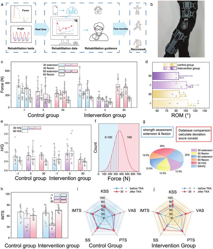
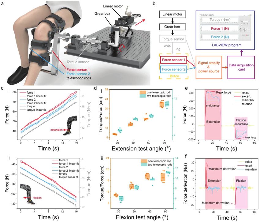
www.advancedsciencenews.com                                                                                       www.advancedscience.com

patients cannot do daily activities independently. Apart from in-        surement module is characterized via a standard torque assess-
convenience, the treatment is expensive too. Research statistics         ment platform. The relationship between the loaded torque and
indicate that the cost of treating OA has accounted for 1.0 to 2.5%      the muscle forces measured by our strength module of the tele-
of the gross national product GDP of developed countries.[9] Knee        scopic rods is recorded. As for the angle measurement module,
osteoarthritis (KOA),[10–12] the most common OA, not only causes         liquid lubrication[59] and sponge buffering are introduced for en-
ache and dysfunction,[13,14] but also induces psychosocial anxiety,      suring its long-term working stability. Clinical experiments are
helplessness, depression, and social disorders.[15,16] For advanced      performed, and demonstrate that the brace realizes the quantifi-
degenerative KOA, total knee arthroplasty (TKA) is now generally         cation of the TKA patient’s recovery process, showing a grad-
regarded as a safe and effective treatment.[17,18] The annual num-        ual increasing trendy of the myodynamia after surgery. More-
ber of total knee replacement in the United States is predicted to       over, facilitated by the conjoint rotation sensing module and cell-
increase progressively from 1 065 000 to 1 921 000.[19] Although         phone application program, we can assess the daily activities of
millions of surgeries are completed, after surgery, these individ-       patients after surgery, and give out home training instructions. It
uals are always plagued with quadriceps muscle impairments               is found that, the intervention group shows an apparent enhance-
and functional limitations,[20] which might be a combination of          ment both in muscle force and joint bending range, compared to
muscle atrophy and neuromuscular activation deficits.[21,22] To           the control group. Furthermore, we establish a new rehabilitative
avoid side effect and ensure desirable long-term functional gains,        indicator, isometric muscle test score (IMTS), for quantitatively
which would influence the life quality of individuals, chronic            evaluating TKA rehabilitation. We anticipate that, as the cloud
quadriceps muscle impairments should be tackled adequately.              database is employed and more rehabilitation data are collected,
Therefore, during the recuperative period, longitudinal and accu-        the brace system could not only facilitate rehabilitations of TKA
rate rehabilitation assessment, composed of quantitative medical         patients, but also improve life quality for geriatric patients, and
indicators, is indispensable and critical for valid treatment.[23,24]    even open a new space for remote AI medical.
   For now, two typical muscle testing systems are developed,
that is, isokinetic muscle strength assessment and training
system,[25–27] and manual muscle testing systems.[28,29] These two       2. Results
systems can provide sufficient medical examination for doctors
during hospitalization, but they are either bulky or requires doc-       2.1. Overall Flow
tors’ operation experience. Therefore, they cannot be widely ap-
plied to postoperative and long-term rehabilitation evaluation of        We developed two sensing modules that can be mounted onto
patients. To achieve postoperative and longitudinal rehabilitation       current braces (Figure 1), including isometric myodynamia mea-
monitoring of patients, wearable technologies, mainly based on           surement (real-time force analysis) and active range of joint
inertial measurement units (IMUs) with accelerometers and gy-            movement measurement (real-time angle analysis). Figure 1a de-
roscopes, are proposed.[30–34] IMUs are able to measure the knee         picts the donning and exploded views of the brace system, which
bending motions, but cannot assess myodynamia. Additionally,             mainly consists of a force gauge, a telescopic rod, and an active
the IMUs measure indirectly, requiring complicated body param-           angle sensor (the total weight of them is merely 71.0 g, see Figure
eters for post computational modeling to calculate out the mo-           S3, Supporting Information). Via the brace system, force and an-
tions, and extra corrections, from time to time, to reduce the mis-      gle information can be recorded and displayed on the cellphone,
alignment error that grows as a function of time.[35] Thus, new          accessible to patients and doctors, benefiting for rehabilitation
wearable long-term monitoring technology is in demand.                   monitoring (Figure 1b). Figure 1c shows the photo of the brace
   To date, many patients choose to use knee brace. As global            and how to perform the measurements.
knee braces market report states, the worldwide quantity demand
dramatically increases in the recent, and the market size is ex-
pected to reach $1.9 billion by 2025.[36] Knee brace, a device for re-   2.2. Isometric Myodynamia Measurement Module
placing cumbersome and airtight plaster in post-hospital protec-
tion, is widely used in hospital and domestic environment among          Since traditional muscle function analysis is normally based on
patients with tendonitis, OA, and other injuries,[37–39] making          torque, in this proof-of-concept research, we explore the relation-
it an ideal carrier of personalized intelligent healthcare[40] and       ship between torque and force via a standardized test platform,
conductive to the recuperation of orthopedic patients.[41] Tri-          as shown in Figure 2a. The standardized test platform is aimed
boelectric nanogenerator (TENG), first invented by Zhong Lin              to gain mechanical response of the brace system under linear
Wang et al in 2012, is a promising technology that might facil-          torque stimulus, which is provided by a linear motor and a gear
itate IoT,[42–45] artificial intelligence (AI),[46–50] and personalized   box. The procedure of the mechanical characterization is shown
medicine[51–55] via self-powered motion sensors. Compared to tra-        in Figure 2a,b. To verify the positive/negative correlation between
ditional sensors, TENG based angle sensors have the advantages           torque and tension, one participant, who wear the brace, is in-
of light weight, wide selection of materials, and self-powered           structed to sit right beside the platform and keep lower limb mus-
ability.[56,57] In our previous work, a self-powered angle sensor        cles relaxed during the test. We assure that the knee brace is coax-
based on TENG is reported and embedded in a knee brace for               ial with the transmission and no residual torque left. Under this
bending detection.[58] However, only information of movement             configuration, the torque measured by the torque sensor is the
detection is not sufficient for rehabilitation assessment.                 torque applied to the brace. Furthermore, a three-channel electri-
   Here, we report a portable, modular, and wearable brace for           cal acquisition and analysis system is developed to display values
self-assessment of TKA patients’ rehabilitation. A strength mea-         of torque and forces in the experiment. In the isometric extension

Adv. Sci. 2022, 2105219                                       2105219 (2 of 10)   © 2022 The Authors. Advanced Science published by Wiley-VCH GmbH
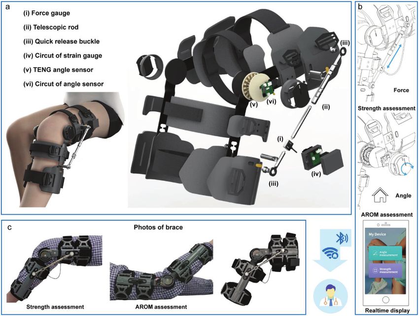
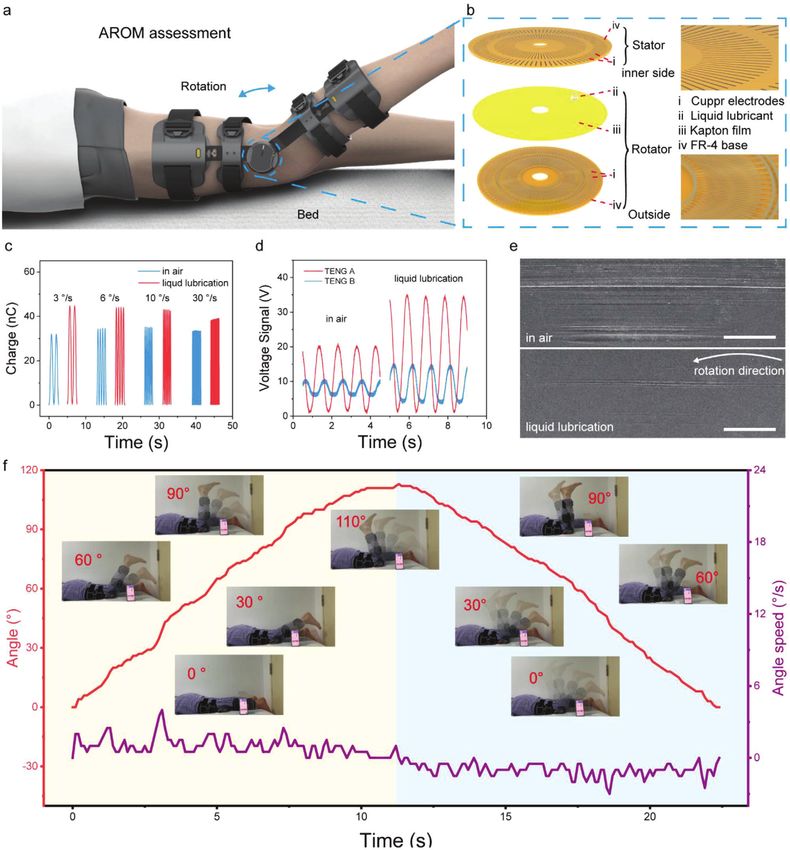
www.advancedsciencenews.com                                                                                              www.advancedscience.com

Figure 1. Schematic illustration of the rehabilitation brace system. a) A perspective view of a brace with modular device for measuring isometric my-
odynamia and joint ROM (range of motion). The brace system includes i) a force gauge; ii) telescopic rod corresponding to brace bending angle of
30°, 50°, 60°, 90°; iii) quick release buckle for telescopic rod fixation and disassemble; iv) data processing circuit of force gauge for isometric muscle
measurement; v) active angle sensor with resolution of 1°; vi) data processing circuit of angle sensor for joint movement. b) Sketches of data collecting
and transmitting. c) Measurement performing.

simulation experiment, extension force generated by muscle ex-                myodynamia strength assessment. One testing curve of myody-
tension is obtained from force transducers on the telescopic rods,            namia is illustrated in Figure 2e,f, from which, we can obtain
while the applied torque is detected by the torque sensor. The                the dynamic myodynamia information, such as peak force, en-
positive/negative correlation between force and toque in exten-               durance time, maximum derivation of force, representing maxi-
sion/flexion simulation experiment is shown in Figure 2c. The                  mum muscle strength, muscle power, and endurance in medical
red, blue, and grey lines represent the indications of force trans-           application, respectively.
ducer 1 (at the outer side of brace), force transducer 2 (at the in-
ner side of brace), and torque transducer (installed on the stan-
dard characterize platform), respectively. Notably, transducer 2              2.3. Active Rotation Sensing Module
and the inner-side rod are normally replaced with an angle limiter
in daily use. Herein, they are assembled for the contrast experi-             The active range of movement measurement, supported by the
ment. An apparent linear relationship between the torque and                  brace, is demonstrated by Figure 3a. Exploded view of the active
the force measured by transducer 1,2 can be obtained from Fig-                angle sensor is shown in Figure 3b. The sensor (60 mm in di-
ure 2c. The ratio of torque to force under various extension and              ameter) mainly consists of FR-4 substrates (1 mm in thickness),
flexion angles are shown in Figure 2d. The trend is consistent                 grid electrodes (≈0.1 mm in width), Kapton film (40 μm in thick-
with the theoretical analysis (see in Note S1 and Figures S1 and              ness), and liquid lubricant (20 μL in volume). Working princi-
S2, Supporting Information). It also indicates that the results ob-           ple can be found in Figure S7, Supporting Information, and our
tained from one telescopic rod corresponds well with that from                previous work.[58] To improve the stability and reliability of the
two telescopic rods, therefore, one telescopic rod is sufficient for            sensors, we investigated the effect of different lubricants on the

Adv. Sci. 2022, 2105219                                           2105219 (3 of 10)     © 2022 The Authors. Advanced Science published by Wiley-VCH GmbH
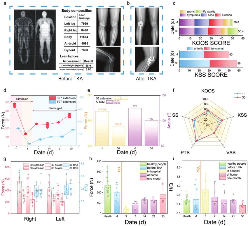
www.advancedsciencenews.com                                                                                               www.advancedscience.com

Figure 2. Characterization of the strength measurement module. Relationship between torque and force is explored through a precise concentric trans-
mission. a) Schematic of the configuration of a transmission system for calibrating the strength module. The direction of torque transmission is shown by
red arrow. b) Flow chart demonstrating the key steps of the signal acquisition and visualization. Once the linear motor slides, rotations of shaft and arm
will be triggered, respectively. At the meanwhile, two force gauges will detect the force applied on the brace through leg. c) Measured relationship among
real-time extension and flexion forces and torque in the test system. d) Calculated torque/force ratios at 30°, 50°, 60°, 90° under one-telescopic-rod
condition and two-telescopic-rod condition. e,f) Medical convalescent information derived from acquired isometric muscle strength test.

signal (see in Figure S8, Supporting Information). The introduc-               Bluetooth and display the angle information through the applica-
tion of the lubricant (Figure 3d,e), here preferably squalene, en-             tion program (APP) on the cellphone. Detail information about
hances the voltage signals that generated by two sets of TENGs                 the APP is shown in Figure S8, Supporting Information. Angle
(Figure 3c,d), which is beneficial for the rotation angle detection.            sensing with a resolution of 1° is achieved. Demonstration of the
More importantly, the durability of the TENG angle sensors is                  real-time angle sensing is plotted in Figure 3f. Original derived
significantly improved by the liquid lubrication. Compared with                 signals is angles, while derivation of angle, rotation speed is the
air lubricant, liquid modification can increase the durability of               blue line. In the measurement, the patient bends his knee from
TENG during 100 000 continuous cycles of rotation regarding                    0° to 110° and move back to 0°.
transfer charge signals.[59] Additionally, liquid lubrication signifi-
cantly minimized Kapton film surface scratches as shown in SEM
images of Figure 3e.                                                           2.4. Quantification of the TKA Patient’s Recovery Process
   For the purpose of detecting real-time angular information,
we developed a circuit to acquire the signal generated by TENG                 Pre-operative information, dual energy X-ray body composition
angle sensors and then, transmitted to a cellphone terminal via                analysis and X-ray images, of a patient is shown in Figure 4a.

Adv. Sci. 2022, 2105219                                            2105219 (4 of 10)     © 2022 The Authors. Advanced Science published by Wiley-VCH GmbH
www.advancedsciencenews.com                                                                                              www.advancedscience.com

Figure 3. The response of brace angle measurement module. Schematic illustration of a) active range of movement test and b) TENG liquid-enhanced
angle sensor with interface liquid enhancement c) Transferred charge of a TENG angle sensor in air and with interface liquid lubrication. d) Open-circuit
voltage of TENG angle sensor in air and with interface liquid lubrication. e) SEM images results of Kapton film surface after 100 000 cycles in air and
with interface liquid lubrication (scale bar, 100 μm). f) The real-time angle signals, angle speed, and photos during the active range of movement test.

After the TKA, X-ray images (Figure 4b) of the patient is taken to            logue scale (VAS), psychological test score (PTS), and step speed
validate the success of the procedure. It is found that the surgery           (SS) of the patient before and after the operation are recorded.
is successful and body composition information is shown in the                Figure 4c presents the patient’s KOOS and KSS score composi-
chart of Figure 4a. Apart from these messages, other medical in-              tion (−1 and 30 represent the day before and one month after
dicators, including knee injury and osteoarthritis outcome score              surgery, respectively). To increase comparability among all these
(KOOS), American knee society knee score (KSS), visual ana-                   scores, we rescaled the total number of scores to 100 without

Adv. Sci. 2022, 2105219                                           2105219 (5 of 10)     © 2022 The Authors. Advanced Science published by Wiley-VCH GmbH
www.advancedsciencenews.com                                                                                              www.advancedscience.com

Figure 4. Clinical analysis of TKA patients’ rehabilitation through brace system. a) Dual energy X-ray body composition analysis images and data; as well
as X-ray images of a patient before TKA. b) X-ray images of the patient after TKA. c) Composition of KOOS and KSS scores before and after TKA: the day
performing TKA is regarded as 0, −1 represents the day before TKA and 30 represents the 30th day after TKA. d) Isometric muscle tests of the patient
during one month (extension peak force) e) 30° extension peak force and active range of motion of the patient before TKA and one month after TKA. f)
Rehabilitation indicators comparison between pre-operation and post-operation. g) Isometric muscle tests data of 10 healthy participants. A group of
patients’ 60° isometric muscle tests in one-month scale: h) Extension peak force and i) hamstring to quadriceps (H/Q) ratio.

changing the composition proportions of the scores. To a certain              clined by 24.87% and 28%, respectively (Figure 4e). To establish a
extent, the patient seems to have recovered partially after the first          reference myodynamia library measured by the brace, 10 healthy
month of operation, although the rehabilitation is not completed.             candidates (5 males and 5 females) participated the measurement
Meanwhile, we applied the myodynamia measurement mod-                         and their lower limp muscle strength data at two typical angles
ule to the patient for one-month longitudinal monitoring, and                 were collected. In Figure 4g, relevant clinical data, including peak
quantitatively determined the rehabilitation level of muscular                force and hamstring to quadriceps (H/Q) ratio, are extracted from
strength. From the 30° and 60° extension results (Figure 4d), we              the isometric muscle tests which repeated at least three times for
can find that muscle strength (force) of the patient did increase              a participant at one angle. The average right/left limb peak forces
gradually after surgery, yet fail to reach to the preoperative sta-           of isometric myodynamia measurements, which are manipu-
tus on the 30th day. Other traditional medical indicators, KOOS,              lated under 30° extension, 30° flexion, 60° extension, and 60° flex-
KSS, VAS, PTS, and SS, are shown in Figure 4f. Variation of                   ion, are 562.56/440.81 N, 299.74/256.45 N, 446.35/357.81 N, and
these indicators in percentage are 9.48%, 16%, 0%, −3.80%, and                247.25/208.86 N, respectively (Figure 4g red part). The average
13.54%, respectively. However, 30° extension force and ROM de-                right/left leg H/Q ratios, tested under 30° and 60°, are 0.55/0.60

Adv. Sci. 2022, 2105219                                           2105219 (6 of 10)     © 2022 The Authors. Advanced Science published by Wiley-VCH GmbH
www.advancedsciencenews.com                                                                                          www.advancedscience.com

Figure 5. Brace-based personalized medicine contributing to convalescent. a) Schematic images of brace system’s effect on TKA patients over the
rehabilitation period. b) Photo of exercise with brace. c) Myodynamia data of control and intervention group under 30 extensions, 30 flexions, 60
extensions, as well as 60 flexion isometric tests. d) ROM of controlled group and intervention group tested on day 1, day 3, and day 30. e,f) Normal
distribution of force from 60 extension isometric test. g) Components of IMTS and score assignment principal. h) IMTS scores of controlled group and
intervention group tested on day 1, 3 30. i,j) rehabilitation indicators of controlled group and intervention group.

Adv. Sci. 2022, 2105219                                         2105219 (7 of 10)    © 2022 The Authors. Advanced Science published by Wiley-VCH GmbH
www.advancedsciencenews.com                                                                                        www.advancedscience.com

and 0.56/0.59, respectively (Figure 4g blue part). Additionally,       3. Conclusion
isometric myodynamia data of patients (three) are investigated
in one-month duration, and plotted in Figure 4h,i and Figure           Here, we report a portable, modular, and wearable brace for self-
S10, Supporting Information. Here, the trend of isometric mus-         assessment of TKA patients’ rehabilitation. The brace consists of
cle strength of patients was consistent with the tendency of the       a force transducer for isometric myodynamia measurement and
patient mentioned above, showing a decline on the third postop-        an active angle sensor for knee bending detection. In addition,
erative day as well as a slow post-surgery increase, however, nei-     the force and angle resolutions are 0.01 N and 1°, respectively.
ther myodynamia nor H/Q ratio returned to preoperative status.         Clinical experiments on TKA patients (n = 14) and healthy peo-
                                                                       ple (n = 10) demonstrate the system feasibility. Key advances are
                                                                       as follows: 1) the capability in quantifying the TKA patients’ re-
                                                                       habilitation process in terms of myodynamia; 2) the definite re-
2.5. Rehabilitation Enhancement                                        habilitation enhancement due to the conjoint myodynamia as-
                                                                       sessment and knee bending sensing; 3) new quantified standard
Subsequently, two groups of patients (5 patients each group) were      indicators, that is, IMTS, for evaluating TKA rehabilitation is pro-
randomly selected for verifying the effects of the brace on the         posed based on the brace-assisted approach.
rehabilitation. Overall procedure of brace-assisted rehabilitation        Notably, patients’ rehabilitation data including myodynamia
healthcare is shown in Figure 5a. The intervention group are           and knee bending activities can be recorded and further uploaded
monitored through the brace, as shown in Figure 5b. Therefore,         onto a cloud database for TKA rehabilitation. Doctors can expe-
their daily bending motions are able to be sent to the doctor, who     diently give out rehabilitation advices to patients on line, facili-
will then give exercise suggestions back. The isometric muscle         tating patients to reach ideal prognosis after surgery. Addition-
strength data of the intervention and control groups are shown         ally, we anticipate that, combing the database with the machine-
in Figure 5c. Compared with the preoperative myodynamia re-            learning analysis, personal rehabilitation advices can be gener-
sults, the data on the third day of both the intervention and con-     ated directly, which will improve life quality for geriatric patients
trol groups dropped dramatically, but after one month, the recov-      and even open a new space for AI medical consulting.
ery of intervention group, with brace intervention, was apparently
enhanced compared to that of control group in terms of muscle          4. Experimental Section
strength. For instance, on average, the muscle strength measured
under 30° extension was recovered to 68.35% compared to the               Fabrication of Telescopic Rods: To begin with, range of torque for hu-
preoperative status for the control group, whereas, that was re-       man isometric rods was investigated and converted to relevant force on
                                                                       telescopic rods. Then proper positions on the brace for fixed points were
covered to above 98.41% for the intervention group. Figure 5d
                                                                       selected which means the length of telescopic rods at different angles.
shows the data of patients’ knee bending ability before and af-        Mechanical draws and 3D model were created based on upper principals.
ter TKA in both groups, which is evaluated by ROM. And the             Traditional manufacturing processes, which included laser cutting, oxida-
ROM shows a reduction induced by the surgery on the 30th day.          tion coating, and gridding, were applied in manipulating the telescopic
Specifically, the reduction was found to be lower in the interven-      rods. Here, the materials for the rods were high young’s modules metal,
tion group (−11.99%) than that in the control group (−12.18%).         preferably chosen aluminum and carbon fiber for lightweight.
Moreover, the intervention group also recuperated better regard-          Fabrication of Standard Platform: The torque sensor with a range of
                                                                       200 N was selected first, and the corresponding transmission structure,
ing H/Q ratio in comparison to the control group, that is, inter-      components of which were finished by machining and surface corrosion
vention group’s outcomes were closer to the normal human H/Q           treatment, was designed according to this torque sensor.
ratio. To improve the comparability of the measured isometric             Synthesis of TENG Angle Sensors: The fabrication of the TENG based
myodynamia data, we proposed an evaluation standard, named             angle sensor was mainly based on the mature printed circuit board tech-
with IMTS, as follows (Figure 5f,g): the score is a weighted sum of    nology. Thus, schematic illustrations of SPAS’s multilayer structures were
isometric test scores of 30 extension, 30 flexion, 60 extension, 60     necessary before the automatic production. An electronic structural design
                                                                       software named of Altium Designer 16 was used to depict the structural
flexion, 30 H/Q, 60 H/Q; each test score is obtained through com-
                                                                       sketches. Detailed fabrication procedures are revealed as follows: The sub-
paring the testing result with the normal distribution of healthy      strate chosen for both the rotator and stator was FR-4 epoxy glass with a
people’s data; the percentage of each test score is illustrated in     thickness of 1 mm. To transfer patterns of copper to this substrate, first
Figure 5g (detail scoring information see Note S2, Supporting In-      it was covered with a complete copper sheet with a thickness of 50 μm
formation). Subsequently, IMTSs of two groups before and after         on FR-4 base through cold rolling craft followed by laminate a sensitive
TKA is shown in Figure 5h. When compared to the presurgical            dry layer on the top of the copper sheet. After that, the sensitive layer was
                                                                       exposed to patterned UV light via photo tools, and the unexposed part of
IMSTs, the IMSTs of the control group reduced by 12.04% one
                                                                       this layer was removed by the developing solution. Before the strip of the
month after operation, while the IMSTs of the intervention group       dry film layer, redundant copper was etched by ferric chloride solution and
rose by 15.36%. A new comprehensive evaluation standard in the         removed from the patterned copper. Finally, a Kapton film with thickness
form of radar graph, comprising of traditional medical indicators,     ≈50 μm was attached to the stator part to function as a triboelectric layer
and IMTS, are established and illustrated in Figure 5i,j (detail in-   while a layer of gold was deposited to the surface of patterned copper of
formation see Table S2, Supporting Information). It can be found       the rotator part to prevent copper from oxidation.
that the proposed standard shows a clear distinction between the          Integration of ROM Module and Myodynamia Module: On the one
                                                                       hand, the TENG angle sensor was embedded between the friction sub-
two groups. Additionally, the intervention group’ rehabilitation       sets of the brace with squalene lubrication and sponge cushioning. On the
is apparently enhanced. By summing up the scores of multiple           other hand, after the fixed columns of the muscle module were mounted
indicators, of control and intervention group, the variations are      on the outside of the brace, the telescopic rod and other components were
found to be 3.77% and 21.90%, respectively.                            assembled subsequently on fixed columns which is depicted in Figure 1a.

Adv. Sci. 2022, 2105219                                     2105219 (8 of 10)    © 2022 The Authors. Advanced Science published by Wiley-VCH GmbH
www.advancedsciencenews.com www.advancedscience.com
Statistical Analysis: Pre-processing of data: isometric muscle test data [5] D. T. Felson, R. C. Lawrence, P. A. Dieppe, R. Hirsch, C. G. Helmick,
was obtained by subtracting the maximum and minimum values from J. M. Jordan, R. S. Kington, N. E. Lane, M. C. Nevitt, Y. Zhang, Ann.
the baseline. Processing of IMST data is shown in Note S2, Supporting Intern. Med. 2000, 133, 635.
Information. Other data (such as transferred charge, short-circuit cur- [6] N. Arden, M. C. Nevitt, Best Pract. Res., Clin. Rheumatol. 2006, 20, 3.
rent, open-circuit, ROM, force, and torque) are presented without pre- [7] Y. Zhang, J. M. Jordan, Clin. Geriatr. Med. 2010, 26, 355.
processing. Data presentation: mean ± SD; mean. Sample size (n): Fig- [8] A. D. Woolf, B. Pfleger, Bull. W. H. O. 2003, 81, 646.
ure 2d (5); Figure 4d (3); Figure 4g (15); Figure 4h (9); Figure 5c, e, h (30); [9] L. M. March, C. J. M. Bachmeier, Bailliere’s Best Pract. Res., Clin.
Figure 5d (5). Software used for statistical analysis: Origin 2021. Rheumatol. 1997, 11, 817.
Ethics Oversight: All procedures in the tests in healthy individuals and
[10] R. E. Phillips, JAMA, J. Am. Med. Assoc. 2021, 325, 2504.
TKA patients were in accordance with the experimental protocol approved
[11] J. N. Katz, K. R. Arant, R. F. Loeser, JAMA, J. Am. Med. Assoc. 2021,
by the Committee on the Use of Humans as Experimental Subjects of the
325, 568.
Xiangya Hospital, Central South University (COUHES, no. 201 908 798).
[12] G. Peat, R. McCarney, P. Croft, Ann. Rheum. Dis. 2001, 60, 91.
All participants were informed with written consent.
[13] E. M. Roos, W. Herzog, J. A. Block, K. L. Bennell, Nat. Rev. Rheumatol.
2011, 7, 57.
[14] R. Becker, A. Berth, M. Nehring, F. Awiszus, J. Orthop. Res. 2004, 22,
Supporting Information 768.
[15] A. M. Sherman, Soc. Sci. Med. 2003, 56, 247.
Supporting Information is available from the Wiley Online Library or from
the author. [16] P. Creamer, M. Lethbridge-Cejku, M. C. Hochberg, Rheumatology
2000, 39, 490.
[17] A. J. Carr, O. Robertsson, S. Graves, A. J. Price, N. K. Arden, A. Judge,
D. J. Beard, Lancet 2012, 379, 1331.
Acknowledgements [18] A. J. Price, A. Alvand, A. Troelsen, J. N. Katz, G. Hooper, A. Gray, A.
J.L. and Y.L. contributed equally to this work. This work was supported Carr, D. Beard, Lancet 2018, 392, 1672.
by the foundation from National Key R&D Project from Minister of Sci- [19] J. A. Singh, S. Yu, L. Chen, J. D. Cleveland, J. Rheumatol. 2019, 46,
ence and Technology (Grant Nos. 2021YFA1201601, 2019YFAO111900), 1134.
National Natural Science Foundation of China (Grant Nos. 52192610, [20] A. H. Alnahdi, J. A. Zeni, L. J. S. H. Snyder-Mackler, Sports Health
61875015, 81874030, and 82072506), Youth Innovation Promotion Asso- 2012, 4, 284.
ciation of Chinese Academy of Sciences, Beijing Natural Science Founda- [21] W. Meier, R. Mizner, R. Marcus, L. Dibble, C. Peters, P. C. Lastayo, J.
tion (NO. JQ20038), Provincial Natural Science Foundation of Hunan (No. Orthop. Sport Phys Ther. 2008, 38, 246.
2020JJ3060), Provincial Clinical Medical Technology Innovation Project of [22] R. L. Mizner, S. C. Petterson, J. E. Stevens, K. Vandenborne, L. Snyder-
Hunan No. 2020SK53709. The authors thank Leo N.Y. Cao for writing help; Mackler, J. Bone Jt. Surg. 2005, 87, 1047.
Sheng Shu, and Pengfei Chen for fruitful discussions; Jianjun Luo, Xue Shi, [23] S.-M. Park, D. D. Won, B. J. Lee, D. Escobedo, A. Esteva, A. Aalipour, T.
Xin Zhao, Linlin Sun, and Dongli Zhang for laboratory help; Jingfei He for J. Ge, J. H. Kim, S. Suh, E. H. Choi, A. X. Lozano, C. Yao, S. Bodapati,
mechanical fabrication support. F. B. Achterberg, J. Kim, H. Park, Y. Choi, W. J. Kim, J. H. Yu, A. M.
Bhatt, J. K. Lee, R. Spitler, S. X. Wang, S. S. Gambhir, Nat. Biomed.
Eng. 2020, 4, 624.
Conflict of Interest [24] A. Slomski, JAMA, J. Am. Med. Assoc. 2021, 325, 2427.
[25] J. M. Rothstein, R. L. Lamb, T. P. Mayhew, Phys. Ther. 1987, 67, 1840.
The authors declare no conflict of interest. [26] P. Kannus, Int. J. Sports Med. 1994, 15, S11.
[27] G. Sole, J. Hamrén, S. Milosavljevic, H. Nicholson, S. J. Sullivan, Arch.
Phys. Med. Rehabil. 2007, 88, 626.
Data Availability Statement [28] L. Merlini, Lancet Neurol. 2010, 9, 1146.
[29] S. C. Cuthbert, G. J. Goodheart, Chiropr. Osteopat. 2007, 15, 4.
The data that support the findings of this study are available from the cor- [30] S. Bahadori, T. Immins, T. W. Wainwright, J. Rehabil. Assistive Technol.
responding author upon reasonable request. Eng. 2018, 5, 2055668318771816.
[31] S. Follis, Z. Chen, S. Mishra, C. L. Howe, N. Toosizadeh, M. Dohm,
J. Orthop. Res. 2021, 39, 2093.
Keywords [32] S. R. Small, G. S. Bullock, S. Khalid, K. Barker, M. Trivella, A. J. Price,
BMJ Open 2019, 9, e033832.
personalized healthcare, rehabilitation, self-powered sensors, total knee [33] M. Gonzalez-Franco, S. Gilroy, J. O. Moore, in 2014 36th Annual In-
arthroplasty, triboelectric nanogenerators ternational Conference of the IEEE Engineering in Medicine and Biology
Society, IEEE, Piscataway, NJ 2014, 6308.
Received: November 15, 2021 [34] C.-Y. Chiang, K.-H. Chen, K.-C. Liu, S. J.-P. Hsu, C.-T. Chan, Sensors
Revised: December 8, 2021 2017, 17, 418.
Published online: [35] C. Li, D. Liu, C. Xu, Z. Wang, S. Shu, Z. Sun, W. Tang, Z. L. Wang, Nat.
Commun. 2021, 12, 2950.
[36] K. Research Global Knee Braces Market (2019-2025),
https://www.reportlinker.com/p05862268/Global-Knee-Braces-
[1] T. A. Ghebreyesus, Nat. Aging 2021, 1, 865. Market.html?utm_source=PRN (accessed: January 2020).
[2] T. B. L. Kirkwood, Nature 2008, 451, 644. [37] A. Kirkley, S. Webster-Bogaert, R. Litchfield, A. Amendola, S. Mac-
[3] D. J. Hunter, D. Schofield, E. Callander, Nat. Rev. Rheumatol. 2014, Donald, R. McCalden, P. J. J. Fowler, J. Bone Jt. Surg. 1999, 81,
10, 437. 539.
[4] S. Glyn-Jones, A. J. R. Palmer, R. Agricola, A. J. Price, T. L. Vincent, H. [38] L. Sharma, J. Song, D. T. Felson, S. Cahue, E. Shamiyeh, D. D. J. J.
Weinans, A. J. Carr, Lancet 2015, 386, 376. Dunlop, JAMA, J. Am. Med. Assoc. 2001, 286, 188.
Adv. Sci. 2022, 2105219 2105219 (9 of 10) © 2022 The Authors. Advanced Science published by Wiley-VCH GmbHwww.advancedsciencenews.com www.advancedscience.com
[39] K. S. Brooks, J. Prosthet. Orthot. 2014, 26, 2. [49] S. Xiang, D. Liu, C. Jiang, W. Zhou, D. Ling, W. Zheng, X. Sun, X. Li,
[40] J. R. J. Greenfield, H. F. Hwang, C. Davies, A. J. McDaid, in 2017 Int. Y. Mao, C. Shan, Adv. Funct. Mater. 2021, 31, 2100940.
Conf. on Rehabilitation Robotics (ICORR), IEEE, Piscataway, NJ 2017, [50] Y. Liu, B. Chen, W. Li, L. Zu, W. Tang, Z. L. Wang, Adv. Funct. Mater.
352. 2021, 31, 2104770.
[41] B. Weinberg, J. Nikitczuk, S. Patel, B. Patritti, C. Mavroidis, P. Bonato, [51] R. Hinchet, H.-J. Yoon, H. Ryu, M.-K. Kim, E.-K. Choi, D.-S. Kim, S.-W.
P. Canavan, in Proc. of 2007 IEEE Int. Conf. on Robotics and Automa- Kim, Science 2019, 365, 491.
tion, IEEE, Piscataway, NJ 2007, pp. 4126–4133. [52] H. Ouyang, Z. Liu, N. Li, B. Shi, Y. Zou, F. Xie, Y. Ma, Z. Li, H. Li, Q.
[42] J. Luo, Z. Wang, L. Xu, A. C. Wang, K. Han, T. Jiang, Q. Lai, Y. Bai, W. Zheng, Nat. Commun. 2019, 10, 1821.
Tang, F. R. Fan, Nat. Commun. 2019, 10, 5147. [53] M. Zhu, Z. Sun, T. Chen, C. Lee, Nat. Commun. 2021, 12, 2692.
[43] Y. Tang, H. Zhou, X. Sun, N. Diao, J. Wang, B. Zhang, C. Qin, E. Liang, [54] S. Gao, T. He, Z. Zhang, H. Ao, H. Jiang, C. Lee, Adv. Sci. 2021, 8,
Y. Mao, Adv. Funct. Mater. 2020, 30, 1907893. 2101834.
[44] N. Zhang, C. Qin, T. Feng, J. Li, Z. Yang, X. Sun, E. Liang, Y. Mao, X. [55] B. Zhang, Y. Tang, R. Dai, H. Wang, X. Sun, C. Qin, Z. Pan, E. Liang,
J. N. R. Wang, Nano Res. 2020, 13, 1903. Y. Mao, Nano Energy 2019, 64, 103953.
[45] M. Wang, J. Zhang, Y. Tang, J. Li, B. Zhang, E. Liang, Y. Mao, X. J. A. [56] Q. Jing, Y. Xie, G. Zhu, R. P. S. Han, Z. L. Wang, Nat. Commun. 2015,
n. Wang, ACS Nano 2018, 12, 6156. 6, 8031.
[46] H. Guo, X. Pu, J. Chen, Y. Meng, M.-H. Yeh, G. Liu, Q. Tang, B. Chen, [57] G. Zhu, J. Chen, T. Zhang, Q. Jing, Z. L. Wang, Nat. Commun. 2014,
D. Liu, S. J. S. R. Qi, Sci. Rob. 2018, 3, eaat2516. 5, 3426.
[47] J. Yu, G. Gao, J. Huang, X. Yang, J. Han, H. Zhang, Y. Chen, C. Zhao, [58] Z. Wang, J. An, J. Nie, J. Luo, J. Shao, T. Jiang, B. Chen, W. Tang, Z. L.
Q. Sun, Z. L. Wang, Nat. Commun. 2021, 12, 1581. Wang, Adv. Mater. 2020, 32, 2001466.
[48] J. An, P. Chen, Z. Wang, A. Berbille, H. Pang, Y. Jiang, T. Jiang, Z. L. [59] L. Zhou, D. Liu, Z. Zhao, S. Li, Y. Liu, L. Liu, Y. Gao, Z. L. Wang, J.
Wang, Adv. Mater. 2021, 33, 2101891. Wang, Adv. Energy Mater. 2020, 10, 2002920.
Adv. Sci. 2022, 2105219 2105219 (10 of 10) © 2022 The Authors. Advanced Science published by Wiley-VCH GmbHYou can also read