Reactive Pigmentation of Skin Graft Mimicking a Lentigo Maligna Recurrence: a Case Report
←
→
Page content transcription
If your browser does not render page correctly, please read the page content below
Reactive Pigmentation of Skin Graft Mimicking a
     Lentigo Maligna Recurrence: a Case Report
     Matías Gárate1, Valentina Vera1, Nadia Vega1, Jonathan Stevens2, Verónica Sanhueza3

1 Department of Dermatology, Faculty of Medicine, Universidad de Chile, Santiago, Chile
2 Dermatology Section, Oncology Institute, Fundación Arturo López Pérez (FALP), Santiago, Chile
3 Pathology Section, Oncology Institute, Fundación Arturo López Pérez (FALP), Santiago, Chile

Key words: dermoscopy, neoplasm recurrence, skin pigmentation, scar
Citation: Gárate M, Vera V, Vega N, Stevens J, Sanhueza V. Reactive pigmentation of skin graft mimicking a lentigo maligna recurrence:
A Case Report. Dermatol Pract Concept. 2022;12(2):e2022054. DOI: https://doi.org/10.5826/dpc.1202a54
Accepted: August 30, 2021; Published: April 2022
Copyright: ©2022 Gárate et al. This is an open-access article distributed under the terms of the Creative Commons Attribution-
NonCommercial License (BY-NC-4.0), https://creativecommons.org/licenses/by-nc/4.0/, which permits unrestricted noncommercial use,
distribution, and reproduction in any medium, provided the original authors and source are credited.
Funding: None.
Competing interests: None.
Authorship: All authors have contributed significantly to this publication
Corresponding author: Valentina Vera Giglio, Servicio de Dermatología, Hospital Clínico Universidad de Chile, Santos Dumont 999,
Independencia, Santiago, Chile. Postal code: 8380456. E-mail: draveragiglio@gmail.com

Introduction                                                            skin graft. Histopathologic examination confirmed the diag-
                                                                        nosis of LM, without ulceration, mitosis, nor lympho-vascu-
There is limited information on dermoscopy of recurrent                 lar invasion, and with clear surgical margins. At a 3-month
hyperpigmentation in skin grafts following lentigo maligna              follow-up, the patient reported recurrent pigmentation
(LM) excision, especially in facial locations. We present a             within the scar. Physical examination revealed an erythem-
case with highly suspicious dermoscopic features of local re-           atous scar with a brown pigmented border, 13 x 10 mm
currence of LM within the skin graft.                                   in diameter (Figure 1C). Dermoscopic evaluation showed
                                                                        brown pigmented areas extending slightly beyond the edge

Case Presentation                                                       of the graft, with a pseudo-network pattern, asymmetric
                                                                        pigmented follicular openings and circles within circles, pre-
A 49-year-old woman consulted with a 3-year history of hy-              dominantly in the upper lateral area (Figure 1D). Recurrent
perpigmented lesion on the left cheek, with gradual growth              LM versus reactive graft pigmentation were the proposed
and darkening in the last 11 months. Physical examina-                  diagnosis. A new biopsy was performed, with complete
tion revealed phototype IV and an 15-mm diameter brown                  excision of the scar, and histopathologic study ruled out
macule. Dermoscopic evaluation (3Gen – DermLite DL4®)                   malignant melanocytic neoplasia, with findings of dermal
showed a pseudo-network with asymmetric pigmented fol-                  scarring, foreign body-type granulomas and dermal melano-
licular openings (Figure 1, A and B). Incisional biopsy was             sis. SOX-10 staining showed normotypic melanocytes, ade-
performed and confirmed the diagnosis of LM. Wide local                 quate in number and size (Figure 2, A- C). Patient remains
excision with 5-mm margin was performed, followed by im-                without signs of recurrence or pigmentation at the 6-months
mediate reconstruction with a retro-auricular split-thickness           follow-up.

Research Letter | Dermatol Pract Concept. 2022;12(2):e2022054                                                                            1
A B
Figure 1. (A) Clinical appearance pretreatment. Hyperpigmented macula, 15 x 11 mm in diameter, on the left cheek. (B) Dermoscopy
pretreatment. Brown macula, with pseudo-network structure. Loss of follicular openings is observed isolated in the periphery (arrows).
Arboriform telangiectasias at the bottom of the lesion. (C) Clinical appearance after surgery. Erythematous plaque with a scar-like aspect and
a brown pigmented border measuring 13 x 10 mm in diameter, on the left cheek. (D) Dermoscopy after surgery. In the center of the lesion:
follicular openings can be seen forming whitish circles. In the periphery: brown pigmentation which exceed the edge of the scar with the
appearance of a pseudo-network. In superior lateral region: structures in a double concentric circle (arrow).
Conclusions In our case, the pattern of double circles associated
with hyperpigmentation that exceeds the edge of the graft
The most frequently described dermoscopic features of reac- scar was observed, leading to suspect a recurrence of LM,
tive pigmentations includes: a homogeneous radial band-like which was histologically ruled out. We suggest that in grafts
and continuous brownish lines that extends perpendicularly of the facial area in patients with darker skin phototypes,
to the scar [1]. This case report describes unusual dermo- the underlying inflammation related to scarring would lead
scopic characteristics of reactive pigmentation within a skin to reactive melanosis with a double circle pattern on der-
graft, resembling a recurrence of LM. Circle within circle moscopy as seen in LM recurrence in a scar. As reported by
sign is associated with LM with an odds ratio of 6.32 [2]. In Navarrete-Dechent et al. in patients with scar tissue from
addition, hyperpigmentation exceeding the edge of the scar previous treatment of LM, dermoscopy of melanoma-spe-
is considered one of the most important criteria to suspect cific features has limitations [3], therefore, histopathological
recurrent melanoma. confirmation is essential for the differential diagnosis.
2 Research Letter | Dermatol Pract Concept. 2022;12(2):e2022054C
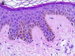
Figure 2. (A) Histopathology examination (H&E, 4x) shows a slightly atrophic epidermis with basal hypermelanosis. Proliferation of fibro-
blasts in the dermis associated with a perivascular lymphohistiocytic inflammatory infiltrate and the formation of granulomas with multi-
nucleated giant cells. (B) Higher magnification (H&E, 20x) shows basal hypermelanosis without proliferation of melanocytes. (C) SOX-10
staining shows melanocytes of adequate number and size, equidistant and normotypic.
References keratosis and solar lentigines. Acta Dermatovenerol Croat.
2019:27(3):146–152. PMID: 31542057
1. Moscarella E, Argenziano G, Lallas A, Longo C, Al Jalbout S, 3. Navarrete-Dechent C, Cordova M, Liopyris K, et al. Reflectance
Zalaudek I. Pigmentation in a scar: use of dermoscopy in the man- confocal microscopy and dermoscopy aid in evaluating repig-
agement decision. J Am Acad Dermatol. 2013;69(3):e115–e116. mentation within or adjacent to lentigo maligna melanoma sur-
DOI: 10.1016/j.jaad.2013.03.008. PMID: 23957988. gical scars. J Eur Acad Dermatol Venereol. 2020;34(1):74–81.
2. Ozbagcivan O, Akarsu S, Ikiz N, Semiz F, Fetil E. Dermoscopic DOI: 10.1111/jdv.15819. PMID: 31325402. PMCID:
differentiation of facial lentigo maligna from pigmented actinic PMC7592341.
Research Letter | Dermatol Pract Concept. 2022;12(2):e2022054 3You can also read


