Quad Convolutional Layers (QCL) CNN Approach for Classification of Brain Stroke in Diffusion Weighted (DW) - Magnetic Resonance Images (MRI) - INASS
←
→
Page content transcription
If your browser does not render page correctly, please read the page content below
Received: October 22, 2021. Revised: November 22, 2021. 414

 Quad Convolutional Layers (QCL) CNN Approach for Classification of Brain
 Stroke in Diffusion Weighted (DW) - Magnetic Resonance Images (MRI)

 Andi Kurniawan Nugroho1,4 Terawan Agus Putranto2 I Ketut Eddy Purnama1,3
 Mauridhi Hery Purnomo1,3,5*

 1
 Electrical Engineering Department, Institut Teknologi Sepuluh Nopember, Surabaya, Indonesia.
 2
 RSPAD Gatot Subroto Presidential Hospital, Jakarta, Indonesia.
 3
 Department of Computer Engineering, Institut Teknologi Sepuluh Nopember, Surabaya, Indonesia.
 4
 Electrical Engineering Department, Universitas Semarang, Semarang, Indonesia.
 5
 University Center of Excellence on Artificial Intelligence for Healthcare and Society (UCE AIHeS), Indonesia.
 * Corresponding author’s Email: ketut@ee.its.ac.id; hery@ee.its.ac.id

 Abstract: Commonly, clinicans have problems for recognising brain stroke injury images. However, with the
 advantages of Information technology it is expected that will be a new method that can support the clinicans’ opinion
 for recognising the brain stroke injury for type of stroke (hemorrhagic, ischemic, and normal). Therefore, this study
 aim is to discovery a new model to classify hemorrhagic, ischemic and normal based on Diffusion Weighted (DW)-
 Magnetic Resonance (MR) images. This study argues by using Qual Convolutional Layers (QCL-CNN) which applied
 in CNN can classified type of stroke. For this study experiment, this research conducted two experiment to asses the
 performance of QCL-CNN. The first experiments partitioned the MR image dataset into 20 percent testing and 80
 percent training sets. Then, the second testing performed ten-fold cross-validation on the image dataset. The result
 from the first experiment of the classification accuracies obtained 93.90 percent (1st dataset) and 94.96 percent (2nd
 dataset). As for the second experiment, the results shows that the classification accuracies obtained 95.91 percent ( 1st
 data set) and 97.31 percent ( 2nd data set). The data source for this study gained from Indonesian hospital and the web
 sources dataset public from Ischemic Stroke Lesion Segmentation (ISLES). This study also compared, the QCL-CNN
 model with other architecture model such as AlexNet, ResNet50, and VGG16. The result of the comparison experiment
 shows that QCL-CNN architectures model has excellent performance than the others model.

 Keywords: QCL-CNN, DW-MRI, Brain stroke injury, Image, Preprocessing, Classification.

 to an appropriate health staff diagnosis for the brain
1. Introduction stroke injury lead to the appropriate and proper
 treatment for the patient. The treatment and diagnosis
 For the brain stroke injury, health staff generally
 of stroke are carried out by clinical examination,
used radiological modality of Computerized
 followed by assessing radiological modalities, such
Tomography Scanning (CT scan), for examining the
 as CT scan [2].
patient. The health staff identified stroke based on the
 CT is the primary mode of diagnosis in the early
caused of the brain stroke injury. Based on causes,
 stages of stroke for separating hemorrhagic from
stroke is classified into two, namely ischemic (in
 ischemic disorders. Nevertheless, CT has less ability
which the blood supply stops flowing to the brain due
 for detect the stroke lesions during the patient’s acute
to blockage) and hemorrhagic (where there is
 period. Conventional CT or Medical Resonance
bleeding in the brain tissue) [1]. Based on the
 Image (MRI) occasionally ineffective at predicting
classification, the health staff is essential to carry out
 the presence and amount of acute damage [3].
an appropriate diagnosis before starting stroke
 To address for ineffective of CT and MRI, the
treatment due to different disease conditions. It is due
 health staff using Diffusion Imaging as hyper-
International Journal of Intelligent Engineering and Systems, Vol.15, No.1, 2022 DOI: 10.22266/ijies2022.0228.38
Received: October 22, 2021. Revised: November 22, 2021. 415

intensity (DWI).The reduction in water diffusion is Brain strokes injury is caused by hemorrhage
reflected in the DW-MRI mode by a drop in the occur when a blood artery rupture and spills into the
Apparent Diffusion Coefficient (ADC) trace map [4], surrounding brain tissue. Hypertension, trauma,
[5], which is visible in Diffusion Imaging as hyper- aberrant blood arteries (for instance, arteriovenous
intensity (DWI). The usage of DWI has been proven malformation (AVM)), bleeding problems,
in previous animal research, and the investigations aneurysms, and drug use all contribute to this.
showed, the DWI has an ability of showing ischemic Ischemic stroke, on the other hand, happens when the
brain alterations within five minutes and one to three brain's blood supply is cut off due to a clot. After an
hours after the patients feel the symptoms brain ischemic stroke, a brain hemorrhage can occur,
stroke injury. In medical diagnosis for human, these resulting in significant consequences [7].
alterations are noticed as early as two to six hours Fig. 1 illustrates a human brain stroke injury
after the beginning of brain stroke injury symptoms. image. The first ( a) and second (b) row indicate
Moreover, DWI has a low rate of false-negative hemorrhage and ischemic stroke, respectively,
investigations (5 %), with a clear distinction between whereas the third (c) row normal instances. The part
ischemic and hemorrhagic lesions. Thus, DWI of the brain where a brain stroke injury develops is
enables the early identification of the kind, location, highlighted in red’s colours and it is to assist
and size of a brain stroke and aids in the prediction of inexperienced readers. The intensity of the core
the patients' clinical outcomes [6]. infarct is what differentiates suspected hemorrhagic
 To address the problem, the hospital create a and ischemic strokes. Additionally, bleeding is
procedures, for the clinicans treating the patient’s indicated with darker in color than the ischemic core.
brain stroke injury. Firstly, before spotting a stroke, Based on the problem, the prior research showed
clinicians need to confirm the symptoms with the that Computer-Aided Diagnosis (CAD) system can
patient or the patient’s family members. This help supporting the manual diagnostic procedure in
identification process is essential since it helps the hospital in identifiying patient with brain stroke
clinician to have clearly and accurately injury. However, in fact the doctor and clinicans
clinical’sjudgement for determining the patient's rarely used the CAD system for diagnosing the
condition. patient with brain stroke injury [8, 9]. So, , the
 Next, the clinicians is analyzing the neurological researcher Peixoto and Filho [8] suggested further
imaging recordings of the patient using the CT scan. studies to automate the CAD system's performance.
By using the CT scan, it is the first technique for From the prior research, there are a litte gap for
diagnosing brain stroke. The reason for using The CT research in detection in brain stroke injury image
scan for detecting the brain stroke injury is the tool is using DWI- MRI. Since, there was no research that
affordable in price compare to the others medical employs the DWI- MRI for data set for experiment in
tools. detection of brain stroke injury. Futhermore, there
 Then, the patient brain’s image is sent to the was no research that suggesting method to detect
radiologist. The radiologist will determine the brain stroke injury using DW-MRI.
patient brain stroke injury type. Therefore, this paper proposed a new method to
 To support the radiologist decision, another tools detect brain stroke injury on DW-MRI. As many
used which called MRI. MRI is used by the research that already used deep learning to aid the
radiologist to obtain detailed changes in patient’s study of medical image processing. This study also
brain structure anatomy. With the advantages of seek the possibility for using deep learning to slasify
technology DW mode applied in MRI to help detect the brain stroke injury on DW-MRI.
the beginning of a patient’s brain stroke injury, Deep learning is a technique that support in a
particulary for the brain stroke injury with the classification techniques. [10, 11]. Deep learning also
ischemic type. support an automation for calculating deep
 After , the first patient's physical examination is convulational system [11]. The key advantage of this
performed. The clinicians give the patients methodology is that it outperforms other picture
treatmentent according to the clinical procedure. classification techniques. [12]. Since the beginning of
 However, the patients with brain stroke injury, is development, numerous deep learning methods have
not only once patient. The hospitals are receveid been formed, for intances recurrent neural networks,
numerous patients with stroke symptoms on the same Long Short-Term Memory (LSTM) [13], CNN [14],
day. It also become problematic for the hospital and Deep Belief Net (DBN) [15], all of which are
clinician to provide a suit treatment for patient’s brain based on the neural network concept. In addition to
stroke injury if to many patient that need to be handle deep learning methods, other classification
by the clinican algorithms [16, 17, 18]. Although SVM works
International Journal of Intelligent Engineering and Systems, Vol.15, No.1, 2022 DOI: 10.22266/ijies2022.0228.38
Received: October 22, 2021. Revised: November 22, 2021. 416

properly on linear data, it is difficult to process high- Then, Mosqueda et al. [23] examined the concept
dimensional information [19, 20]. of clinical data classification on acute ischemic
 Meanwhile, KNN is a simple method that requires patients using CT or MR angiography images to
a large storage capacity to accommodate millions of detect large and small strokes (Boston Acute Stroke
objects in the dataset [20]. When training CNN Imaging Scale/ BASIS). This research argues, that
models with larger datasets, deeper architectures are BASIS classification instrument is effective and
recommended over shallow architectures. Bansal et appears superior to Alberta Stroke Program Early CT
al. [21]. However, this study also found that for larger Score (ASPECTS) in predicting outcomes in acute
datasets that used to performs the experiment is , ischemic stroke. However, the ASPECTS has
shallow architectures outperform deeper weakness because this method employs scoe based
architectures. on the health staff opinion.
 In this study, a stroke classification method was Next, S. Anbumozhi [24] developed a technique
proposed Quad Convolutional layers (QCL-CNN) for for detecting and diagnosing brain stroke. A
stroke classification on DW-MRI images, using the directional filtering method is used to minimize
CNN architecture comprising two main blocks, each impulse noise in brain MRI pictures. Oriented local
consisting of two convolutions with max-pooling. histogram equalization (OLHE) approaches are used
The goal is that reduction in size causes less to improve the quality of the noise-reduced brain
computational overhead for subsequent network image. After that, the skull is eliminated from the
layers and prevents excessive over-fitting. improved brain image. A k-means classifier is used
Moreover,an automatic classification method was to extract features and segment the stroke region.
proposed in this study to predict the category of DW- Based on the location, a segmented stroke is
MRI brain images, including hemorrhage, ischemic, classified as mild, moderate, or severe. Nevertheles,
and normal categories. this OLHE only detects small infact in brain stroke
 The contributions of this study are : injury.
 Other researchers, Gautam et al. [25] illustrate
 • Classifying the particular features of brain stroke how to segment hemorrhagic strokes from CT scan
 injury using DW-MRI imaging. images using a fuzzy clustering variation called
 • Adapting CNN architecture with QCL to Modified Robust Fuzzy C-Means Clustering
 produce shallow architure to gain high accuracy (MRFCM) and a segmentation technique called
 brain stroke injury classification. Distance Regularization Level Set Evolution
 (DRLSE). However, this approach was unable to
 This paper further ordered as follows: The Section identify a very small lesion from a CT scan image.
2 discusses the preliminary studies that similar with Similar with the first experiment conducted by
the QCL-CNN, Section 3 explains the classifies the Gautam et al [26], also presented a system for
proposed methodology for this study, and Section 4 classifying CT scan images of the brain into three
provides details of the experiment to test the QCL- categories: hemorrhage, ischemia, and normal. Local
CNN with MR image datasets and the results. Gradient of Gradient Pattern was offered as a new
Meanwhile, the Section 5 discusses the result of the feature descriptor (LG2P). Using 900 image datasets
QCL-CNN and Section 6 summarises the result of with fine kNN and cubic SVM, the greatest
this study. classification accuracy of 83.11 percent and 86.11
 percent was attained. However, this precision was
2. Preliminary studies insufficient and needed to be improved in order to
 achieve better outcomes.
 This section explores the preliminary studies From the explanation on the prior studies above
related to an approach of brain classification. First, [22-26], it can be summarized that none of them
the study conducted by Saatman et al. [22]. This doing research to classify ischemic stroke, stroke
study classified traumatic brain injury to seek the best haemorrhage, and normal conditions of the human
treatment for patient. This paper used the degree of brain using deep learning methods with limited layer
brain injury to classify the traumatic of brain injury. convolutions. Therefore, the research in this paper is
Saatman et.al[22] research employs the Glasgow the first to proposes the use of four-CNN convolution
Coma Scale (GCS) and other criteria. However, this layers in deep learning for brain stroke classification,
study only used the expert judgement from health and the author’s name it as Quad Convolutional
staff to clasify the brain injury. Therefore, this study Layers (QCL)-CNN.
has a weakness on the data accuration. The data is
only used the health staff opinion.

International Journal of Intelligent Engineering and Systems, Vol.15, No.1, 2022 DOI: 10.22266/ijies2022.0228.38
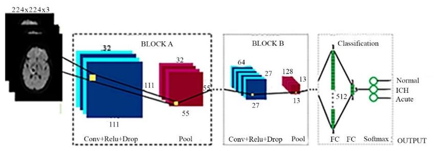
Received: October 22, 2021. Revised: November 22, 2021. 417

3. Proposed method Numerous preprocessing processes were
 conducted for this experiment the images were
3.1 Dataset for the experiment/ preparation attained using a variety of scanners and protocols.
 Therefore, this study applied homogeneous linear
 The study used DW MRI scans from two hospitals
 sampling method to gain DW-MRI Image in uniform
in Indonesian and the web sources dataset public
 physical size.
from Ischemic Stroke Lesion Segmentation (ISLES)
 Moreover, because the image is anisotropic in the
to gather 1742 hemorrhage, acute ischemic, and
 axial (or z-axis) direction, the resampling operation
normal strokes for every class. This expemerint
 introduced interpolation errors. As a result, a 2D slice
employed the 5226 DW MR images.
 analysis was done rather than a 3D volume study.
 This research worked based on the ethical
 Data preparation and data augmentation was done
approval from Health Research and Development
 to eliminate superfluous contextual information and
Agency Indonesia (Badan Penelitian dan
 to equalize the pixel counts of normal and lesion
Pengembangan Kesehatan - Balitbangkes) No.
 pixels. As a result it is an effective form of data
LB.02.01/2/KE.289/2018. Moreover, all clinical
 augmentation, as all pixels were classified as being
images were labelling by number, so the patients’
 part of the obstruction and bleeding. A patch was
identity did not recognised in this study.
 extracted around each of these pixels and placed in
 The MRI were acquired with the following
 random spots. Each patch contains pixels from
parameters: field strength 1.5 T, slice thickness 5 mm,
 infarct/hemorrhage and general tissue/background.
slice distance 0.7 mm, pixel size 320x320, echo
 When a pixel was located in the center of an
duration 71 ms, repetition time 4000 ms, flip angle
 infarct/bleed, the patch extracted included only that
150 °, and step coding phase 287. All DW-MRI
 pixels.
images were labeled appropriately for acute ischemic,
hemorrhagic, and normal strokes.
 3.3 Pre-processing
3.2 Data preparation and data augmentation A multi-layer design was proposed to perform the
 brain's DW-MRI classification. In this experiment, a
 Each DWI scan is limited in terms of the number
 2D slice of the DW-MRI image was considered on
of lesions. Additionally, whether training data were
 the section's axial side. The hospital radiologist ( as
created at the cut-out level or lesion sample stage,
 expert fo brain stroke injury) is help aid this study in
only a small number of patches were accessible. Due
 the process of labelling and selecting the images to be
of the high number of parameter files and the
 separated into three classes. The classes of images
requirement to generate a large number of pictures
 was grouping to hemorrhage, ischemic, and normal.
(patches), different methods were used to replenish
the training data based on a finite amount of DWI.
 3.4 Image classification process
 To begin, the extracted picture (patch) was split,
zoomed, and flipped horizontally. Second, the patch 1st Phase, is input Layer, which consist of
extraction procedure was also used to supplement training, validating, and testing data.
data. 324 DWI were utilized for training, 36 for 2nd Phase is Convolution extracts features from
validation, and 90 for testing in the initial data set. images by convolving each element with a filter that
 The solution for this study, the researcher employs has the same depth as the image[27] . The last detail
the data augmentation. before implementing CNN defined the end-to-end
 The algorithm can seen below, design and the Convolutional layer’s dimensions to
 construct the building blocks mentioned above.
Algorithm: Pseudocode for preparing the data and To calculate the spatial dimensions of the
augmentation Convolutional Layer, a formula that functions from
 the input volume and hyperparameters is needed as
1. Load the original input image from the disk.
 follows:
2. The original image is changed randomly with the For each ( ( )) input volume dimension:
 technique of sliding series, series, zoom, flip
 horizontally.
 ( ) − + 2
3. images that have been processed, used and written ( ) = 1 + (1)
 back to disk.
4. the second and third steps are repeated continuously
 according to the number of N Where, ( ) is the input dimension, R is the
 value of the receptive field, P is the padding value,

International Journal of Intelligent Engineering and Systems, Vol.15, No.1, 2022 DOI: 10.22266/ijies2022.0228.38

Received: October 22, 2021. Revised: November 22, 2021. 418

 hemorrhage

 Ischemic

 Normal

 Figure. 1 DWI mode that differentiates hemorrhage, ischemic and normal conditions

and S is the stride value. This formula does not The 4th phase Pooling Layer . Convolutional is
depend on the depth of the input. Therefore, to obtain placed before the Pooling Layer, serves to reduce the
the volume, the dimension value is explained in the spatial dimensions (Width x Height) of the Input
following steps: Volume. this does not affect the volume depth
 Suppose the input volume dimension is dimension. Down sampling is done at layer 4.
224x224x3, and the stride value is 2 along with the Reducing the size will also cause a reduction in
horizontal and vertical directions, then, WIn = 224 information.
and S = 2, (2.P - R) need to be the integer for the The 5th phase is Dropout layer. This is a
calculated value. When the padding is 0 and R = 4, regularization strategy for preventing network
the results obtained are Wout = (224-4 + 2.0) / 2+ 1 overfitting [28].
= 284/2 + 1 = 111. The 6th phase layer is Fully-Connected. n layer 6,
 3rd phase The third layer is ReLU. ReLU layer is it functions as an output link and a layer that regulates
to the Unit Rectifier, the most commonly used the amount of output. The number of inputs for the
activation function for CNN neuron output. It is 6th layer is multiplied by the matrix, and the bias
mathematically, explained as follows: vector is added.The range [0 1] [27] is then
 normalized using layer softmax.
 ≥0 In Fig. 2, this study proposes the QCL-CNN
 ( ) = { (2) architecture, where there are convolution, max-
0 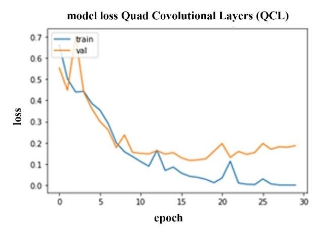
Received: October 22, 2021. Revised: November 22, 2021. 419

 Table 1. Properties of QCL-CNN
 Numbers Layer Properties
 1 Input Layer 1st layer of input size 224 x 224 x 3
 2 Convolution Layer 1st layer contains 32 filters of [2,2]
 3rd layer contains 32 filters of [2,2]
 5th layer contains 64 filters of [2,2]
 7th layer contains 128 filters of [2,2]
 3 ReLU Layer ReLU is used in 1st , 3rd 5th , 7th , 10th.
 4 Max Pooling Layer 2x2 max pooling with stride[2 2] has been used
 2th, 4th, 6th, 8th.
 5 Dropout Layer 11th er with dropout probability 0.5
 12th layer with 512 output value and 3 or 2 output
 Fully Connected Layer value-dependent of the dataset
 Softmax layer 12th layer
 6 Classification Layer 12th layer for image classification

 Figure. 2 Proposed research block diagram

 The batch size used for the AlexNet proposal Rate (TPR), false-positive rate (FPR), F-measure,
and network is 128, while for ResNet50, it is 12. accuracy, and ACC as an evaluation measure. This
 is calculated as follows:
3.5 Implementation details and performance
measures
 = (5)
 The experiment was carried out in 2 methods. In +
the first, the hemorrhage and ischemic data were
divided into 80percent training and 20percent = (6)
testing. The second experiment added normal DW- +
MRI data by dividing it into 20percent testing and
80percent training. The image preprocessing = (7)
techniques were used to experiment by adding the +
data limitations with augmentation and image
resizing methods at the beginning of the input = 2. (8)
image from the CNN process. Based on the trained +
model, the recommended QCL-CNN model were +
placed on the training dataset. As illustrated in Fig. = (9)
 + + +
2, it delivers classification results on the test dataset.
The properties of all layers used are shown in Table FN, FP, TN, and TP are False Negative, False
1. Furthermore, the following precisions were used Positive, True Negative, and True Positive.
to check the method's effectiveness: True Positive
International Journal of Intelligent Engineering and Systems, Vol.15, No.1, 2022 DOI: 10.22266/ijies2022.0228.38
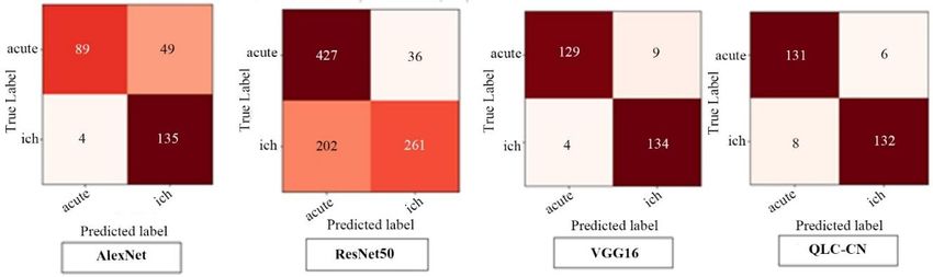
Received: October 22, 2021. Revised: November 22, 2021. 420

 QCL-CNN

 (a) (b) (c) (d)
 Figure. 3 Confusion matrix 1st dataset of: (a) AlexNet, (b) ResNet50, (c) VGG16, (d) QCL-CNN, with 80 percent
 training and 20 percent testing.

 Table 2. Comparison of the first data classification accuracy using 80 percent training and 20 percent testing
 Method Precision TPR FPR F-measure ACC (%)
 AlexNet 0.94 0.65 0.04 0.77 80.23
 ResNet50 0.90 0.71 0.08 0.79 81.52
 VGG16 0.94 0.90 0.01 0.94 94.27
 QCL-CNN 0.96 0.96 0.04 0.96 95.91

 K-fold cross-validation divides the sample
randomly into k equal-sized sets. Each of the k 4.1 Results of the 1st dataset
shares contains a single set of validation data for The 1st dataset processed with two experiments.
testing the model, while the remaining k -1 shares The first experiment was carried out in this section to
contain training data [29]. The procedure of cross- see if the proposed classification approach was
validation is then performed k times, with each of effective. The image classification studies were
the k sets being validated exactly once. The mean determined by dividing DW-MRI images into 20
performance is then utilized to evaluate the method percent testing and 80 percent training sets for
under consideration. This strategy is ischemic (acute) and hemorrhage (ich) conditions.
computationally intensive, but it fully exploits the Each category has 1742 images, which were further
entire collection of data, which is especially separated into 697 images for testing and 2787
important when the sample size is very small. images for training. The confusion matrix was
Additionally, this approach demonstrates how the created after several techniques of classification were
trained model is generalizable to previously used, as shown in Fig. 3, with ich and acute in the
unknown data, avoiding the purposeful selection of hemorrhage and ischemic classifications,
data with superior test results [30]. respectively.
 The numbers of correct and incorrect cases in the
4. Results and discussion confusion matrix were shown in brown, and beige.
 This study used two datasets for the experiment The resulting confusion matrix of commonly used
consisting of two and three classes that has been CNN architectures, namely AlexNet, ResNet50, and
classified. The first dataset has ischemic and VGG16, are shown in Fig. 3 (a), 3 (b), and 3 (c). The
hemorrhage DW-MRI images. Meanwhile, in the various evaluation steps discussed in the previous
second, another class was included together with section were also calculated for the classification
these two image types, which contain DW-MRI of method and are shown in Table 2. The mean
the normal brain. The image dataset classification classification accuracy obtained by AlexNet,
using AlexNet and ResNet50 was only possible using ResNet50, and VGG16 was 80.3percent,
a size of 224 × 224 × 3. 81.52percent, and 94.27percent, respectively.
 However, the QCL-CNN accuracy classified is 95.91
 percent.

International Journal of Intelligent Engineering and Systems, Vol.15, No.1, 2022 DOI: 10.22266/ijies2022.0228.38
Received: October 22, 2021. Revised: November 22, 2021. 421

 QCL-CNN

 (a) (b) (c) (d)
 st
 Figure. 4 Confusion matrix 1 dataset of: (a) AlexNet, (b) ResNet50, (c) VGG16, (d) QCL-CNN, with 10-fold cross-
 validation

 Table 3. Comparison of Classify accuracy of 1st dataset using 10-fold cross-validation
 Method Precision TPR FPR F-measure ACC (%)
 AlexNet 0.91 0.78 0.07 0.84 85.20
 ResNet50 0.88 0.56 0.08 0.69 74.30
 VGG16 0.94 0.96 0.06 0.95 95.04
 QCL-CNN 0.98 0.97 0.03 0.97 97.31

 QCL-CNN

 (a) (b) (c) (d)
 Figure. 5 Confusion matrix 2nd dataset of: (a) AlexNet, (b) ResNet50, (c) VGG16, (d) QCL-CNN, with 80 percent
 training and 20 percent testing

 In the second experiment with 1st dataset , 10-fold by these performance measures. On a 20 percent
cross-validation was employed to determine the testing dataset, the approach achieved average
average categorization accuracy overall folds. The accuracy, TPR, FPR, and F-measure of 0.96, 0.96,
classification algorithms' confusion matrices are 0.04, and 0.96, respectively. Their values in 10 fold
depicted in Fig. 4. Table 3 shows that QCL-CNN cross-validation are 0.98, 0.97, 0.03, and 0.97,
worked well in this trial, with an average accuracy of respectively.
98.77 percent. However, accuracies of 85.20 percent,
74.30 percent, 95 percent, and 97.31 percent were 4.2 Result of the 2nd Dataset
obtained using AlexNet, ResNet50, VGG18, and The ischemic (acute), hemorrhage (ich), and
QCL-CNN. The confusion matrices make it easy to normal DW MR images of the brain were used in this
identify the classification outcomes for both part, with each category containing 1742 images.
experiments in terms of precision, TPR, FPR, and F- Two experiments were also carried out by the authors.
measure. Tables 2 and 3 illustrate the values derived

International Journal of Intelligent Engineering and Systems, Vol.15, No.1, 2022 DOI: 10.22266/ijies2022.0228.38
Received: October 22, 2021. Revised: November 22, 2021. 422

 Table 4. Comparison of Classify accuracy of 2nd dataset with 80 percent training and 20 percent testing
 Method Precision TPR FPR F-measure ACC(%)
 AlexNet 0.95 0.85 0.04 0.90 90.17
 ResNet50 0.88 0.53 0.07 0.66 72.93
 VGG16 0.95 0.92 0.05 0.93 93.25
 QCL-CNN 0.97 0.91 0.03 0.94 93.90

 QCL-CNN

 (a) (b) (c) (d)
 Figure. 6 Confusion matrix 2nd dataset of: (a) AlexNet, (b) ResNet50, (c) VGG16, (d) QCL-CNN, with 10-fold cross-
 validation.

The first was based on dividing the image dataset into respectively. However, as seen in Table 5, QCL-CNN
an 80:20 training and testing ratio, while the others provides substantially superior accuracy.
used ten-fold cross-validation on image datasets.
 The first experiment for 2nd dataset used 80 5. Discussion
percent and 20 percent of the images obtained by
dividing for training and testing, respectively. In Fig. The numbers of successfully categorized cases
5, confusion matrices show the correctly and utilizing the first experiment are shown in Fig. 3. The
incorrectly classified images of the dataset using AlexNet classification revealed that 190 cases of
various classification methods. A normal DW MR hemorrhage (ich) stroke were accurately identified
image of the brain is represented by N in the matrix. with a 54.44 percent accuracy. On the other hand,
AlexNet, ResNet50, VGG16, and QCL-CNN after correctly classifying 340 instances out of 349,
achieved 90.17 percent, 72.93 percent, 93.25 percent, the classification accuracy of ischemic (acute) stroke
and 93.90 percent classification accuracy on the is 97.42 percent. Both categories had average
testing dataset, respectively, as shown in Table 4. accuracy, TPR, FPR, and F-measure of 0.94, 0.65,
Table 4 also includes other performance indicators 0.04, and 0.77, respectively. The accuracy of image
such as precision, TPR, FPR, and F-measure. The categorization with ResNet50 is 71.06 percent and
proposed CNN delivers the best classification 91.98 percent, respectively. The accuracy of image
accuracy on three category datasets, with an accuracy categorization using VGG16 for ich patients is 99.71
of 93.90 percent, according to the experiment. percent, whereas the accuracy for acute stroke cases
Furthermore, just 36 DW MR images were is 86.82 percent.
incorrectly detected, compared to 190, 408, and 52 The other evaluation metrics are 0.96, 0.90, 0.01,
for AlexNet, ResNet50, and VGG16, respectively. and 0.0.94 for average precision, TPR, FPR, and F-
 The second test using ten fold cross-validation measure for this dataset, respectively. For ich
technique of the 2nd dataset of DW MRI. Fig. 6 instances, image classification accuracy is 88.43
shows the confusion matrices created after percent, whereas, for acute stroke cases, accuracy is
categorizing the three-category picture dataset. 96.66 percent. For dataset 1, the QCL-CNN precision,
AlexNet, ResNet50, VGG16, and QCL-CNN have TPR, FPR, and F-measure evaluations are 0.96, 0.96,
classification accuracy of 86.45 percent, 71.43 0.04, and 0.96, respectively. The total accuracy of
percent, 87.90 percent, and 94.96 percent, QCL-CNN classification was 1.65percent higher

International Journal of Intelligent Engineering and Systems, Vol.15, No.1, 2022 DOI: 10.22266/ijies2022.0228.38
Received: October 22, 2021. Revised: November 22, 2021. 423

than VGG16, 14.39 percent higher than ResNet50, performance indicators, with the proposed
and 15.18 percent higher than AlexNet. approaches (QCL-CNN) having lower FPRs than the
 In the second experiment, ten-fold cross- other three ways.
validation was used to classify images from dataset The second dataset describes the form of image
one, as shown in Fig. 4. Furthermore, the classification with three categories of lower
classification accuracies for ICH and acute stroke classification accuracy. The fundamental reason for
images generated by AlexNet, ResNet50, and this is that some of the situations are natural, such as
VGG16 are 97.12 percent and 65.49 percent, ich, because white regions are comparable and
respectively. The ich and acute stroke accuracies of contribute to the ich stroke's properties. Acute cases,
ResNet50 are 56, 37, and 92.22 percent, respectively. on the other hand, are identified by the similarity of
The ich and acute stroke accuracies in Model VGG16 dark gray level pixels within the image. In confusion
are 97.10 percent and 92.75 percent, respectively. matrices, the number of cases incorrectly recognized
The accuracy of ich and acute strokes, when as the other kind is represented by a deep brown color.
classified using QCL-CNN, is 96.35 percent and 95.6 In the first experiment, assuming the number of
percent, respectively. When classification is done accurately categorized images of each stroke is taken
with QCL-CNN, however, the system's overall into account, a total of 104 (54percent) will be
accuracy is better than the other three approaches, achieved. The chart also shows that 18 ich stroke
with improvements of 12.11 percent, 23.01 percent, images are classified as acute and 78 as normal,
and 2.27 percent over AlexNet ResNet50 and VGG16, accounting for 9 percent and 39 percent of the 200
respectively. Table 3 shows the results of all other

 Table 5. Comparison of Classify accuracy of 1st dataset with after 10-fold cross- validation of image
 Method Precision TPR FPR F-measure ACC(%)
 AlexNet 0.83 0.92 0.19 0.87 86.45
 ResNet50 0.67 0.83 0.40 0.74 71.43
 VGG16 0.90 0.85 0.09 0.87 87.90
 QCL-CNN 0.96 0.94 0.04 0.95 94.96

 Figure. 7 Performance of the classification model on 224 × 224 × 3 data size ( 1st dataset )

 Figure. 8 Performance of the classification model on 224 × 224 × 3 data size ( 2nd dataset )

International Journal of Intelligent Engineering and Systems, Vol.15, No.1, 2022 DOI: 10.22266/ijies2022.0228.38
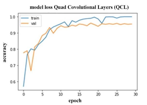
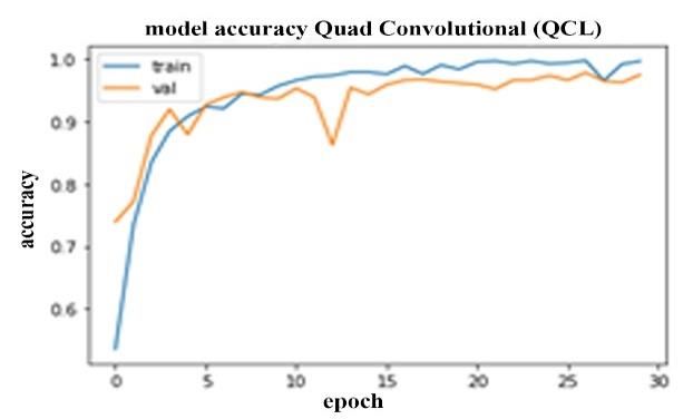
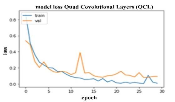
Received: October 22, 2021. Revised: November 22, 2021. 424

 Table 6. Experiment results with DW-MRI researcher data (2nd dataset, n = 5226)
 Number of
 Prior Work Network Convolution ACC (%)
 Layers
 Do et al. [31] Recurrent Residual Convolutional
 12 92.31
 Neural Network (RRCNN)
 Zhu, H et al. [32] Cross-Modal Convolutional Neural
 7 91.78
 Network
 H. Kim et al [33] 3D Convolutional Neural Network 13 90.30
 Quad Convolutional
 Quad Convolutional Layers 4 93 - 97
 Layers (QCL)-CNN

images, respectively. The percentage of acute and Methods including data augmentation, learning
normal DW MR pictures properly classified is 53.50 rate variation, and annealing were used to help fit the
percent and 100 percent, respectively. Similarly, large dataset into the deep convolutional neural
when employing the ResNet50 approach, the network architecture, as discussed above. This was
classification accuracy for ich and acute strokes is done to produce significant results, as shown in Fig.
5percent and 0.5 percent, respectively, whereas the 8. Training loss = 0.0098, training accuracy = 0.9970,
classification accuracy for normal pictures is 90.5 validation loss: 0.0946, and validation accuracy:
percent, as shown in Fig. 4. (b). In Fig. 4 (c), the 0.9749 are the final results (1st dataset). For 2nd
VGG16 method shows that the ich and acute strokes dataset, the training accuracy was 1.0000, the training
accuracies are 77.5percent, and 96.5percent with loss was 5.4236e-04, the validation loss was 0.1861,
normal images classification of 100percent. The and the validation accuracy was 0.9552.
images classified by the authors using the proposed The proposed method is also compared to Do et
layers (QCL-CNN) have ich and acute strokes of al.[31] research, which similarly classified stroke
91.44percent and 95.34percent, with 100percent images. In stroke care, the proposed early diagnosis
correctly classified normal images as shown Fig. 4 and rapid quantification of acute ischemic lesions are
(d). AlexNet, ResNet50, and VGG16 all performed critical. DWI datasets are showing acute anterior
better than AlexNet, ResNet50, and VGG16 in circulation stroke. The classification method used in
classifying ich, acute, and normal images. As this study is Repeated Residual Convolution Neural
demonstrated in Table 4, the average accuracy of Network (RRCNN). A pre-trained VGG16 and
QCL-CNN is higher than that of others. Inception V3 employs twelve convolution layers.
 Experiments performed on three categories of Zhu, H et al.[32] suggested an automatic machine
image datasets using the 10 fold cross-validation in learning technique. They first create a cross-modal
2nd dataset, shows that the ich stroke is 89.87percent convolutional neural network that can accurately
by identifying 71 images correctly out of 79. The detect stroke lesions from DWI and FLAIR images,
number of acute images correctly identified is 63 employing seven convolution layers. Kim et al. [33]
(79.75percent) of total acute images, and 100percent The 3D Convolutional neural network method was
of normal images. When performed with ResNet50, used in this study using Fluid-Attenuated Inversion
the classification accuracies of ich and acute strokes Recovery (FLAIR) MRI data with employs thirteen
are 67.08percent, and 65.83percent, while the normal convolution layers.
images are 89.87percent. Likewise, when using the The dataset used in the study, with a total of 5226
VGG16 method, ich and acute strokes' classification images (2nd dataset). Then, the data was tested on the
are 83.54percent and 91.14 percent, while convolution layer model from the previous
98.73percent was obtained for the normal images, as researchers (Table 6).
shown in Fig. 6 (c). When classified by the QCL- The results of these experiments are written in
CNN, the classification accuracies obtained for ich Table 6. Do et al. [31] adopted the VGG16 and
and acute strokes are 97.12percent, and 95.65percent, Resnet, employing 12 convolution layers, and the
while 100percent was used for normal images. Based accuracy result shows 92.31 percent. As for Zhu, H
on these accuracies, the overall classification of the et al. [32], which employs seven convolution layers,
QCL-CNN has also been improved by 8.51percent, the accuracy result is 91.78 percent. The research
23.53percent, and 7.06percent over AlexNet, conducted by H. Kim et al. [33] adopted 13
ResNet50, and VGG16 respectively, as shown in convolution layers, and the accuracy result has a
Table 5. value of 90.30 percent. In this study, we used only
International Journal of Intelligent Engineering and Systems, Vol.15, No.1, 2022 DOI: 10.22266/ijies2022.0228.38
Received: October 22, 2021. Revised: November 22, 2021. 425

four convolution layers in the proposed Quad Nugroho; Writing original draft preparation: Andi
Convolutional Layers (QCL)-CNN, and the Kurniawan Nugroho; Writing review and editing:
experiment deliver the highest accuracy value 93.90 Terawan Agus Putranto, I Ketut Eddy Purnama and
percent. Mauridhi Hery Purnomo;Visualization: Mauridhi
 Hery Purnomo; Supervision: Mauridhi Hery
6. Conclusion Purnomo;Funding acquisition: I Ketut Eddy Purnama.
 This study introduces a new classification on brain References
stroke injury at DW MR images, by employing Quad
Convolution Layer adapted in CNN. [1] J. T. Marbun, Seniman, and U. Andayani,
 The new QCL-CNN architecture model can be “Classification of stroke disease using
used for recognising the dissimilar between the first convolutional neural network”, J. Phys. Conf.
brain stroke injury type ( ICH and acute)/ 1st data set Ser., Vol. 978, No. 1, 2018, doi: 10.1088/1742-
and second brain stroke injury type ( ICH, acute and 6596/978/1/012092.
normal)/ 2nd data set . [2] U. R. Acharya, K. M. Meiburger, O. Faust, J. E.
 The performance of QCL-CNN model W. Koh, S. L. Oh, E. J. Ciaccio, A. Subudhi, V.
architecture assessed into two testing. The first Jahmunah, and S. Sabut, “Automatic detection
experiment used an image dataset split into 20 of ischemic stroke using higher order spectra
percent testing and 80 percent training. Then, ten-fold features in brain MRI images”, Cognitive
cross-validations were performed in the second Systems Research, Vol. 58, pp. 134–142, 2019,
experiment. doi: 10.1016/j.cogsys.2019.05.005.
 This study also comparing the QCL-CNN with [3] K. J. V. Everdingen, J. V. D. Grond, L. J.
others CNN architectures such as AlexNet, ResNet50, Kappelle, L. M. P. Ramos, and W. P. T. M. Mali,
and VGG16. The results showed that QCL-CNN is “Diffusion-weighted magnetic resonance
performing excellent from those method on data set imaging in acute stroke”, Stroke, Vol. 29, No. 9,
DW MR image. pp. 1783–1790, 1998, doi:
 10.1161/01.STR.29.9.1783.
Acknowledgments [4] W. Reith, Y. Hasegawa, L. L. Latour, B. J.
 Dardzinski, C. H. Sotak, and M. Fisher,
 The author would like to thank the Ministry of “Multislice diffusion mapping for 3D evolution
Research, Technology and Higher Education of the of cerebral ischemia in a rat stroke model”,
Republic of Indonesia for supporting this research Neurology, Vol. 45, No. 1, pp. 172–177, 1995,
through Indonesian Education Scholarship (BPPDN). doi: 10.1212/WNL.45.1.172.
Furthermore, the authors also gratefull to the Gatot [5] T. Back, M. H. Berlage, K. Kohno, and K. A.
Subroto Army Hospital, the Department of Hossmann, “Diffusion nuclear magnetic
Radiology, the Universitas Airlangga Hospital for resonance imaging in experimental stroke
providing MRI data for stroke patients, and the correlation with cerebral metabolites”, Stroke,
University Center of Excellence on Artificial Vol. 25, No. 2, pp. 494–500, 1994, doi:
Intelligence for Healthcare and Society (UCE 10.1161/01.STR.25.2.494.
AIHeS). In addition, this study was partially funded [6] A. G. Sorensen , F. S. Buonanno, R. G. Gonzalez,
by the Education Fund Management Institute (LPDP) L. H. Schwamm, M. H. Lev, F. R. H. Helbinger,
under the Innovative Productive Research Grant T. G. Reese, R. M. Weisskoff, T. L. Davis, MS,
(RISPRO) scheme - Invitation 2019, contract N. Suwanwela, U. Can, J. A. Moreira, W. A.
number: PRJ-41/LPDP/2019. Copen, R. B. Look, B. A. S. P. Finklestein, B. R.
 Rosen, W. J. Koroshetz, “Hyperacute stroke:
 evaluation with combined multisection
Conflicts of interest diffusion-weighted and hemodynamically
 The authors declare no conflict of interest. weighted echo-planar MR imaging”, Radiology,
 Vol. 199, No. 2, pp. 391–401, 1996, doi:
Author contributions 10.1148/radiology.199.2.8668784.
 [7] M. Matešin, L. Sven, and D. Petravić, “A Rule-
 Conceptualization: Andi Kurniawan Nugroho; Based Approach to Stroke Lesion Analysis from
Methodology: Andi Kurniawan Nugroho; Software: CT Brain Images”, Int. Symp. Image Signal
Andi Kurniawan Nugroho; Validation: Terawan Process. Anal. ISPA, Vol. 2001-Janua, pp. 219–
Agus Putranto; Formal analysis: Andi Kurniawan 223, 2001, doi: 10.1109/ISPA.2001.938631.

International Journal of Intelligent Engineering and Systems, Vol.15, No.1, 2022 DOI: 10.22266/ijies2022.0228.38
Received: October 22, 2021. Revised: November 22, 2021. 426

[8] S. A. Peixoto and P. P. R. Filho, “Neurologist- Network and Feature Selection Method”, Expert
 Level Classification of Stroke Using a Structural Syst. Appl., Vol. 149, p. 113274, 2020, doi:
 Co-Occurrence Matrix Based on The Frequency 10.1016/j.eswa.2020.113274.
 Domain”, Comput. Electr. Eng., Vol. 71, No. [19] I. Guyon, “Gene Selection for Cancer
 April, pp. 398–407, 2018, doi: Classification”, pp. 389–422, 2002.
 10.1016/j.compeleceng.2018.07.051. [20] S. R. Amendolia, G. Cossu, M. L. Ganadu, B.
[9] W. L. Nowinski, G. Qian, and D. F. Hanley, “A Golosio, G. L. Masala, and G. M. Mura, “A
 CAD System for Hemorrhagic Stroke”, comparative Study of K-Nearest Neighbour,
 Neuroradiol. J., Vol. 27, No. 4, pp. 409–416, Support Vector Machine and Multi-Layer
 2014, doi: 10.15274/NRJ-2014-10080. Perceptron for Thalassemia screening”,
[10] Z. Mousavi, T. Y. Rezaii, S. Sheykhivand, A. Chemom. Intell. Lab. Syst., Vol. 69, pp. 13–20,
 Farzamnia, and S. N. Razavi, “Deep 2003, doi: 10.1016/S0169-7439(03)00094-7.
 Convolutional Neural Network for [21] A. Bansal, C. Castillo, R. Ranjan, and R.
 Classification of Sleep Stages from Single- Chellappa, “The do’s and don’ts for CNN-Based
 Channel EEG Signals”, J. Neurosci. Methods, Face Verification”, In: Proc. of 2017 IEEE
 Vol. 324, No. December 2018, 2019, doi: International Conference on Computer Vision
 10.1016/j.jneumeth.2019.108312. Workshops, pp. 2545–2554, 2017, doi:
[11] E. Başaran, Z. Cömert, and Y. Çelik, 10.1109/ICCVW.2017.299K.
 “Convolutional neural network approach for [22] K. E. Saatman, A. C. Duhaime, R. Bullock, A. I.
 automatic tympanic membrane detection and R. Maas, A. Valadka, and G. T. Manley,
 classification”, Biomed. Signal Process. Control, “Classification of Traumatic Brain Injury for
 Vol. 56, 2020, doi: 10.1016/j.bspc.2019.101734. Targeted Therapies”, Journal of Neurotrauma,
[12] H. I. Suk, S. W. Lee, and D. Shen, “Hierarchical Vol. 25, No. 7, pp. 719–738, 2008, doi:
 Feature Representation and Multimodal Fusion 10.1089/neu.2008.0586.
 with Deep Learning for AD/MCI Diagnosis”, [23] F. T. Mozqueda, J. He, I. B. Yeh, L. H.
 Neuroimage, Vol. 101, pp. 569–582, 2014, doi: Schwamm, M. H. Lev, P. W. Schaefer, and R. G.
 10.1016/j.neuroimage.2014.06.077. González, “An Acute Ischemic Stroke
[13] S. Hochreiter, “Long Short-Term Memory”, Classification Instrument That Includes CT or
 Neural Comput., Vol. 1780, pp. 1735–1780, MR Angiography: The Boston Acute Stroke
 1997. Imaging Scale”, AJNR Am J Neuroradiol, Vol.
[14] F. Ouhmich, V. Agnus, V. Noblet, F. Heitz, and 29, No. 6, pp. 1111–1117, 2008, doi:
 P. Pessaux, “Liver Tissue Segmentation in 10.3174/ajnr.A1000.
 Multiphase CT Scans Using Cascaded [24] S. Anbumozhi, “Computer-aided detection and
 Convolutional Neural Networks”, Int. J. Comput. diagnosis methodology for brain stroke using
 Assist. Radiol. Surg., Vol. 14, No. 8, pp. 1275– adaptive neuro-fuzzy inference system
 1284, 2019, doi: 10.1007/s11548-019-01989-z. classifier”, Int. J. Imaging Syst. Technol., Vol.
[15] G. E. Hinton, S. Osindero, and Y. W. Teh, “A 30, No. 1, pp. 196–202, 2020, doi:
 Fast Learning Algorithm for Deep Belief Nets”, 10.1002/ima.22380.
 Neural Comput., Vol. 18, pp. 1527–1554, 2006. [25] A. Gautam, B. Raman, and S. Raghuvanshi, “A
[16] C. H. Wan, L. H. Lee, R. Rajkumar, and D. Isa, hybrid approach for the delineation of brain
 “A Hybrid Text Classification Approach with lesion from CT images”, Biocybern. Biomed.
 Low Dependency on Parameter by Integrating Eng., Vol. 38, No. 3, pp. 504–518, 2018, doi:
 K-Nearest Neighbor and Support Vector 10.1016/j.bbe.2018.04.003.
 Machine”, Expert Syst. Appl., Vol. 39, No. 15, [26] A. Gautam and B. Raman, “Local Gradient of
 pp. 11880–11888, 2012, doi: Gradient Pattern: a Robust Image Descriptor for
 10.1016/j.eswa.2012.02.068. The Classification of Brain Strokes from
[17] A. Subudhi, M. Dash, and S. Sabut, “Automated Computed Tomography Images”, Pattern Anal.
 Segmentation and Classification of Brain Stroke Appl., Vol. 23, No. 2, pp. 797–817, 2020, doi:
 Using Expectation-Maximization and Random 10.1007/s10044-019-00838-8.
 Forest Classifier”, Biocybern. Biomed. Eng., Vol. [27] A. Krizhevsky, I. Sutskever, and G. E. Hinton,
 40, No. 1, pp. 277–289, 2020, doi: “ImageNet Classification with Deep
 10.1016/j.bbe.2019.04.004. Convolutional Neural Networks”, Commun.
[18] M. Toğaçar, Z. Cömert, and B. Ergen, ACM, Vol. 60, No. 6, pp. 84–90, 2017, doi:
 “Classification of Brain MRI Using Hyper 10.1145/3065386.
 Column Technique with Convolutional Neural [28] G. E. Hinton, “Rectified Linear Units Improve
International Journal of Intelligent Engineering and Systems, Vol.15, No.1, 2022 DOI: 10.22266/ijies2022.0228.38
Received: October 22, 2021. Revised: November 22, 2021. 427

 Restricted Boltzmann Machines”, No. 3.
[29] I. B. Santoso, Y. Adrianto, A. D. Sensusiati, D.
 P. Wulandari, and I. K. E. Purnama, “Epileptic
 EEG Signal Classification Using Convolutional
 Neural Network Based on Multi-Segment of
 EEG Signal”, Int. J. Intell. Eng. Syst., Vol. 14,
 No. 3, pp. 160–176, 2021, doi:
 10.22266/ijies2021.0630.15.
[30] K. Men, H. Geng, C. Cheng, H. Zhong, M.
 Huang, Y. Fan, J. P. Plastaras, A. Lin, and Y.
 Xiao, “Technical Note: More accurate and
 efficient segmentation of organs‐at‐risk in
 radiotherapy with convolutional neural networks
 cascades”, Med. Phys., p. mp.13296, 2018, doi:
 10.1002/mp.13296.
[31] L. N. Do, B. H. Baek, S. K. Kim, H. J. Yang, I.
 Park, and W. Yoon, “Automatic Assessment of
 ASPECTS Using Diffusion-Weighted Imaging
 in Acute Ischemic Stroke Using Recurrent
 Residual Convolutional Neural Network”,
 Diagnostics, Vol. 10, No. 10, 2020, doi:
 10.3390/diagnostics10100803.
[32] H. Zhu, L. Jiang, H. Zhang, L. Luo, Y. Chen, and
 Y. Chen, “An Automatic Machine Learning
 Approach for Ischemic Stroke Onset Time
 Identification Based on DWI and FLAIR
 Imaging”, NeuroImage Clin., Vol. 31, p. 102744,
 2021, doi: 10.1016/j.nicl.2021.102744.
[33] H. Kim, Y. Lee, Y. H. Kim, Y. M. Lim, J. S. Lee,
 J. Woo, S. K. Jang, Y. J. Oh, H. W. Kim, E. J.
 Lee, D. W. Kang, and K. K. Kim, “Deep
 Learning-Based Method to Differentiate
 Neuromyelitis Optica Spectrum Disorder From
 Multiple Sclerosis”, Front. Neurol., Vol. 11, p.
 599042, 2020, doi: 10.3389/fneur.2020.599042.

International Journal of Intelligent Engineering and Systems, Vol.15, No.1, 2022 DOI: 10.22266/ijies2022.0228.38
You can also read