Protection from T cell-dependent colitis by the helminth-derived immunomodulatory mimic of transforming growth factor-β, Hp-TGM
←
→
Page content transcription
If your browser does not render page correctly, please read the page content below
Discovery Immunology, 2023, 2, 1–12
https://doi.org/10.1093/discim/kyad001
Advance access publication 18 January 2023
Research Article

Research Article
Protection from T cell-dependent colitis by the helminth-
derived immunomodulatory mimic of transforming growth

                                                                                                                                                              Downloaded from https://academic.oup.com/discovimmunology/article/2/1/kyad001/6991351 by guest on 05 February 2024
factor-β, Hp-TGM
Danielle J. Smyth1,3, Madeleine P. J. White1, Chris J. C. Johnston2, Anne-Marie Donachie1,
Marta Campillo Poveda1, , Henry J. McSorley3, and Rick M. Maizels1,*,
1
 Wellcome Centre for Integrative Parasitology, School of Infection and Immunity, University of Glasgow, Glasgow, UK
2
 Department of Clinical Surgery, University of Edinburgh, Edinburgh, UK
3
 Division of Cell Signalling and Immunology, University of Dundee, Dundee, UK
*
 Correspondence: Rick M. Maizels, Wellcome Centre for Integrative Parasitology, School of Infection and Immunity, University of Glasgow, Glasgow, UK.
Email: rick.maizels@glasgow.ac.uk

Abstract
In animal models of inflammatory colitis, pathology can be ameliorated by several intestinal helminth parasites, including the mouse nematode
Heligmosomoides polygyrus. To identify parasite products that may exert anti-inflammatory effects in vivo, we tested H. polygyrus excretory–se-
cretory (HES) products, as well as a recombinantly expressed parasite protein, transforming growth factor mimic (TGM), that functionally mimics
the mammalian immunomodulatory cytokine TGF-β. HES and TGM showed a degree of protection in dextran sodium sulphate-induced colitis,
with a reduction in inflammatory cytokines, but did not fully block the development of pathology. HES also showed little benefit in a similar acute
trinitrobenzene sulphonic acid-induced model. However, in a T cell transfer-mediated model with recombination activation gene (RAG)-deficient
mice, HES-reduced disease scores if administered throughout the first 2 or 4 weeks following transfer but was less effective if treatment was
delayed until 14 days after T cell transfer. Recombinant TGM similarly dampened colitis in RAG-deficient recipients of effector T cells, and was
effective even if introduced only once symptoms had begun to be manifest. These results are a promising indication that TGM may replicate,
and even surpass, the modulatory properties of native parasite HES.

Graphical Abstract

Keywords: dextran sodium sulphate, Heligmosomoides polygrus, inflammatory bowel disease
Abbreviations: DAI: disease activity index; DSS: dextran sodium sulphate; GFP: green fluorescent protein; HES: Heligmosomoides polygrus excretory/secretory
products; HTAB: hexadecyltrimethylammonium bromide; IBD: inflammatory bowel disease; IFN: interferon; MLN: mesenteric lymph node; MPO: myeloperoxidase;
NBF: neutral buffered formalin; OVA: ovalbumin; PBS: phosphate-buffered saline; RAG: recombination activation gene; TGF-β: transforming growth factor-β;
TGM: transforming growth factor mimic; TNBS: trinitrobenzene sulphonic acid; TNF: tumour necrosis factor.

Introduction                                                                    by T-cell subsets reacting to intestinal antigens such as those
Inflammatory bowel diseases (IBDs) are immunological                            presented from commensal microorganisms and is attributed to
disorders with increasing prevalence in humans and limited                      dysregulation of the natural control mechanisms that prevent
therapeutic options [1–4]. Inflammation is largely mediated                     inappropriate immune responses to self- and environmental

Received 4 August 2022; Revised 7 December 2022; Accepted for publication 17 January 2023
© The Author(s) 2023. Published by Oxford University Press on behalf of the British Society for Immunology.
This is an Open Access article distributed under the terms of the Creative Commons Attribution License (https://creativecommons.org/licenses/by/4.0/),
which permits unrestricted reuse, distribution, and reproduction in any medium, provided the original work is properly cited.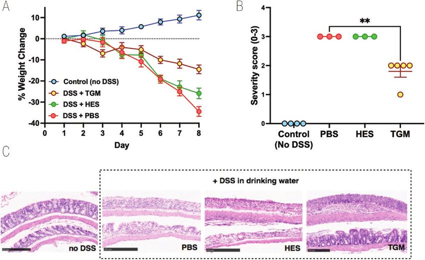
2                                                                                                                     Smyth et al.

antigens. While the cause of such dysregulation has yet to be      Materials and methods
determined, treatment strategies that boost natural regulatory
                                                                   Animals
pathways may offer transformational new therapies for IBD
[5, 6].                                                            Transgenic mouse strains (all on C57BL/6 background)
   Among many drivers of immune regulation, helminth               RAG1−/− [36] and Foxp3-GFP [37] for in vivo experiments
parasites have engendered much interest as they have adapted       were aged 6–12 weeks old and bred in-house or purchased
over long evolutionary time to target and dampen the im-           from Envigo (UK). BALB/c mice for TNBS colitis experiments
mune system of their host [7, 8]. In the setting of colitis, a     were bred in-house. No randomization was used in setting up
range of helminths—in particular those involving the gastro-       experiments. Animals were housed in individually ventilated
intestinal tract—have been found to alleviate inflammation in      cages. All transgenic animal breeding and procedures were
rodent models [9–12], and there is epidemiological evidence        approved by the University ethics committees and performed
that helminth-infected children are less prone to develop IBD      under UK Home Office licence, and all research adhered to

                                                                                                                                     Downloaded from https://academic.oup.com/discovimmunology/article/2/1/kyad001/6991351 by guest on 05 February 2024
[13] In a placebo-controlled primate study, macaques treated       the ARRIVE guidelines.
with the pig whipworm Trichuris suis were protected from
                                                                   Generation of immunomodulators
an idiopathic form of colitis [14]. However, similar trials in
IBD patients infected with whipworms have fallen short of          HES was prepared by culturing adult worms in protein-free
expectations [15, 16]. Although more promising results were        media and diafiltration into phosphate-buffered saline (PBS)
found in Coeliac disease patients given the human hookworm         as described in detail elsewhere [38]. Only HES batches with
Necator americanus [17], concerns of limited efficacy and          bacterial endotoxin lipopolysaccharide (LPS) measuring
practicality of live helminth infections have led to greater em-   below 1 U/µg of protein were used for experiments, measured
phasis on molecular mediators derived from helminths that          using the endpoint chromogenic limulus amebocyte lysate
could be configured into pharmacological products for IBD          assay (Lonza). Recombinant mammalian codon-optimized H.
therapy [18–20].                                                   polygyrus TGM (corresponding to TGM-1 in the gene family
   One route to discovering anti-inflammatory products             described by Smyth et al. [39] was expressed using mamma-
from helminths is through mouse models in which ther-              lian HEK293T cells and purified using affinity chromatog-
apeutic candidates can be readily identified and tested for        raphy, as previously described [26]. Either PBS or ovalbumin
efficacy in a homologous system. An ideal such model is            (OVA) in PBS were used as controls in in vivo experiments.
Heligmosomoides polygyrus, a natural murine intestinal par-        OVA was Triton-X114 treated as previously described [40,
asite related to the human hookworms, which is known to            41] to remove any LPS from the commercial product prior
alleviate colitis in a range of experimental settings [21–24].     to use in vivo.
The immunomodulatory activity of parasite infection can be
                                                                   Continuous infusion via osmotic minipump
recapitulated in many respects by molecules released by adult
worms in vitro (the ‘Excretory-Secretory’ or ES products),         ALZET osmotic minipumps (supplied by Charles River UK)
that have been well characterized in H. polygyrus. One ob-         of 100 μl capacity were selected according to the duration of
jective of the current study was to ascertain if H. polygyrus      infusion required for individual experiments (model 1002—
ES (HES) products could protect mice from colitic inflam-          14 days; model 1004—28 days). In a sterile Class II hood
mation.                                                            minipumps were filled with the substance for infusion (sterile
   Within the complex of proteins that are present in HES, we      HES, sterile TGM, or sterile PBS/OVA protein control) and
recently identified a novel protein secreted by H. polygyrus       primed overnight by incubation in sterile PBS at 37°C. Under
which acts as a functional mimic of TGF-β, which we have           general anaesthesia (isoflurane) abdominal fur was removed
designated as transforming growth factor mimic (TGM).              by shaving and the skin was prepared with chlorhexidine so-
Despite no sequence similarity to the mammalian cytokine,          lution (Hydrex Clear). The peritoneal cavity was accessed
TGM binds to host TGF-β receptors and like TGF-β itself,           through an upper midline incision and the minipump was
activates the pathway that induces regulatory T cells (Tregs)      placed in the right paracolic gutter. Closure was in two layers
[25–28]. TGF-β signalling is known to play a critical role         with 5-0 undyed Vicryl (Ethicon UK).
in controlling intestinal inflammation [29–31] and indeed
recently proposed therapeutic for IBD sought to enhance            Purification of naïve CD4+ T cells from Foxp3-
TGF-β activation by inhibiting its negative regulator Smad7,       reporter mice
an inhibitor of this pathway [32]. Moreover, activation or ex-     Naïve CD4+ T cells required for the induction of the T cell
pansion of the Tregs compartment represents a potential ther-      transfer model of colitis were purified from the spleen and
apeutic strategy for IBD [5].                                      peripheral lymph nodes of Foxp3-GFP reporter mice. Isolated
   In recent studies, we have shown that TGM can mediate           cells were firstly pre-sorted by magnetically activated cell
suppression of immune inflammation, e.g. in models of              sorting (MACS) selection for CD4+ using a depletion isola-
airway allergy [33]. We also established the ability of TGM        tion strategy that leaves CD4+ cells untouched, performed ei-
to abate the severity of dextran sodium sulphate (DSS) colitis     ther on manual LD columns (Miltenyi) or on an AutoMACS
in mice, using high doses delivered orally through admixture       Pro Separator (Miltenyi). Cells were then further purified by
with drinking water [34]. Focusing on colitis, we now eval-        fluorescence-activated cell sorting (FACS) by staining with
uate in more depth the relative potencies of HES and TGM,          anti-CD4-APC (clone RM4-5/BioLegend) and anti-CD25-PE
using several different murine models of colitis [35], including   (clone PC61.5/eBioscience) antibodies and then sorted for
inflammation induced with trinitrobenzene sulphonic acid           CD4+CD25– Foxp3–(GFP) on a BD FACSAria III. Following
(TNBS), and a T cell transfer model in which adaptive im-          FACS purification, live cells were counted using 0.4% trypan
mune responses to intestinal antigens more closely represents      blue and a haemocytometer. Cells were next washed three
human disease.                                                     times in sterile PBS before finally resuspending in sterile PBS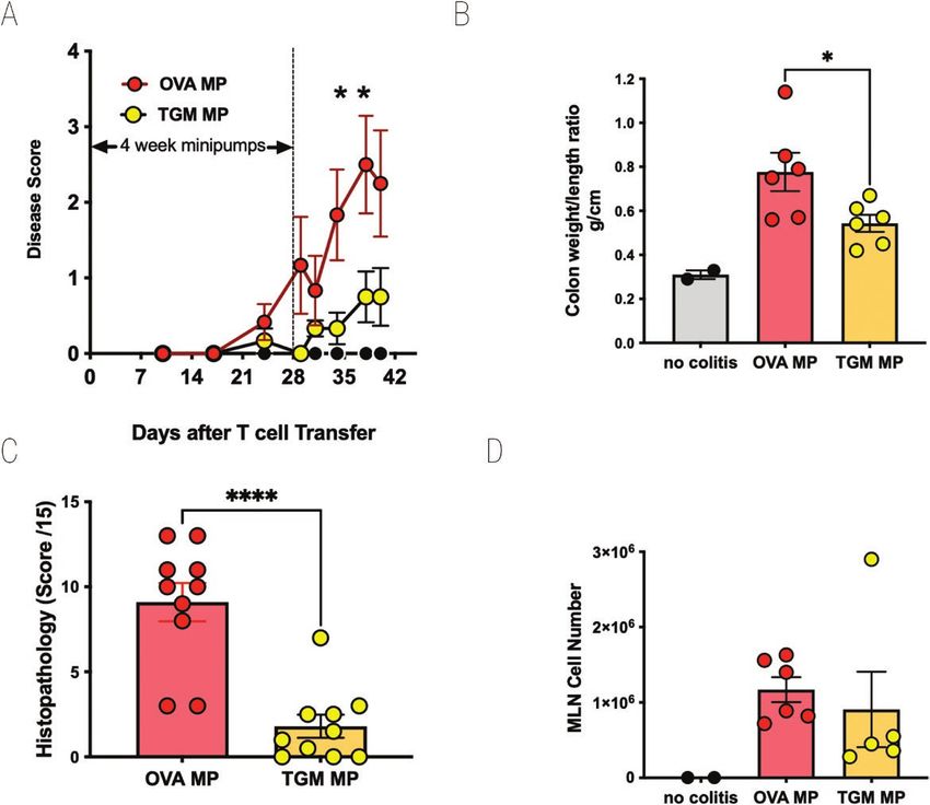
TGF-β mimic suppresses colitis, 2023, Vol. X, No. X                                                                              3

to a concentration of 2.5 × 10⁶ cells/ml. Mice received 200 µl       Scoring of histological specimens for all T cell transfer
of this suspension (5 × 10⁵ cells) for the induction of T cell    experiments was performed in a blinded fashion using a ma-
transfer colitis.                                                 trix of six parameters: (i) crypt architecture; (ii) ulceration;
                                                                  (iii) crypt abscesses; (iv) goblet cell loss; (v) mucosal inflam-
Induction and monitoring of colitis                               matory infiltration; and (vi) submucosal inflammatory infil-
To induce colitis in the DSS model [42, 43], mice were placed     tration. Each parameter was given a score of 0–3 with the
on 2% or 5% DSS (36,000—50,000 MW, MP Biochemical)                final value being the sum of these, the maximum score being
in tap water and allowed to drink ad libitum for the dura-        18. Scoring of histological specimens for DSS experiments
tion of the experiment (4–7 days). Mice were monitored and        was performed in a blinded fashion using a severity of tissue
scored daily for the duration of the experiment using a dis-      disruption score range of 0–3: 0 = no damage; 3 = severe
ease activity index (DAI) score matrix (Supplementary Table       damage.
1) and graphed as Disease Score vs Days.

                                                                                                                                      Downloaded from https://academic.oup.com/discovimmunology/article/2/1/kyad001/6991351 by guest on 05 February 2024
   To induce colitis in the TNBS model [44], BALB/c mice were     Cellular immunology assays
anaesthetized using inhalational isoflurane and intrarectally     Single cell suspensions were made from murine mesenteric
administered with 150 µl of a solution of 2.5% TNBS in 50%        lymph nodes (MLN) by maceration through 70 μm filters
ethanol or 50% ethanol alone (control) using flexible tubing      (BD) into complete RPMI 1640 (cRPMI) medium containing
(ID 0.75 mm/OD 1.6 mm) attached onto a 1 ml syringe which         HEPES (Gibco), supplemented with 2 mM l-glutamine, 100
was inserted 4 cm proximal to the anus. The mouse was held        U/ml penicillin, and 100 μg/ml streptomycin (Gibco), 10%
upside down for 1 min after administration to ensure absorp-      heat-inactivated foetal calf serum (Gibco). Contaminating
tion. Mice given HES were intraperitoneally injected with         red blood cells were removed by resuspending the cells from
10 µg of HES in 200 µl PBS on days 0 and 1. Mice were             one spleen in 2 ml of red blood cell lysis buffer (Sigma) and
monitored and scored daily for the duration of the experi-        incubating at RT for 2 min. Cells were then washed with
ment using the DAI score matrix (Supplementary Table 1) and       cRPMI and counted on a haemocytometer by trypan blue ex-
graphed as Disease Score vs Days.                                 clusion.
   For induction of colitis in the T cell transfer model [45,     For flow cytometry analysis of murine blood taken at 21 days
46], following cell isolation and MACS/FACS purification, 5       post naïve T cell transfer, a few drops of blood were collected
× 105 naïve CD4+CD25−GFP− T cells isolated from the spleen       into several millilitres of FACS buffer containing EDTA (to
and peripheral lymph nodes of Foxp3-GFP reporter mice. T          prevent coagulation), centrifuged for 5 min at 1500 g and
cells were adoptively transferred by intravenous injection into   pellets resuspended in 3 ml of red blood cell lysis buffer
each RAG1−/− recipient. Mice were monitored regularly and         (Sigma), incubated for 5 min at room temperature, topped up
scored for the duration of the experiment using the DAI score     with FACS buffer and recentrifuged for 5 min at 1500 g. Cells
matrix (Supplementary Table 1) and graphed as Disease Score       were then used for subsequent flow cytometry staining and
vs Days (post cell transfer).                                     analysis (see below).

Colon tissue processing                                           Flow cytometry staining/analysis
The colon was firstly dissected from the mouse just below         Cells were washed in PBS and stained for viability with
the caecum and then at the rectum, with its length measured       LIVE/DEAD® fixable blue (Life Technologies) which was
using a ruler before being flushed with ice cold PBS (to re-      diluted 1:1000 in PBS; 200 μl was added to each sample of
move faeces) and having its weight measured using a fine bal-     cells, which were then incubated in the dark for 20 min at
ance. Results were graphed either as a ratio of colon weight/     4°C and washed twice in FACS buffer (PBS containing 0.5%
length (mg/cm), or just colon length (mm) if the weight meas-     BSA, 5 μM EDTA, and 0.05% sodium azide). To prevent
urement was not taken.                                            non-specific antigen binding, cells were incubated with ei-
   Tissue required for analysis of myeloperoxidase (MPO)          ther 50 μl of polyclonal IgG (Sigma) or anti-CD16/32 (clone
activity and tissue cytokines was a 0.5 cm section from the       93; BioLegend) (diluted 1:50 in FACS buffer) for 10 min
proximal colon, nearest the caecum and adjacent to the            at 4°C and then washed twice in FACS buffer. Single stain
tissue used for histology; tissue was dissected, blotted dry,     controls were individually added to one drop of UltraComp
and rapidly frozen in dry ice before storage −80°C until          eBeads (eBioscience). Samples were incubated with a sur-
assayed.                                                          face stain combination, as indicated in each figure legend,
                                                                  for 20 min at 4°C, washed twice in FACS buffer and then
Intestinal tissue processing and histological                     resuspended in 200 μl FACS buffer for acquisition of surface
scoring                                                           marker data directly, or further processed for intracellular
Dissected colon and small intestine tissues were washed in        staining. Samples were acquired on a BD Biosciences LSR II
cold PBS, inverted onto PBS pre-soaked wooden skewers             or Celesta flow cytometer and analysed using FlowJo soft-
(brochettes) and semi-fixed in 10% neutral buffered formalin      ware (Tree Star).
(NBF) solution (Sigma) for 3–5 h before being fully cut lon-
gitudinally with a scalpel blade, rolled into so-called ‘Swiss    Cytokine measurements
rolls’ [47], placed in double height histology cassettes and      Serum samples analysed for cytokines were taken at the end
fixed in 10% NBF for a further 14–19 h. The fixed tissue rolls    of experiments by collecting mouse blood into Microtainer
were next rinsed in PBS and transferred into 70% ethanol for      serum collection tubes (BD) which were then centrifuged to
24 h before being processed for histology and embedded in         separate out the serum. Serum was analysed either neat or
paraffin. Cross-sections of 5 μm were cut and stained with        diluted 1 in 10 and cytokines measured by either Cytometric
haematoxylin and eosin.                                           Bead Array Flex Sets (BD) according to manufacturer’s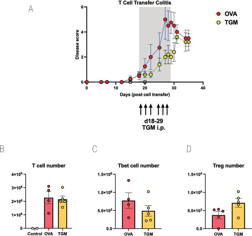
4                                                                                                                                           Smyth et al.

instructions or standard ELISA protocols for individual                         hexadecyltrimethylammonium bromide (HTAB) and then
cytokines. Results are graphed as pg/ml of sera.                                measuring for its activity using a dianisidine-H2O2 assay. In
   Colon tissue samples analysed for cytokines were                             brief, colon tissue samples taken from the area between the
resuspended in 400 μl of 2× Cell Lysis Buffer (Cell Signalling)                 proximal and mid colon (adjacent to the tissue used for his-
supplemented with 1 mM PMSF (Sigma) and homogenized                             tology) were rinsed with cold PBS, blotted dry, and stored at
using a Tissue-Lyzer II (Qiagen) at 30 Hz for 4 min (5 mm                       −80°C. For assaying, tissue samples were weighed, suspended
metal bead included at time of buffer addition). Samples were                   in 0.5 ml cold 50 mM potassium phosphate buffer (pH 6.0)
then centrifuged at 13,400 g for 10 min at 4°C and total pro-                   containing 0.5% HTAB (Sigma) and homogenized using a
tein concentrations of the homogenized tissue supernatant were                  Tissue-Lyzer II (Qiagen) at 30 Hz for 4 min (5 mm metal
quantified using the Bradford Assay (samples being measured                     bead included at time of buffer addition). Samples were then
in duplicate). Colon homogenate supernatant was analysed for                    centrifuged at 13,400 g for 10 min at 4°C. The MPO level
cytokines either neat or diluted 1 in 10 and cytokines measured                 in 10 μl of supernatant was determined by adding 190 μl

                                                                                                                                                            Downloaded from https://academic.oup.com/discovimmunology/article/2/1/kyad001/6991351 by guest on 05 February 2024
by either Cytometric Bead Array Flex Sets (BD) according to                     of o-dianisidine dihydrochloride (0.167 mg/ml; Sigma) and
manufacturer’s instructions or standard ELISA protocols for                     H2O2 solution (0.0005%) and measuring the change in ab-
individual cytokines. Results are graphed as pg/mg tissue.                      sorbance over 1 min at 450 nm using a 96-well microtitre
                                                                                plate reader. One unit of MPO activity was defined as the
Determination of tissue MPO activity                                            quantity able to convert 1 μmol of H2O2 to water in 1 min
Determination of tissue MPO, primarily produced by                              at RT and was expressed as units/mg protein. Total protein
neutrophils, was based on a previously published pro-                           concentrations of homogenized tissue were quantified using
tocol [48] and performed by solubilizing MPO with                               the Bradford Assay. Samples were measured in triplicate.

Figure 1: effect of HES in the DSS model of Colitis. DSS was administered as a 2% solution in drinking water to induce acute colitis in C57BL/6 mice
for the duration of the 10-day experiment. A 10 µg amount of HES was administered daily from day 0 to day 7 by intraperitoneal injection to one of
the DSS groups; the other DSS group received nothing or PBS; a third group (control) received neither DSS nor HES. Results are one of four similar
experiments, with n = 5 for the DSS groups presented. (A) Weight change over time course; data represent means ± SE. (B) Comparison of individual
weights at day 10; horizontal solid line represents weight at the start of DSS treatment. (C) Time course of Disease Scores (Disease Activity Indices) in
the three groups.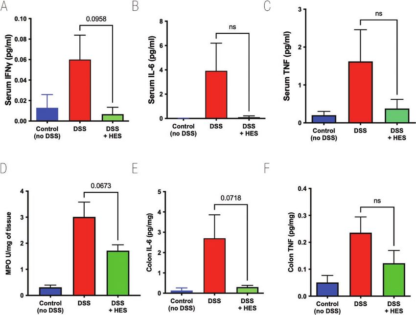
TGF-β mimic suppresses colitis, 2023, Vol. X, No. X                                                                                                        5

Results                                                                          so in those also receiving HES (Fig. 2D). We also found high
                                                                                 levels of IL-6 and TNF in DSS-treated colonic tissue, but these
HES dampens the inflammatory response in DSS-
                                                                                 cytokine responses were subdued in HES recipients (Fig. 2E
induced colitis
                                                                                 and F). Colonic IFN-γ levels were variable and did not differ
To assess the effects of H. polygyrus HES, we first used the well                between control and DSS groups (data not shown).
characterized model of DSS administration to induce acute co-                       In additional experiments, we also tested the ability of
litis in C57BL/6 mice [42, 43]. In initial experiments, HES was                  the recently described TGF-β mimic (TGM) protein from H.
administered (10 µg/day) from day 0 to day 7 of the study,                       polygyrus, which has been shown to prolong allograft survival
and mice were monitored over a 10-day period. HES treatment                      in mice when administered through an osmotic minipump
reduced the degree of weight loss, although this did not reach                   [26]. However, using a similar protocol of 50 ng/day did not
statistical significance (Fig. 1A and B). The overall effects of                 show amelioration of DSS colitis (Supplementary Fig. 1A and
DSS colitis were evaluated by a DAI matrix that scored body                      B), while oral gavage 1 µg/day of TGM was similarly ineffec-

                                                                                                                                                                Downloaded from https://academic.oup.com/discovimmunology/article/2/1/kyad001/6991351 by guest on 05 February 2024
weight, stool consistency and blood content, and motility (see                   tive (Supplementary Fig. 1C and D). In parallel studies, we
Supplementary Table 1); again HES-treated animals tended to                      had succeeded in ameliorating DSS colitis by administering
show lower disease scores throughout the experiment (Fig. 1C).                   high-oral doses in a curtailed (4-day) regimen [34], while
    Cytokine levels in serum were measured in samples taken                      also finding that intraperitoneal injection of TGM strongly
at day 10 of DSS-induced colitis. DSS provoked high levels of                    suppressed inflammation in an airway allergy model [33].
IFN-γ, IL-6, and tumour necrosis factor (TNF), each of which                     We therefore tested i.p. administration of HES and TGM in a
were reduced to control levels in mice receiving HES, although                   4-day DSS protocol and found prolonged protection by TGM
effects were statistically marginal at best (Fig. 2A–C). No sig-                 as evaluated by reduced weight loss (Fig. 3A) and significantly
nificant quantities of serum IL-5, IL-10, IL-12p70, IL-13, or                    lower levels of tissue damage when histological sections were
IL-17A were detected in any group (data not shown). To eval-                     evaluated (Fig. 3B and C). As previously, HES restrained DSS-
uate tissue damage in the colon, we measured MPO activity,                       induced colitis to only a minor degree.
which was elevated in all DSS-treated animals, although less

Figure 2: effect of HES on Cytokine levels in DSS Colitis. Serum and colon samples taken from mice at day 10 of DSS treatment, as shown in Fig. 1, for
measurement of cytokines by ELISA, and MPO in colon homogenates as detailed in Materials and Methods. Data represented as means ± SE, and P
values calculated by unpaired t tests are shown if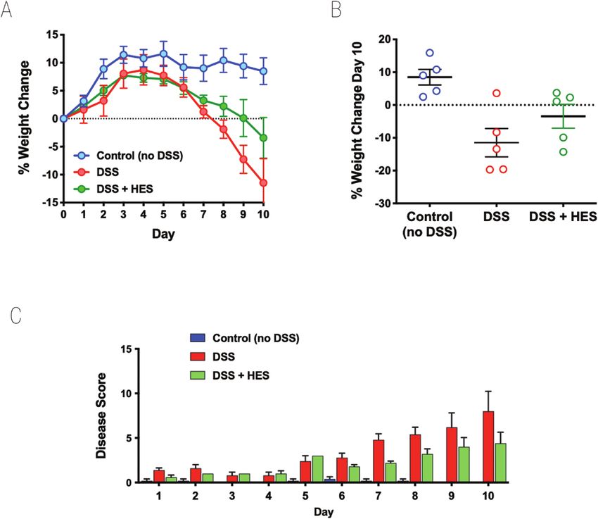
6                                                                                                                                            Smyth et al.

   We then assessed whether HES could inhibit colitic inflam-                   and peripheral lymph nodes of Foxp3-GFP reporter mice.
mation in another model, in which TNBS is administered                          Recipient RAG1–/– mice were monitored on a regular basis for
intrarectally to BALB/c mice, provoking a rapid innate inflam-                  34 days. Beyond day 23, control mice began to lose weight
matory reaction within 2–3 days [35]. In this system, however,                  while HES-treated animals maintained or even gained weight
we found HES did not alter outcome measured by weight loss                      (Fig. 4A), a difference which reached significance at day 34
(Supplementary Fig. 2A), colonic shortening (Supplementary                      (Fig. 4B). From day 23, control animals began to display signs
Fig. 2B), or DAI Scoring (Supplementary Fig. 2C). Taken to-                     of disease, while HES recipients showed no deterioration (Fig.
gether with the results of DSS colitis, we concluded that in                    4C). Histopathological analysis of intestinal tissues at day 34
these acute models of innate immunity-mediated colitis, while                   again showed significantly reduced disease scores in the mice
active infection with H. polygyrus is known to suppress in-                     given HES (Fig. 4D and E). Peripheral blood from recipient
flammation [21, 24], inhibition by parasite products at the                     mice taken at day 21 showed similar levels of CD4+ T-cell en-
doses used was too slight to warrant further investigation.                     graftment (Supplementary Fig. 3A) and of Foxp3+ expression

                                                                                                                                                              Downloaded from https://academic.oup.com/discovimmunology/article/2/1/kyad001/6991351 by guest on 05 February 2024
                                                                                within the donor T-cell population (Supplementary Fig. 3B);
HES suppresses colitis following adoptive transfer                              samples collected at the end of the experiment showed that
of naïve CD4+Foxp3− T cells into RAG1−/− recipients                             HES tended to reduce serum and colonic IL-6 (Supplementary
An established model of intestinal immunopathology is a T cell                  Fig. 3C and D), and increase IL-10 (Supplementary Fig. 3E),
transfer model of colitis, in which lymphocyte-deficient mice                   while IL-17 or IFN-γ were maintained (Supplementary Fig.
seeded with effector T cells develop intestinal inflammation                    3F and G).
over a 3–6 week period [35, 45, 46]. A series of experiments                      Similar protection against colitis and weight loss were
were undertaken using osmotic minipumps to continuously                         obtained with a 28-day minipump (not shown). However, only
release HES for periods of 14–28 days, administering totals                     a modest degree of amelioration of weight loss and disease
of 75–100 µg per animal. First, 14-day minipumps were                           activity was observed when implantation of the minipump
implanted into RAG1–/– mice intraperitoneally, and the fol-                     was delayed until 14 days post cell transfer (Supplementary
lowing day mice received an i.v. infusion of 5 × 10⁵ naïve                     Fig. 4). Taking together the four independent experiments in
CD4+CD25–Foxp3-GFP– T cells isolated from the spleen                            which HES minipumps were implanted the day prior to T cell

Figure 3: effect of TGM on 4 day model of DSS colitis. Mice were injected intraperitoneally with either PBS, HES (5 µg) or TGM (500 ng) at days 0,
2, 4, 6, and placed on 5% DSS drinking water from day 0 to 3 before switching over from day 4 to normal drinking water for the remainder of the
time course (up to 8 days). Mice that were not placed on DSS were injected with PBS as above and were given normal drinking water for the entire
experiment. All mice were weighed daily and at the end of the experiment had colons fixed for histology and scored for severity; two individual mice
in each of the PBS and HES-treated groups showed >25% weight loss at day 7 and were euthanized at that point; histology was evaluated only on the
remaining animals. Female C57BL/6 mice; n = 3–5; data shown are from one of two replicate experiments. (A) Percentage change from body weight
at the start of the experimental period. (B) Histopathology scoring, according to the scoring system described in Materials and Methods; statistical
analysis by unpaired t test; **P < 0.01. (C) Examples of histological staining of colon tissues from control mice and those receiving DSS in drinking water
with or without administration of HES or TGM. 10× magnification, scale bar represents 250 µm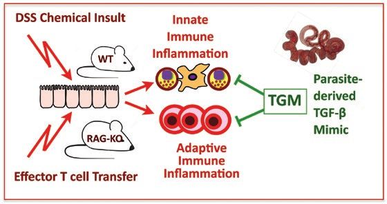
TGF-β mimic suppresses colitis, 2023, Vol. X, No. X 7
Downloaded from https://academic.oup.com/discovimmunology/article/2/1/kyad001/6991351 by guest on 05 February 2024
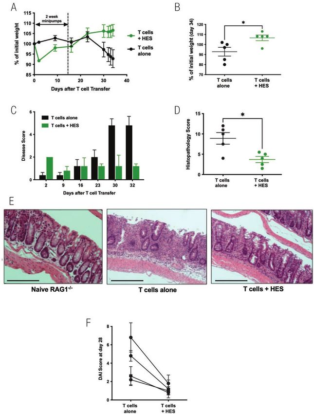
Figure 4: HES suppresses colitis following adoptive transfer of naive CD4+Foxp3− T cells into RAG1−/− recipients. RAG1−/− mice were divided into two
experimental groups—receiving either no treatment, or a continuous infusion of HES via an intraperitoneal osmotic minipump eluting 6.8 μg/day for 14
days. One day after minipump implantation, 5 × 10⁵ sorted naïve CD4+CD25−GFP− T cells from spleens of Foxp3-GFP reporter mice, were adoptively
transferred i.v. into each RAG1−/− recipient mouse. Results are one of four similar experiments, with n = 5 for the groups presented, except for panel F
which presents a summary of mouse weights at day 28 post-T cell transfer in all four experiments. Data were analysed by unpaired t tests; *P < 0.05.
(A) Weight change over time course, (B) Weight change at day 34, (C) Time course of Disease Scores (Disease Activity Indices), (D) Histopathology
scoring, according to the scoring system described in Materials and Methods, (E) Example histological sections (10× magnification, scale bar represents
250 µm), (F) Summary of Disease Scores in four independent experiments at day 28 after T cell transfer.8 Smyth et al.
Downloaded from https://academic.oup.com/discovimmunology/article/2/1/kyad001/6991351 by guest on 05 February 2024
Figure 5: TGM suppresses colitis following adoptive transfer of naive CD4+Foxp3− T cells into RAG1−/− recipients. As above, CD4+CD25−GFP− T cells
from spleens of Foxp3-GFP reporter mice were adoptively transferred into RAG1−/− recipients which then received either no treatment or a 28-day
infusion of TGM via an intraperitoneal minipump (eluting 50 ng/day of TGM). (A) Percentage change from body weight at the start of the experimental
period. (B) Colon weight/length ratio. (C) Histopathology scoring, according to the scoring system described in Materials and Methods. (D) MLN cell
numbers. For A, B, and D, results are one of three similar experiments, with n = 5–6 for the groups presented. For C, data were collected from two
experiments each with n = 5–6 and pooled. Data were analysed by unpaired t tests; *P < 0.05, ****P < 0.0001.
transfer, a significant reduction (P = 0.037) in Disease Score cell transfer, the overall engraftment of donor cells appeared
was observed, as presented in Fig. 4F. to be reduced (Fig. 5D); hence the protection against inflam-
mation might be due to inhibition of systemic T-cell expan-
TGM suppresses T cell transfer colitis sion in the lymphopaenic recipients rather than of effector T
We next evaluated whether a potent immunomodulatory cells in the tissues.
protein within HES, the TGF-β mimic, TGM, could also pro-
tect mice from T cell-mediated inflammatory colitis. Using a TGM modulates colitis following onset of
similar protocol with 4-week infusions from osmotic pumps, symptoms
RAG1−/−-deficient mice receiving CD4+CD25−Foxp3−GFP− To test whether TGM could in fact suppress inflammation,
T cells were given either 50 ng/day TGM (i.e. 1.4 µg total rather than T-cell engraftment following transfer to RAG1−/−
amount of protein over 28 days) or OVA as a control. mice, we therefore repeated these experiments, but delayed
Animals receiving TGM were largely protected from weight administration of TGM until colitic symptoms had become
loss (Fig. 5A), significantly so between days 34 and 38. apparent. Towards the end of week 3, animals began to show
They also showed less colonic thickening as measured in the one or more signs of disease, and at this time one group of
colon weight:length ratio (Fig. 5B) and substantially less- mice received TGM and the others received OVA. As the
histological evidence of inflammation (Fig. 5C). However, we animals were already symptomatic, daily intraperitoneal in-
noted that in mice that had received TGM from the day of T jection was preferred over surgical implantation of osmoticTGF-β mimic suppresses colitis, 2023, Vol. X, No. X 9
Downloaded from https://academic.oup.com/discovimmunology/article/2/1/kyad001/6991351 by guest on 05 February 2024
Figure 6: TGM ameliorates disease if given at the onset of symptoms in T cell-mediated colitis. CD4+CD25−GFP− T cells from spleens of Foxp3-GFP
reporter mice were adoptively transferred i.v. into RAG1−/− recipients. Mice were allowed to progress to showing first signs of mild colitis (at day 18).
Mice then received either OVA or TGM via six intraperitoneal injections of 1 µg between days 18 and 29. Results are one of two similar experiments,
with n = 5 for the groups presented. (A) Time course of Disease Scores (Disease Activity Indices), (B) MLN T-cell numbers, (C) MLN Tbet+ T-cell
numbers, (D) MLN Foxp3+ T-cell numbers. For C and D, single cell suspensions of MLNs prepared as described in the Materials and Methods were
stained for flow cytometry analysis using a panel of antibodies comprising CD3-BV711 (clone 17A2; Biolegend), CD4-FITC (clone GK1.5; Biolegend), and
CD25-BV650 (clone PC61; Biolegend) followed by intracellular staining using the FoxP3-transcription Staining set (Invitrogen) for Foxp3-ef450 (clone FJK-
16s, eBioscience) and Tbet-PerCp-Cy5.5 (clone 4B10; Biolegend).
minipumps, and 1 µg of TGM or OVA was administered in Discussion
PBS each day from day 18 to day 29.
Inflammatory bowel disease is increasing in prevalence in
As shown in Fig. 6A, disease progression was delayed in
many parts of the world with few new therapeutic remedies
recipients of TGM with disease scores reduced to a signifi-
available [49]. While evidence from both human and labora-
cant degree at day 29, the day of the final administration of
tory settings indicate that helminth parasites can ameliorate
TGM. After treatment ceased, the protective effects were lost,
intestinal inflammation, it is important to identify specific
and the TGM-treated group was not significantly different
molecules and mediators that may be able to counter disease.
from OVA-treated animals. At the endpoint, MLN cells were
In this report, we show that H. polygyrus TGM, a secreted
recovered, showing equivalent T-cell repopulation in both
mimic of mammalian TGF-β, can significantly dampen T
groups (Fig. 6B), although following TGM treatment there
cell-mediated colitis, reducing disease score and shifting
was a trend for fewer Tbet+ Th1 cells (Fig. 6C) and a greater
the balance between Th17 and Treg populations. We have
number of Foxp3+ Tregs (Fig. 6D).10 Smyth et al.
recently reported that TGM can exert its effects systemically, the importance of ensuring appropriate dose and delivery, the
in suppressing airway allergy when delivered intraperitoneally results confirm a striking protective effect of TGM in acute
[33]. We now find that intraperitoneal TGM administration colitis. Moreover, we show that at low doses TGM is also
can also suppress intestinal inflammation, in addition to protective in the T cell transfer model of colitis, which, since
our earlier report in which high doses of TGM delivered in the T cell transfer model is considered to more closely reflect
drinking water attenuated DSS-induced colitis [34]. IBD in humans, indicates promising possibilities for the treat-
H. polygyrus is not unique in ameliorating intestinal in- ment of intestinal inflammation. Further understanding of the
flammation. ES from the human hookworm Ancylostoma targets of TGM immune modulation in vivo, and finer defini-
caninum, given to mice daily i.p. during chemically induced tion of the optimal conditions for its delivery, will be required
colitis, has been shown to exert a modest reduction in path- to realize this potential as a novel therapeutic for intestinal
ological scores as well as the inflammatory cytokines, IFN-γ, inflammatory disorders.
and IL-17 [50], and also to raise IL-4 and IL-10 responses
Downloaded from https://academic.oup.com/discovimmunology/article/2/1/kyad001/6991351 by guest on 05 February 2024
[51]. Extracellular vesicles within helminth ES products have
been found to ameliorate colitic pathology [52–54], but to de- Supplementary data
velop novel therapies many investigators have isolated defined Supplementary data is available at Clinical and Experimental
molecular products. Thus, an immunomodulatory protein in Immunology online.
the ES of A. caninum was identified as the hookworm an-
ti-inflammatory protein-1 that blocked TNBS-induced colitis
[55] and a homologous product from the human hookworm Acknowledgements
N. americanus protected against T cell transfer-mediated co- We thank Yvonne Harcus, Nicola Britton, and Claire Ciancia
litis [56]. Other helminth products with reported anti-colitic for excellent technical assistance, the Wolfson Research Facility
activity include cystatins from the filarial nematode Brugia staff, and the Flow Core Facility, University of Glasgow for
malayi [57] and the trematode Clonorchis sinensis [58], and their support. The Editor-in-Chief, Simon Milling, and hand-
a serine protease from Trichinella spiralis [59]. An interesting ling editor, Kathy McCoy, would like to thank the following
development has been to express another helminth cystatin, reviewers, Graham Le Gros and Markus Geuking, for their
from Acanthocheilonema viteae, in a modified probiotic E. contribution to the publication of this article.
coli strain, that protects mice against DSS colitis [60]. In
addition, modest attenuation of DSS colitis was reported
with a peptide from Schistosoma japonicum [61] while the Conflict of interest
glutathione-S-transferase from Schistosoma haematobium, None declared.
P28GST, reduces TNBS-induced colitic inflammation in mice
[62, 63] and has entered trials in Crohn’s disease patients [64].
Mouse models are invaluable for indicating the likely Funding
mechanisms by which helminths and their products can in- This work was supported by the Kenneth Rainin Foundation
hibit colitis. H. polygyrus can generate dendritic cells (DCs) through Synergy and Innovator Grants (Refs 2015-964 and
that confer protection in a T cell transfer model of colitis 2016-3067), the Wellcome Trust through Investigator Awards
[65], while DCs exposed to the tapeworm Hymenolepis to RMM (Ref 106122 and 219530), and the Wellcome Trust
diminuta can protect recipients against DNBS-induced co- core-funded Wellcome Centre for Integrative Parasitology
litis [66]. Many different helminths have been reported to (Ref: 104111).
modulate macrophages towards an anti-colitic state [67–70],
and helminth-conditioned macrophages can activate CD25+
T cells to suppress colitis [71]. Studies are now under way Data availability
to establish the effects of TGM on DCs and macrophages, All data presented in this manuscript are available from the
two populations known to be heavily influenced by TGF-β author in the form of Excel and Prism files, and original
signalling [72]. photomicrographs.
TGM, like TGF-β, is a powerful inducer of Foxp3+ Tregs in
vitro [25, 28], and the role of Tregs in the control of intestinal
inflammation is very well established [73]. However, in our Author contributions
in vivo studies, changes to the Treg compartment were rela- D.J.S., M.P.J.W., C.J.C.J., A.M.D., M.C.P., and H.J.M.
tively modest. Our finding that protection against T cell-me- performed experiments; D.J.S., H.J.M., and R.M.M. designed
diated colitis waned once TGM administration ended (Fig. 6) experiments and analysed data; R.M.M. drafted the paper
might suggest that it acts directly on effector cells (Th17 or which was edited by all authors.
M1 macrophages) rather than indirectly through induction
of Tregs, as the latter may be expected to have longer-lasting
effects. This question is similarly under further investigation. References
In our previous report, high doses of TGM delivered in 1. Baumgart DC, Sandborn WJ. Crohn’s disease. Lancet 2012, 380,
drinking water were able to dampen DSS colitis, albeit in 1590–605. doi:10.1016/s0140-6736(12)60026-9
a moderated model with shorter DSS exposure [34]; in the 2. Chang JT. Pathophysiology of inflammatory bowel diseases. N
current study, parenteral TGM (at lower doses) was simi- Engl J Med 2020, 383, 2652–64. doi:10.1056/nejmra2002697
3. Danese S, Fiocchi C. Ulcerative colitis. N Engl J Med 2011, 365,
larly effective, although low-dose gavage or osmotic pump
1713–25. doi:10.1056/nejmra1102942
modes of administration could not ameliorate the effects of 4. Kaser, A, Zeissig S, Blumberg, RS. Inflammatory bowel disease.
a full-course DSS model. While these disparities emphasises Annu Rev Immunol 2010, 28, 573–621.TGF-β mimic suppresses colitis, 2023, Vol. X, No. X 11
5. Clough JN, Omer OS, Tasker S, Lord GM, Irving PM. Regulatory colitis in a murine model. Infect Immun 2008, 76, 4772–82.
T-cell therapy in Crohn’s disease: challenges and advances. Gut doi:10.1128/iai.00744-07
2020, 69, 942–52. doi:10.1136/gutjnl-2019-319850 25. Cook L, Reid K, Hakkinen E, de Bie B, Tanaka S, Smyth DJ, et al.
6. Neurath MF. Current and emerging therapeutic targets for IBD. Induction of stable human FOXP3+ T regs by a parasite-derived
Nat Rev Gastroenterol Hepatol 2017, 14, 269–78. doi:10.1038/ TGFβ mimic. Immunol Cell Biol 2021, 99, 833–47.
nrgastro.2016.208 26. Johnston CJC, Smyth DJ, Kodali RB, White MPJ, Harcus Y, Filbey
7. Maizels, RM, McSorley HJ, Smyth, DJ. Helminths in the hygiene KJ, et al. A structurally distinct TGF-β mimic from an intestinal
hypothesis - sooner or later? Clin Exp Immunol 2014, 177, 38–46. helminth parasite potently induces regulatory T cells. Nat Commun
8. Weinstock JV, Elliott DE. Helminths and the IBD hygiene hypoth- 2017, 8, 1741.
esis. Inflamm Bowel Dis 2009, 15, 128–33. doi:10.1002/ibd.20633 27. Mukundan A, Byeon CH, Hinck CS, Cunningham K, Campion T,
9. Elliott DE, Li J, Blum A, Metwali A, Qadir K, Urban JF, et al. Expo- Smyth DJ, et al. Convergent evolution of a parasite-encoded com-
sure to schistosome eggs protects mice from TNBS-induced colitis. plement control protein-scaffold to mimic binding of mammalian
Am J Physiol Gastrointest Liver Physiol 2003, 284, G385–391. TGF-beta to its receptors, TbetaRI and TbetaRII. J Biol Chem
Downloaded from https://academic.oup.com/discovimmunology/article/2/1/kyad001/6991351 by guest on 05 February 2024
10. Heylen M, Ruyssers NE, Gielis EM, Vanhomwegen E, Pelckmans 2022, 298, 101994. doi:10.1016/j.jbc.2022.101994
PA, Moreels TG, et al. Of worms, mice and man: an overview of 28. White MPJ, Smyth DJ, Cook L, Ziegler SF, Levings M, Maizels RM.
experimental and clinical helminth-based therapy for inflammatory The parasite cytokine mimic Hp-TGM potently replicates the regu-
bowel disease. Pharmacol Ther 2014, 143, 153–67. latory effects of TGF-β on murine CD4+ T cells. Immunol Cell Biol
11. Khan WI, Blennerhasset PA, Varghese AK, Chowdhury SK, Omsted 2021, 99, 848–64.
P, Deng Y, et al. Intestinal nematode infection ameliorates ex- 29. Feagins LA. Role of transforming growth factor-β in inflammatory
perimental colitis in mice. Infect Immun 2002, 70, 5931–7. bowel disease and colitis-associated colon cancer. Inflamm Bowel
doi:10.1128/iai.70.11.5931-5937.2002 Dis 2010, 16, 1963–8. doi:10.1002/ibd.21281
12. Varyani, F, Fleming JO, Maizels, RM. Helminths in the gastrointes- 30. Ihara, S, Hirata Y, Koike, K. TGF-β in inflammatory bowel disease:
tinal tract as modulators of immunity and pathology. Am J Physiol a key regulator of immune cells, epithelium, and the intestinal mi-
Gastrointest Liver Physiol 2017, 312, G537–49. crobiota. J Gastroenterol 2017, 52, 777–87.
13. Chu KM, Watermeyer G, Shelly L, Janssen J, May TD, Brink K, et 31. Troncone E, Marafini I, Stolfi C, Monteleone G. Transforming
al. Childhood helminth exposure is protective against inflamma- growth factor-beta1/Smad7 in intestinal immunity, inflammation,
tory bowel disease: a case control study in South Africa. Inflamm and cancer. Front Immunol 2018, 9, 1407.
Bowel Dis 2013, 19, 614–20. doi:10.1097/mib.0b013e31827f27f4 32. Ardizzone S, Bevivino G, Monteleone G. Mongersen, an oral
14. Broadhurst MJ, Ardeshir A, Kanwar B, Mirpuri J, Gundra UM, Smad7 antisense oligonucleotide, in patients with active Crohn’s
Leung JM, et al. Therapeutic helminth infection of macaques with disease. Therap Adv Gastroenterol 2016, 9, 527–32. doi:10.1177/1
idiopathic chronic diarrhea alters the inflammatory signature and 756283x16636781
mucosal microbiota of the colon. PLoS Pathog 2012, 8, e1003000. 33. Chauché C, Rasid O, Donachie A-M, McManus C, Löser S, Cam-
doi:10.1371/journal.ppat.1003000 pion T, et al. Suppression of airway allergic eosinophilia by TGM-
15. Garg SK, Croft AM, Bager, P. Helminth therapy (worms) for induc- 1, a helminth mimic of TGF-β. Immunology 2022, 167, 197–211.
tion of remission in inflammatory bowel disease. Cochrane Data- 34. Smyth DJ, Ren B, White M, McManus C, Webster H, Shek V, et al.
base Syst Rev 2014, 1, CD009400. Oral delivery of a functional algal-expressed TGF-β mimic halts
16. Scholmerich J, Fellermann K, Seibold FW, Rogler G, Langhorst J, colitis in a murine DSS model. J Biotechnol 2021, 340, 1–12.
Howaldt S, et al. A randomised, double-blind, placebo-controlled 35. Kiesler, P, Fuss IJ, Strober, W. Experimental models of inflammatory
trial of Trichuris suis ova in active Crohn’s disease. J Crohns Colitis bowel diseases. Cell Mol Gastroenterol 2015, 1, 154–70.
2017, 11, 390–9. 36. Mombaerts P, Iacomini J, Johnson RS, Herrup K, Tonegawa
17. Croese J, Giacomin P, Navarro S, Clouston A, McCann L, S, Papaioannou VE. RAG-1-deficient mice have no mature B
Dougall A, et al. Experimental hookworm infection and gluten and T lymphocytes. Cell 1992, 68, 869–77. doi:10.1016/0092-
microchallenge promote tolerance in celiac disease. J Allergy Clin 8674(92)90030-g
Immunol 2015, 135, 508–516.e5. doi:10.1016/j.jaci.2014.07.022 37. Fontenot JD, Rasmussen JP, Williams LM, Dooley JL, Farr AG,
18. Kahl, J, Brattig N, Liebau, E. The untapped pharmacopeic potential Rudensky AY. Regulatory T cell lineage specification by the
of helminths. Trends Parasitol 2018, 34, 828–42. forkhead transcription factor Foxp3. Immunity 2005, 22, 329–41.
19. Maizels, RM, Smits, HH, McSorley, HJ. Modulation of host im- doi:10.1016/j.immuni.2005.01.016
munity by helminths: the expanding repertoire of parasite effector 38. Johnston CJC, Robertson E, Harcus Y, Coakley G, Smyth DJ,
molecules. Immunity 2018, 49, 801–18. McSorley HJ, et al. Cultivation of Heligmosomoides polygyrus: an
20. Ryan SM, Eichenberger RM, Ruscher R, Giacomin PR, Loukas A. immunomodulatory nematode parasite and its secreted products. J
Harnessing helminth-driven immunoregulation in the search for Vis Exp 2015, 98, e52412.
novel therapeutic modalities. PLoS Pathog 2020, 16, e1008508. 39. Smyth DJ, Harcus Y, White MPJ, Gregory WF, Nahler J, Stephens
doi:10.1371/journal.ppat.1008508 I, et al. TGF-β mimic proteins form an extended gene family in the
21. Donskow-Łysoniewska K, Majewski P, Brodaczewska K, Jowicka murine parasite Heligmosomoides polygyrus. Int J Parasitol 2018,
K, Doligalska M. Heligmosmoides polygyrus fourth stages induce 48, 379–85.
protection against DSS-induced colitis and change opioid expres- 40. Aida Y, Pabst MJ. Removal of endotoxin from protein solutions by
sion in the intestine. Parasite Immunol 2012, 34, 536–46. phase separation using Triton X-114. J Immunol Methods 1990,
22. Elliott DE, Setiawan T, Metwali A, Blum A, Urban JF, Jr, et al. 132, 191–5. doi:10.1016/0022-1759(90)90029-u
Heligmosomoides polygyrus inhibits established colitis in IL-10- 41. Liu S, Tobias R, McClure S, Styba G, Shi Q, Jackowski G. Re-
deficient mice. Eur J Immunol 2004, 34, 2690–8. doi:10.1002/ moval of endotoxin from recombinant protein preparations. Clin
eji.200324833 Biochem 1997, 30, 455–63. doi:10.1016/s0009-9120(97)00049-0
23. Hang L, Setiawan T, Blum AM, Urban J, Stoyanoff K, Arihiro S, et 42. Chassaing, B, Aitken, JD, Malleshappa, M, and Vijay-Kumar, M.
al. Heligmosomoides polygyrus infection can inhibit colitis through (2014). Dextran sulfate sodium (DSS)-induced colitis in mice. Curr
direct interaction with innate immunity. J Immunol 2010, 185, Protoc Immunol 104, 15.25.1–15.25.14.
3184–9. doi:10.4049/jimmunol.1000941 43. Wirtz S, Popp V, Kindermann M, Gerlach K, Weigmann B, Fichtner-
24. Sutton TL, Zhao A, Madden KB, Elfrey JE, Tuft BA, Sullivan CA, Feigl S, et al. Chemically induced mouse models of acute and
et al. Anti-Inflammatory mechanisms of enteric Heligmosomoides chronic intestinal inflammation. Nat Protoc 2017, 12, 1295–309.
polygyrus infection against trinitrobenzene sulfonic acid-induced doi:10.1038/nprot.2017.04412 Smyth et al.
44. Abad C, Martinez C, Juarranz MG, Arranz A, Leceta J, Delgado 59. Long SR, Liu RD, Kumar DV, Wang ZQ, Su CW. Immune protec-
M, et al. Therapeutic effects of vasoactive intestinal peptide in the tion of a helminth protein in the DSS-induced colitis model in mice.
trinitrobenzene sulfonic acid mice model of Crohn’s disease. Gas- Front Immunol 2021, 12, 664998.
troenterology 2003, 124, 961–71. doi:10.1053/gast.2003.50141 60. Whelan RA, Rausch S, Ebner F, Gunzel D, Richter JF, Hering NA,
45. Eri R, McGuckin MA, Wadley R. T cell transfer model of colitis: a et al. A transgenic probiotic secreting a parasite immunomodulator
great tool to assess the contribution of T cells in chronic intestinal for site-directed treatment of gut inflammation. Mol Ther 2014, 22,
inflammation. Methods Mol Biol 2012, 844, 261–75. 1730–40.
46. Ostanin DV, Bao J, Koboziev I, Gray L, Robinson-Jackson SA, 61. Shan W, Zhang W, Xue F, Ma Y, Dong L, Wang T, et al. Schistosoma
Kosloski-Davidson M, et al. T cell transfer model of chronic co- japonicum peptide SJMHE1 inhibits acute and chronic colitis in-
litis: concepts, considerations, and tricks of the trade. Am J Physiol duced by dextran sulfate sodium in mice. Parasit Vectors 2021, 14,
Gastrointest Liver Physiol 2009, 296, G135–146. 455.
47. Moolenbeek C, Ruitenberg EJ. The “Swiss roll”: a simple technique 62. Driss V, El Nady M, Delbeke M, Rousseaux C, Dubuquoy C,
for histological studies of the rodent intestine. Lab Anim 1981, 15, Sarazin A, et al. The schistosome glutathione S-transferase P28GST,
Downloaded from https://academic.oup.com/discovimmunology/article/2/1/kyad001/6991351 by guest on 05 February 2024
57–60. doi:10.1258/002367781780958577 a unique helminth protein, prevents intestinal inflammation in ex-
48. Jawhara S, Thuru X, Standaert-Vitse A, Jouault T, Mordon S, Sendid perimental colitis through a Th2-type response with mucosal
B, et al. Colonization of mice by Candida albicans is promoted by eosinophils. Mucosal Immunol 2016, 9, 322–35.
chemically induced colitis and augments inflammatory responses 63. Sarazin A, Dendooven A, Delbeke M, Gatault S, Pagny A, Standaert
through galectin-3. J Infect Dis 2008, 197, 972–80. A, et al. Treatment with P28GST, a schistosome-derived enzyme,
49. Ng SC, Shi HY, Hamidi N, Underwood FE, Tang W, Benchimol EI, after acute colitis induction in mice: decrease of intestinal inflam-
et al. Worldwide incidence and prevalence of inflammatory bowel mation associated with a down regulation of Th1/Th17 responses.
disease in the 21st century: a systematic review of population- PLoS One 2018, 13, e0209681. doi:10.1371/journal.pone.0209681
based studies. Lancet 2017, 390, 2769–78. doi:10.1016/s0140- 64. Capron M, Beghin L, Leclercq C, Labreuche J, Dendooven A,
6736(17)32448-0 Standaert A, et al. Safety of P28GST, a protein derived from a schis-
50. Cançado GG, Fiuza JA, de Paiva NC, Lemos LD, Ricci ND, tosome helminth parasite, in patients with Crohn’s disease: a pilot
Gazzinelli-Guimarães PH, et al. Hookworm products ameliorate study (ACROHNEM). J Clin Med 2019, 9, 41.
dextran sodium sulfate-induced colitis in BALB/c mice. Inflamm 65. Blum AM, Hang L, Setiawan T, Urban JP, Stoyanoff KM, Leung
Bowel Dis 2011, 17, 2275–86. J, et al. Heligmosomoides polygyrus bakeri induces tolerogenic
51. Ferreira I, Smyth D, Gaze S, Aziz A, Giacomin P, Ruyssers N, et dendritic cells that block colitis and prevent antigen-specific gut
al. Hookworm excretory/secretory products induce interleukin-4 T cell responses. J Immunol 2012, 189, 2512–20. doi:10.4049/
(IL-4)+ IL-10+ CD4+ T cell responses and suppress pathology jimmunol.1102892
in a mouse model of colitis. Infect Immun 2013, 81, 2104–11. 66. Matisz CE, Leung G, Reyes JL, Wang A, Sharkey KA, McKay
doi:10.1128/iai.00563-12 DM. Adoptive transfer of helminth antigen-pulsed dendritic cells
52. Eichenberger RM, Ryan S, Jones L, Buitrago G, Polster R, Montes protects against the development of experimental colitis in mice.
de Oca M, et al. Hookworm secreted extracellular vesicles interact Eur J Immunol 2015, 45, 3126–39. doi:10.1002/eji.201545579
with host cells and prevent inducible colitis in mice. Front Immunol 67. Hunter MM, Wang A, Parhar KS, Johnston MJG, Van Rooijen N,
2018, 9, 850. Beck PL, et al. In vitro-derived alternatively activated macrophages
53. Gao X, Yang Y, Liu X, Wang Y, Yang Y, Boireau P, et al. Extra- reduce colonic inflammation in mice. Gastroenterology 2010, 138,
cellular vesicles derived from Trichinella spiralis prevent colitis 1395–405.
by inhibiting M1 macrophage polarization. Acta Trop 2021, 213, 68. Johnston MJG, Wang A, Catarino MED, Ball L, Phan VC, Mac-
105761. doi:10.1016/j.actatropica.2020.105761 Donald JA, et al. Extracts of the rat tapeworm, Hymenolepis
54. Roig J, Saiz ML, Galiano A, Trelis M, Cantalapiedra F, Monteagudo diminuta, suppress macrophage activation in vitro and alleviate
C, et al. Extracellular vesicles from the helminth Fasciola hepatica chemically induced colitis in mice. Infect Immun 2010, 78, 1364–
prevent DSS-induced acute ulcerative colitis in a T-lymphocyte in- 75. doi:10.1128/iai.01349-08
dependent mode. Front Microbiol 2018, 9, 1036. 69. Ledesma-Soto Y, Callejas BE, Terrazas CA, Reyes JL, Espinoza-Jimenez
55. Ferreira IB, Pickering DA, Troy S, Croese J, Loukas A, Navarro S. A, Gonzalez MI, et al. Extraintestinal helminth infection limits pathol-
Suppression of inflammation and tissue damage by a hookworm ogy and proinflammatory cytokine expression during DSS-induced
recombinant protein in experimental colitis. Clin Transl Immu- ulcerative colitis: a role for alternatively activated macrophages and
nology 2017, 6, e157. prostaglandins. Biomed Res Int 2015, 2015, 563425.
56. Buitrago G, Pickering D, Ruscher R, Cobos Caceres C, Jones L, 70. Smith P, Mangan NE, Walsh CM, Fallon RE, McKenzie ANJ, van
Cooper M, et al. A netrin domain-containing protein secreted by Rooijen N, et al. Infection with a helminth parasite prevents exper-
the human hookworm Necator americanus protects against CD4 T imental colitis via a macrophage-mediated mechanism. J Immunol
cell transfer colitis. Transl Res 2021, 232, 88–102. doi:10.1016/j. 2007, 178, 4557–66. doi:10.4049/jimmunol.178.7.4557
trsl.2021.02.012 71. Reyes JL, Lopes F, Leung G, Jayme TS, Matisz CE, Shute A,
57. Bisht N, Khatri V, Chauhan N, Kalyanasundaram R. Cystatin from et al. Macrophages treated with antigen from the tapeworm
filarial parasites suppress the clinical symptoms and pathology of Hymenolepis diminuta condition CD25+ T cells to suppress colitis.
experimentally induced colitis in mice by inducing T-regulatory cells, FASEB J 2019, 33, 5676–89. doi:10.1096/fj.201802160r
B1-cells, and alternatively activated macrophages. Biomedicines 72. Kelly A, Houston SA, Sherwood E, Casulli J, Travis MA. Regu-
2019, 7, 85. doi:10.3390/biomedicines7040085 lation of innate and adaptive immunity by TGFβ. Adv Immunol
58. Jang SW, Cho MK, Park MK, Kang SA, Na B-K, Ahn SC, et al. Par- 2017, 134, 137–233.
asitic helminth cystatin inhibits DSS-induced intestinal inflamma- 73. Maloy KJ, Powrie F. Intestinal homeostasis and its breakdown
tion via IL-10+F4/80+ macrophage recruitment. Korean J Parasitol in inflammatory bowel disease. Nature 2011, 474, 298–306.
2011, 49, 245–54. doi:10.3347/kjp.2011.49.3.245 doi:10.1038/nature10208You can also read