DNA Vaccines Targeting Novel Cancer-Associated Antigens Frequently Expressed in Head and Neck Cancer Enhance the Efficacy of Checkpoint Inhibitor
←
→
Page content transcription
If your browser does not render page correctly, please read the page content below
ORIGINAL RESEARCH
                                                                                                                                            published: 18 October 2021
                                                                                                                                      doi: 10.3389/fimmu.2021.763086

                                          DNA Vaccines Targeting Novel
                                          Cancer-Associated Antigens
                                          Frequently Expressed in Head and
                          Edited by:
                          Sufang Qiu,
    Fujian Provincial Cancer Hospital,
                                          Neck Cancer Enhance the Efficacy
                               China

                      Reviewed by:
                                          of Checkpoint Inhibitor
                      Yingming Sun,
      Fujian Medical University, China    Chuan Wang 1†, Nur Syafinaz Zainal 2†, San Jiun Chai 2, James Dickie 3, Chai Phei Gan 2,
                   Ravindra Uppaluri,     Natasha Zulaziz 2, Bryan Kit Weng Lye 2, Ruhcha V. Sutavani 3, Christian H. Ottensmeier 1,
 Brigham and Women’s Hospital and         Emma V. King 3, Mannil Thomas Abraham 4, Siti Mazlipah binti Ismail 5, Shin Hin Lau 6,
Harvard Medical School, United States     Thomas George Kallarakkal 5,7, Kein Seong Mun 8, Rosnah binti Zain 7,9,
                *Correspondence:          Zainal Ariff Abdul Rahman 10, Gareth J. Thomas 3, Sok Ching Cheong 2,
                      Kue Peng Lim        Natalia Savelyeva 1*‡ and Kue Peng Lim 2*‡
     kuepeng.lim@cancerresearch.my
                                          1 Head and Neck Cancer Center, Institute of Systems, Molecular and Integrative Biology, University of Liverpool, Liverpool,
                   Natalia Savelyeva
         N.Savelyeva@liverpool.ac.uk      United Kingdom, 2 Cancer Immunology and Immunotherapy Unit, Cancer Research Malaysia, Selangor, Malaysia, 3 Cancer
                                          Sciences, University of Southampton, Southampton, United Kingdom, 4 Ministry of Health Malaysia, Department of Oral &
     †
      These authors have contributed      Maxillofacial Surgery, Tengku Ampuan Rahimah Hospital, Klang, Malaysia, 5 Department of Oral and Maxillofacial Clinical
       equally to this work and share     Sciences, Faculty of Dentistry, University of Malaya, Kuala Lumpur, Malaysia, 6 Stomatology Unit, Cancer Research Centre,
                       first authorship    Institute for Medical Research, Kuala Lumpur, Malaysia, 7 Oral Cancer Research and Coordinating Centre, Faculty of
     ‡                                    Dentistry, University of Malaya, Kuala Lumpur, Malaysia, 8 Department of Pathology, Faculty of Medicine, University of
      These authors have contributed
       equally to this work and share     Malaya, Selangor, Malaysia, 9 Faculty of Dentistry, Malaysian Allied Health Sciences Academy (MAHSA) University, Selangor,
                        last authorship   Malaysia, 10 Department of Oral and Maxillofacial Clinical Sciences, Faculty of Dentistry, University Teknologi Majlis Amanah
                                          Rakyat (MARA), Selangor, Malaysia

                   Specialty section:
         This article was submitted to
                                          HPV-independent head and neck squamous cell carcinoma (HNSCC) is a common
                      Cancer Immunity
                  and Immunotherapy,      cancer globally. The overall response rate to anti-PD1 checkpoint inhibitors (CPIs) in
               a section of the journal   HNSCC is ~16%. One major factor influencing the effectiveness of CPI is the level of tumor
              Frontiers in Immunology
                                          infiltrating T cells (TILs). Converting TILlow tumors to TILhigh tumors is thus critical to
        Received: 23 August 2021
     Accepted: 27 September 2021
                                          improve clinical outcome. Here we describe a novel DNA vaccines to facilitate the T-cell
       Published: 18 October 2021         infiltration and control tumor growth. We evaluated the expression of target antigens and
                            Citation:     their respective immunogenicity in HNSCC patients. The efficacy of DNA vaccines
Wang C, Zainal NS, Chai SJ, Dickie J,
                                          targeting two novel antigens were evaluated with or without CPI using a syngeneic
        Gan CP, Zulaziz N, Lye BKW,
       Sutavani RV, Ottensmeier CH,       model. Most HNSCC patients (43/44) co-expressed MAGED4B and FJX1 and their
  King EV, Abraham MT, Ismail SMb,        respective tetramer-specific T cells were in the range of 0.06-0.12%. In a preclinical
    Lau SH, Kallarakkal TG, Mun KS,
         Zain Rb, Abdul Rahman ZA,
                                          model, antigen-specific T cells were induced by DNA vaccines and increased T cell
Thomas GJ, Cheong SC, Savelyeva N         infiltration into the tumor, but not MDSC or regulatory T cells. The vaccines inhibited tumor
   and Lim KP (2021) DNA Vaccines
                                          growth and improved the outcome alone and upon combination with anti-PD1 and
  Targeting Novel Cancer-Associated
    Antigens Frequently Expressed in      resulted in tumor clearance in approximately 75% of mice. Pre-existence of MAGED4B
 Head and Neck Cancer Enhance the         and FJX1-reactive T cells in HNSCC patients suggests that these widely expressed
      Efficacy of Checkpoint Inhibitor.
         Front. Immunol. 12:763086.
                                          antigens are highly immunogenic and could be further expanded by vaccination. The DNA
   doi: 10.3389/fimmu.2021.763086         vaccines targeting these antigens induced robust T cell responses and with the anti-PD1

Frontiers in Immunology | www.frontiersin.org                                     1                                       October 2021 | Volume 12 | Article 763086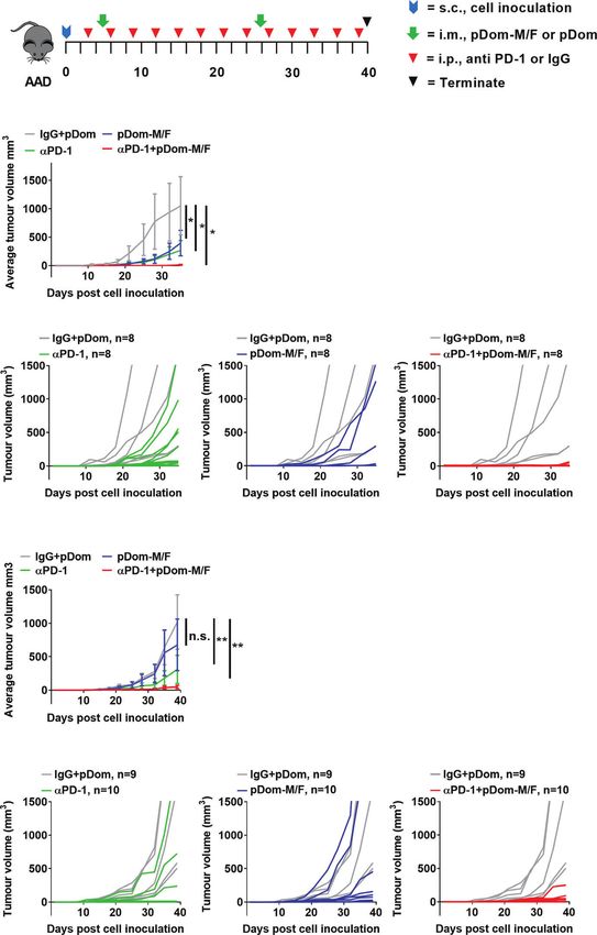
Wang et al.                                                                                                    DNA Vaccines Enhance CPI Responses

                                          antibody conferring excellent tumor control. This opens up an opportunity for combination
                                          immunotherapy that might benefit a wider population of HNSCC patients in an antigen-
                                          specific manner.
                                          Keywords: DNA vaccine, cancer immunotherapy, head and neck cancer, MAGED4B, FJX1, cancer antigens

INTRODUCTION                                                                  in most patients, and some patients had a partial response or
                                                                              stable disease (25).
HNSCC is the sixth most common cancer in the world and is                        We have previously shown that MAGED4B and FJX1 are
more prevalent in South East Asia where it is the second most                 over-expressed in a small cohort of primary HNSCC patients in
common malignancy (1). The incident in some cases is linked to                Malaysia (26, 27). Here we sought to develop a feasible
HPV but for HPV independent (HPVneg) subgroup which                           therapeutic strategy by targeting these antigens in HNSCC
represents 60-80% of all HNSCC cases, the causes are unclear                  globally. To achieve this, we first investigated their expression
and often linked to consumption of alcohol, smoking, smokeless                pattern and immunogenicity in HNSCC patient cohorts from
tobacco and bethel quid [reviewed in (2)]. The HPVneg HNSCC                   Malaysia and the UK. We then designed DNA vaccines targeting
is often difficult to treat; surgery and radiotherapy are associated           MAGED4B and FJX1. We demonstrated that our novel DNA
with significant morbidity and a relatively static 5-year survival             vaccine monotherapy delayed tumor growth in a mouse model
rate of around 50-60% (3).                                                    expressing the target antigens and increased T cell infiltration
    Immune checkpoint inhibitors prolonged survival of some                   into the tumor bed. Promisingly, our vaccine acted
patients with HNSCC and have now gained the approval for                      synergistically when co-administered with anti-PD1 treatment
subsets of patients with advanced disease, however the responses              resulting in tumor clearance in a large proportion of mice.
have only been observed in 14-25% of patients (4–6). While the
response to checkpoint inhibitors is associated with many factors
including expression of PD-L1 (7, 8), tumor mutational burden
(9) and the interferon gene signature (10), one major factor is the           MATERIALS AND METHODS
presence of pre-existing tumor-infiltrating T cells (TIL) (11–15).
High TIL levels correlate with significantly longer survival, and              Patient Sample
many patients who respond to checkpoint inhibitors fall into this             Samples from two cohorts of patients were processed at two
category. TILlow tumors, including most HPVneg HNSCC, are                     separate facilities following surgery. For the Malaysian cohort,
commonly categorized as ‘immune desert’ (absent T cells) or                   blood samples from 28 HNSCC patients were collected from the
‘immune excluded’ (excluded T cells) (13, 16, 17). This                       University Malaya Medical Centre, Tengku Ampuan Rahimah
emphasizes the urgent need of novel immunotherapies that                      Hospital and Hospital Kuala Lumpur. Since HPV positive
can work in these patients. One such intervention is to use                   HNSCC is rare in Malaysia (28), these patients were not tested
cancer vaccines to induce or expand specific T cell responses                  for HPV. For the UK cohort, blood samples from 21 HPVneg
against cancer.                                                               HNSCC patients were collected from patients treated at Poole
    For HPV positive (HPV pos ) HNSCC, cancer vaccines                        Hospital at the time of surgery. The samples were both primary
targeting oncoprotein E6 and E7 from HPV have induced a                       and recurrent tumors. The demographic and clinico-pathological
durable anti-tumor response (18, 19). For HPVneg HNSCC such                   information of 49 HNSCC patients from Malaysia and UK are
viral antigens are not available. Targeting mutated antigens (neo-            presented in Table 1. Peripheral blood mononuclear cells
antigens) is an emerging strategy and has demonstrated clinical               (PBMC) were isolated from whole blood. HLA-A status for the
benefits in late-stage melanoma and newly diagnosed                            Malaysian cohort was determined by direct staining of PBMC
glioblastoma in combination with checkpoint inhibitors (20–                   with mouse anti-human HLA-A2-PE and HLA-ABC-PE (BD
23). A mutanome-targeting DNA vaccine has entered a phase 1/2                 Biosciences, US). HLA status for the UK cohort was determined
trial for patients with locally advanced or metastatic solid tumors           using the AllSet Gold HLA-A SSP Kit (One Lambda, US).
who received checkpoint inhibitor treatment but did not reach a               Formalin-fixed paraffin-embedded (FFPE) tissues from
complete response (24). However, the mutanome-targeting                       HNSCC patients that are in excess of diagnosis were identified
approach is time consuming and costly as it requires the                      and sectioned into 4µm thick for hematoxylin and eosin
generation of personalized vaccines for each patient. We                      (H&E), as well as immunohistochemistry (IHC) staining. For
therefore focused on developing cancer vaccines targeting                     tissues from normal organs, 3-4 independent tissue microarrays
tumor associated antigens (TAAs) in order to generate off-the-                generated in-house were analyzed for the expression of
shelf treatment to a large proportion of HNSCC patients.                      MAGED4B and FJX1.
Recently the feasibility of this approach has been demonstrated                   This project was approved by the Institutional Review Board
in patients with unresectable melanoma post-anti-PD1                          of the Faculty of Dentistry, University of Malaya [DFOS1706/
treatment. Antigen-specific polyfunctional T-cells were induced                0026(L)], Medical Research and Ethics Committee, Ministry of

Frontiers in Immunology | www.frontiersin.org                             2                                October 2021 | Volume 12 | Article 763086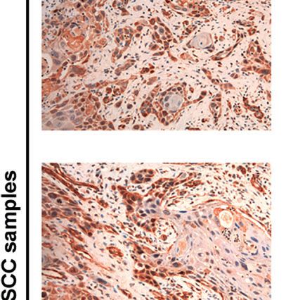
Wang et al.                                                                                                                  DNA Vaccines Enhance CPI Responses

TABLE 1 | The demographic and clinico-pathological information of 49 HNSCC patients included in this study.

Variables                                                                                               Sample size, n                             Percentage, %

Age (Median=64, Range=13-91)
                                            70% purity by high-performance liquid                                     supplemented with 20 U/ml IL-2 every 2-3 days. Stimulated
chromatography (HPLC).                                                                    T cells were subjected to ELISpot.

In Vitro Stimulation of T Cells                                                           Human IFNg ELISpot Assay
PBMCs were thawed and rested at high density (1x10⁷ cells per                             1 x 10⁴ stimulated T cells were incubated with peptide-loaded
well of a 24-well plate) for two days before T cells were isolated                        dendritic cells (DCs) at 1:30 ratio on a 96 well ELISpot plates pre-
using CD4+ and CD8+ microbeads (Miltenyi, UK). The                                        coated with anti-human IFNg antibody (mAb 1-DIK, Mabtech,
remaining T-cell depleted PBMCs were transfected with                                     UK) overnight at 37°C. DC were differentiated from autologous
mRNA encoding individual antigens. DNA fragments                                          CD14+ monocytes, according to Miltenyi manufacturer’s
encoding each antigen (MAGED4B, FJX1 or CEFT) were                                        protocol in ImmunoCult™ DC differentiation media (Stemcell
synthesised through the Invitrogen GeneArt service                                        Technologies, UK) for two weeks. DCs were loaded with 1 µg/ml

Frontiers in Immunology | www.frontiersin.org                                         3                                  October 2021 | Volume 12 | Article 763086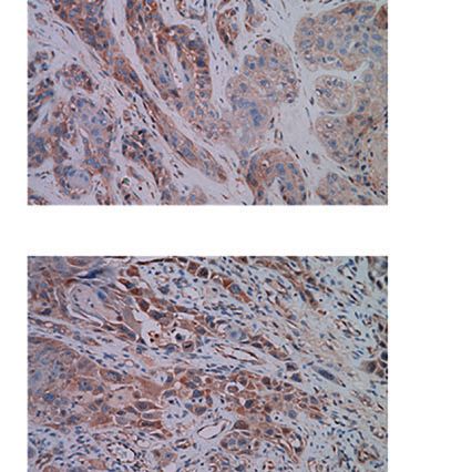
Wang et al.                                                                                               DNA Vaccines Enhance CPI Responses

CEFT, MAGED4B or FJX1 OPP. Samples were plated in                       Kebangsaan Malaysia (Approval No. CRM/2017/KUE PENG/
duplicate or triplicate. Spots were detected with a biotin-             25-JAN./820-APR.-2017-APR.-2020) and Animal Ethics
conjugated human IFNg antibody (mAb 7-B6-1, Mabtech UK)                 Committee of University of Southampton under the Project
followed by incubation with Streptavidin ALP (Mabtech, UK)              Licence (P8969333C). All animal experiments complied with
and BCIP/NBT (Thermo, UK). Plates were scanned using                    the National Institute of Health guide for the care and use of
ImmunoSpots reader (AID, UK). The mean values were                      Laboratory animals (NIH Publications No. 8023, revised 1978).
represented as spot-forming cells (SFCs) per 1 x 10⁴ T cells.
Levels were considered positive if at least two times above             Immunogenicity of pDom-M/F In Vivo
medium control.                                                         C57BL/6 and HLA-A2 tg mice (transgenic for the HLA-A2.1
                                                                        allele, HHD) (31, 32) used in the immunogenicity study were
Flow Cytometry Analysis of                                              bred in animal facility of the University Southampton. C57BL/6
Human Samples                                                           or HLA-A2 tg mice aged 6 -10 weeks (n=5/group) received either
To detect antigen-specific T cells, PBMC from Malaysian HLA-             50mg pDom-M, pDom-F or pDom vector control vaccines
A2 patients were first incubated with FVS780 (fixable viability           intramuscularly (i.m.) into each quadriceps muscles of the
stain) (BD Biosciences, US) for 15 min at room temperature              hind legs. C57BL/6 mice received a booster injection with an
(RT), washed and further incubated with MAGED4B501-509/                 equivalent amount of the vaccine after one week, while HLA-A2
HLA-A2 tetramer-PE for 30 min at RT. After incubation, cells            tg mice received a booster injection after 3 weeks with
were stained with anti-CD3+ (FITC: SK7), CD4+ (PerCP Cy5.5:             electroporation (EP). EP was carried out on mice anaesthetized
SK3) and CD8+ (BV510: RPA-T8) for 30 min at RT. For UK                  by isofluorane, using a custom-made pulse generator from
cohort, similar protocol was used to detect antigen-specific T           Inovio Pharmaceuticals, as described previously (33).
cells. PBMCs were first stained with Live/dead violet stain                  At endpoint, spleens were harvested from experimental mice,
(Invitrogen, US), followed by MAGED4B 501-509 /HLA-A2                   and subsequently mashed though 70 µm strainer to obtain a
tetramer-PE, anti-CD3+ (FITC: OKT3), CD4+ (APC: OKT4),                  single cell suspension. Lymphocytes were then isolated from
CD8+ (PE-Cy7: SK1) for 30 min at RT. MAGED4B501-509/HLA-                splenocytes using Lymphoprep™ (STEMCELL, UK) following
A2 tetramer-PE was generated in house by University of                  manufacturer’s protocol. To determine the presence of IFNg
Southampton Protein Core Facility. Gating strategy is detailed          secreting lymphocytes, anti-mouse IFNg ELISpot kit (BD
in Figure S1A.                                                          Bioscience, UK) was used according to manufacturer’s
    All stained cells were analyzed using LSR Fortessa flow              protocol. Mouse lymphocytes (2.5x10 5 cells/well) were
cytometer or FACSCanto (both BD Biosciences, US) and gated              incubated in complete RPMI media with no peptide
against fluorescence minus one control (FMO) or unstained                (background control), p30 control peptide (1µM), MAGED4B
controls. The flow panels were designed to accommodate                   or FJX1 OPP (1µM each peptide) on pre-coated anti-mouse IFNg
different lasers and FACS machine capacities in both                    ELISpot plates for 40h at 37°C. Samples were plated in triplicate.
institutions. Details of all antibodies used in the flow cytometry       Spots were detected with a biotin-conjugated mouse IFNg
staining are provided in Table S1. Analysis was performed using         antibody (BD Bioscience, UK) followed by incubation with
FlowJo software (BD Biosciences, US).                                   Streptavidin-ALP (Mabtech) and BCIP/NBT (Thermo, UK).
                                                                        Plates were scanned using ImmunoSpots reader (AID, UK).
Generation of pDom Fusion DNA Vaccines                                  The mean values were represented as spot-forming cells (SFCs)
The human wild type sequences of MAGED4B and FJX1 were                  per 106 cells. Levels were considered positive if at least two times
cloned separately into the pcDNA3.1 (Invitrogen, UK) plasmid as         above the background control.
a fusion with the Dom sequence from fragment C of tetanus toxin
as source of CD4+ T cell help (30). The sequence encoding mus           Tumour Models
IgH signal peptide MGWSCIIFFLVATATGVHS was inserted at                  The mouse B16F10 cell line expressing the human HLA-A2 gene
the N-terminus of each construct to enhance secretion. DOM              (B16F10-HLA-A2) was provided by Professor Eric Tartour
fragment and the antigen of interest MAGED4B or FJX1 were               (Sorbonne Paris Cité , University). B16 is one of the most
separated with a seven amino acid linker (AAAGPGP).                     frequently used model for evaluation of the efficacy of anti-
    DNA sequencing was performed to confirm that the plasmids            PD1 antibody and its combinations hence it has been chosen for
contained the correct DNA construct. The generated plasmid              the currently study in favor of less well characterized HNSCC
DNA fusions referred as pDom-MAGED4B (pDom-M) and                       models which has recently emerged (34). It was cultured in
pDom-FJX1 (pDom-F) (plasmid constructs in Figure S2                     RPMI 1640 (Gibco, UK) supplemented with 10% heat
pDom plasmid without the insertion of antigens was used as              inactivated-fetal bovine serum (FBS, Gibco UK), penicillin/
vector control. All plasmids were propagated in DH-5a and               streptomycin (100 U/ml) and 1 mg/ml G418 (EMD Millipore,
purified using QIAGEN Plasmid Plus Giga Kit (QIAGEN, US),                US) at 37°C in a 5% CO2 humidified atmosphere. We generated
according to manufacturer’s instruction.                                two different tumor models expressing our target antigens;
                                                                        B16F10-HLA-A2-MAGED4B (BAM) and B16F10-HLA-A2-
Animal Study                                                            FJX1 (BAF). The expression of HLA-A2 in BAM and BAF cell
All procedures involving the use of animals were reviewed and           lines was confirmed by flow cytometry using human HLA-A2–
approved by the Animal Ethics Committee of Universiti                   PE-conjugated antibody (clone BB7.2, BD Biosciences, US).

Frontiers in Immunology | www.frontiersin.org                       4                                 October 2021 | Volume 12 | Article 763086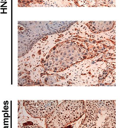
Wang et al. DNA Vaccines Enhance CPI Responses
MAGED4B and FJX1 expression levels were confirmed by additional tubes were stained with viability dye (FVS780), anti-
western blotting using custom made anti-MAGED4B (Dundee CD4, CD45.2, CD11b, and GR1. Cells were subjected to fixation
Cell Products Ltd, UK) and anti-FJX1 (HPA059220, Sigma and permeabilization with Mouse FoxP3 Buffer Set (BD
Aldrich, US) antibodies respectively. Biosciences, US), and stained with anti-FOXP3 (eBioscience,
US). Details of all antibodies used in the flow cytometry
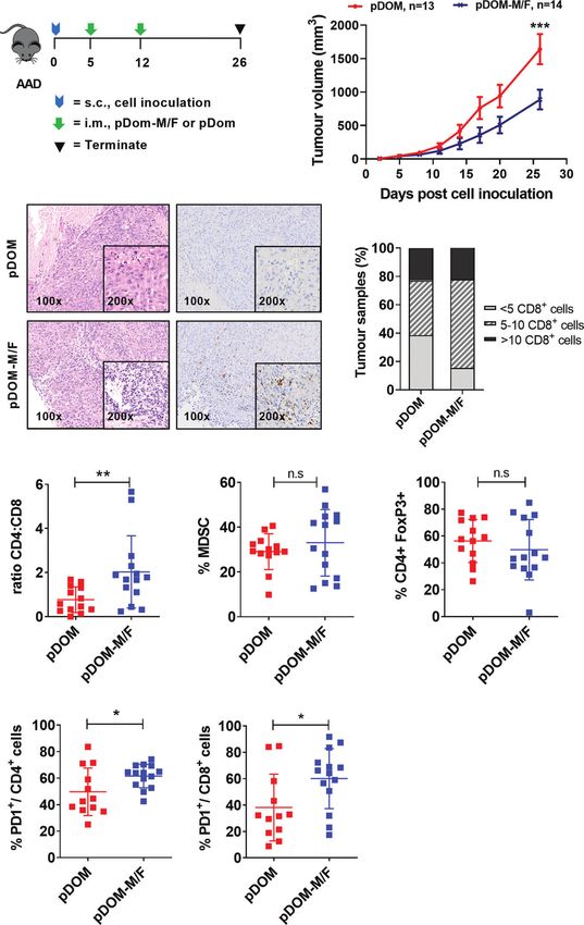
Efficacy of pDom-M/F In Vivo staining is provided in Table S1. PD1-positive CD4+ or CD8+
AAD mice (transgenic for HLA-A2.1/H2-Dd allele) used in the T cells were quantitated based on the relative expression of PD1,
efficacy study were bred in the animal facility of National gating strategy is detailed in Figure S3.
University Malaysia. AAD mice (6-10 weeks) were inoculated
with 1x10⁶ BAM cell line (n=27 mice) at the right flank at day 0. Immunohistochemistry
These mice were randomized to receive vaccination of 100 µg To assess the expression level of MAGED4B and FJX1 in HNSCC
pDom-M/F DNA vaccines (50 µg of each vaccine) or 50 µg samples from the UK cohort, anti-MAGED4B polyclonal
pDom DNA vaccine i.m into each quadriceps muscles of the antibody (1:200, Novus-Bio) and anti-FJX1 polyclonal antibody
hind legs on day 5 after palpable tumors were observed (1:200, Novus-Bio) were used. Deparaffinization, rehydration,
(~25cm 3 ). A booster vaccination at the same dose was antigen retrieval, and IHC staining were performed using a
administered at day 12 (the vaccination schedule is depicted in Dako PT Link Autostainer using EnVision FLEX Target
Fig. 3A). Tumour sizes were evaluated every 3-4 days and Retrieval Solution, High pH (Agilent Dako, UK) and DAKO
volumes were calculated using the formula: volume = ½ Auto-stainer Link48™ in the Cellular Pathology Department of
(length X width 2). Tumour growth inhibition (TGI) was the University Hospital of Southampton NHS Trust. Images were
calculated using the formula: TGI=(Vc-Vt)/Vc X 100%; where captured using ZEISS Axio scanner.
Vc and Vt are the average tumor volume of control and For the Malaysian cohort, IHC was performed on patient
treatment group respectively at endpoint. On day 26, mice FFPE samples using anti-MAGED4B polyclonal antibody (1:100,
were sacrificed and tumors were harvested for analysis of T Sigma Aldrich, US) and anti-FJX1 polyclonal antibody (1:200,
cell infiltration by IHC and flow cytometry as described below. Sigma Aldrich, US). Antigen retrieval was performed in citrate
To evaluate the efficacy of combination treatment of pDom- buffer pH 6 and Tris-EDTA pH 9 for MAGED4B and FJX1
M/F vaccine with anti-PD-1 antibody, AAD mice were respectively using microwave heating method. IHC staining were
inoculated with 1x10⁵ BAM (n=32 mice) or BAF cells (n=39 performed using Dako Cytomation Envision+ Dual Link
mice). Subsequently, mice were randomized into four treatment System- HRP (DAB+) kit (Dako,US) following protocol
groups; pDom-M/F, anti-PD-1 antibody, pDom-M/F + anti-PD- recommended by manufacturer. Direct comparisons of
1 antibody, or pDom + IgG isotype control. pDom-M/F antibodies to each antigen used for the UK and the Malaysian
vaccinations were given at day 5 and day 26 post-cell cohorts were performed using 5 independent HNSCC samples
inoculation. One hundred microgram of anti-PD-1 monoclonal and the same data were obtained. The Novus antibodies were
antibody (CD279, Bioxcell, US) or rat IgG2a isotype control chosen in Southampton because they generated less background
(Bioxcell, US) was given intraperitoneally every 3 days from day with the automated system.
3 as indicated in Figure 4A. Tumour sizes were measured every For mouse IHC staining, FFPE sections were stained with
3-4 days and tumor volume were calculated using the formula rabbit anti-mouse CD8a (1:400; clone D4W2Z; Cell Signaling
described above. Technologies, US). All sections from mice were processed after
antibody staining using Dako Animal Research Kit (Dako, US)
Flow Cytometry Analysis of Mouse according to the recommendation by manufacturer (27). The
Tumour Infiltrating Lymphocytes field containing the highest density of CD8-positive cells within
To determine the expression of T cell markers after pDom-M/F the tissue were identified. CD8-positive cells were counted by 3
treatment in mouse models, tumor infiltrating lymphocytes individuals including a board-certified pathologist and graded as
(TILs) were isolated from mouse tumor samples. Briefly, “less than 5 cells”, “5 to 10 cells” and “more than 10 cells”
500mm3 tumors were harvested and minced into smaller (35, 36).
pieces (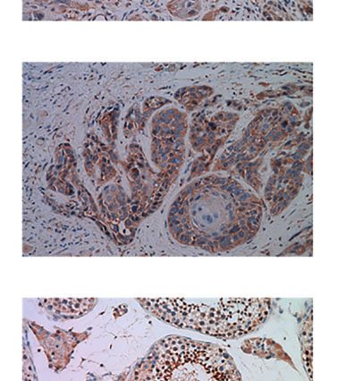
Wang et al.                                                                                             DNA Vaccines Enhance CPI Responses

RESULTS                                                                 DNA Vaccines Targeting MAGED4B and
                                                                        FJX1 Induce T Cell Responses in Mice
MAGED4B and FJX1 Are Expressed in                                       To induce broad CD4/CD8+ anti-tumor responses irrespective
HNSCC Tumour Samples                                                    of HLA subtypes, the full-length MAGED4B or FJX1 sequences
Using TCGA-HNSC project data, we demonstrated that both                 were linked to 3’ Dom sequence and inserted into pcDNA3
MAGED4B and FJX1 gene expression levels in the HPV negative             plasmid to give rise to pDom-MAGED4B (pDom-M,
(n=415) and HPV positive (n=72) tumour tissue samples were              Figure S2A) and pDom-FJX1 (pDom-F, Figure S2B). In vivo
significantly higher than levels in adjacent normal tissues (n=44,       immunogenicity was tested using the HLA-A2 transgenic (tg)
Figure 1A). HPV status of TCGA samples was available from               and wildtype C57BL/6J mice using our previous protocols (31).
cBioportal for the PanCancer and was the result of complete             Splenocytes from immunized mice were evaluated by IFNg
analysis of the cohort for HPV transcripts (37). Importantly,           ELISpot using MAGED4B or FJX1 OPP to stimulate antigen-
either one or both antigens were also found expressed in other          specific immune responses. In wildtype mice, pDom-M showed
cancer types including lung, breast, oesophageal, and stomach           significantly higher antigen specific T cell responses to
cancer at the mRNA level (Figure S4). We subsequently                   MAGED4B OPP than control pDom immunized mice
examined the protein expression of MAGED4B and FJX1 in                  (p=0.012, Figure 2A). Similarly, T cell response against FJX1
HNSCC tumour tissues from both Malaysian and UK cohorts by              OPP was also significantly elevated in mice received pDom-F as
IHC. All tested samples from Malaysian cohort (n=28) expressed          compared to the pDom control group where no responses were
both antigens, except one patient who did not express FJX1              detected (p=0.012). In HLA-A2 tg mice, vaccination with pDom-
(Table 1). All samples from the UK cohort (n=16) expressed              M and pDom-F resulted in a significant T cell response against
both MAGED4B and FJX1 (Table 1). In total 43/44 samples were            MAGED4B (p=0.016, Figure 2B) or FJX1 (p=0.037, Figure 2B)
positive for both antigens (representative images are depicted in       OPP respectively as compared to the pDom control. Both
Figure 1B; testes used as positive control). The evaluation of          vaccines induced robust T-cell responses with lower levels
expression of MAGED4B and FJX1 in five major organs and                  observed in HLA-A2 tg mice because of lower number of
healthy oral epithelia revealed very low levels in basal                T-cells and lower MHC I expression (41). The responses to
keratinocytes of stratified squamous epithelium with no                  pDom vector control were negative in both strains as expected.
expression above the background elsewhere (Figure S5).
                                                                        Vaccination With pDom-M/F Vaccine
MAGED4B and FJX1 Specific T Cells Were                                   Increased T Cell Infiltration and Delayed
Detected in HNSCC Patients at High                                      Tumour Growth
Frequency                                                               We subsequently evaluated efficacy of these two vaccines using
T cells specific for neo-antigens were detected in patients with         the BAM tumor model. This model expresses HLA-A2 (42)
HPVneg HNSCC (38), indicating that the patients’ immune                 (Figure S6A). It expresses FJX1 endogenously and was
system is capable of recognising the tumour antigens in this            engineered to express MAGED4B because the antigen is not
disease. To determine whether HNSCC patients inherently                 expressed in mice (Figure S6B). The HLA-A2 mice were
harbour MAGED4B- and FJX1-specific T cells, we generated                 challenged with 106 BAM tumor cells and once the tumors
MAGED4B501-509/HLA-A2 tetramer using the HLA-A2 epitope                 were palpable, mice were immunized with either combined
we previously identified (39). MAGED4B-specific CD8+T cells               pDom-M and pDom-F (referred as pDom-M/F vaccine) or
were identified in 4/4 HLA-A2pos patients from the Malaysian             pDom control as indicated (Figure 3A). Mice immunized
cohort and 8/11 HLA-A2pos patients from the UK cohort                   with pDom-M/F showed a delay in their tumor growth as
(Figure 1C and Table 2) at a frequency observed at a similar            compared to mice received pDom (average tumor sizes of
level to neoepitopes in melanoma (range 0.06-0.12%) (40).               pDom and pDom-M/F at endpoint (day 26): 1641.22 mm3 and
PBMC samples of HLA-A2neg patients were used to confirm                  887.24 mm3, TGI: 45.94%; Figure 3B).
the specificity (Figure 1C, Figure S1B and Table 2). Since no                Subsequently, we determined whether vaccination could
FJX1 epitope was available for a tetramer, the T cell responses         increase infiltration of T cells into the tumor, as demonstrated
were analysed by IFNg ELISpot using OPP for the entire                  in a DNA vaccine clinical trial targeting E6/E7 in HPVpos
antigenic sequence. In parallel we also analysed the responses          HNSCC (18). Tumours from the pDom-treated group
to MAGED4B OPP to extend the data beyond the HLA-A2                     predominantly displayed an immune deserted phenotype,
positive cohort. We initially performed ex vivo ELISpot using           where immune cells were absent or scarce throughout the
PBMCs from two patients (Figure S1C), which suggested                   tissue (Figure 3C). This phenotype was expected as the
specific T cell responses could be captured without in vitro re-         parental tumor were previously reported to have low TILs (43).
stimulation (IVS) occasionally but not reliably. We therefore           By contrast, tumors from mice vaccinated with pDom-M/F
opted for IVS ELISpot; 3/7 and 5/7 patients responded to FJX1           showed increased levels of T cells infiltrated into the tumor
OPP or MAGED4B OPP respectively with 3/7 demonstrating                  (Figure 3C). Enumeration of CD8+ T cells in the tumors further
responses against both (Figure 1D; representative examples,             indicated that the infiltration of CD8+ T cells was higher in the
Table 2). Collectively, we have confirmed both antigens are              pDom-M/F-vaccinated tumors as compared to pDom-
immunogenic in HPVneg HNSCC patients.                                   vaccinated tumors (Figure 3D). As tumor-specific CD4

Frontiers in Immunology | www.frontiersin.org                       6                               October 2021 | Volume 12 | Article 763086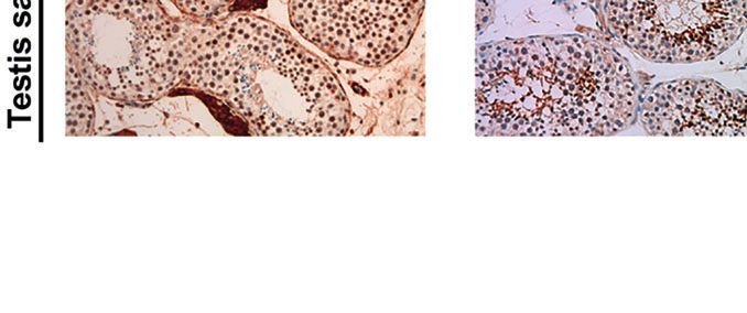
Wang et al.                                                                                                                   DNA Vaccines Enhance CPI Responses

                            A                                                        C

                                                                                              D

                            B

 FIGURE 1 | MAGED4B and FJX1 are immunogenic tumour-associated antigens in HPVneg HNSCC patients. (A) Both HPVneg and HPVpos HNSCC samples from
 TCGA has significant elevation of MAGED4B and FJX1 compared to adjacent normal tissue at transcriptomic level. P values were calculated using Kruskal-Wallis test
 on normalized mRNA expression (****p-value < 0.0001; **p-value < 0.01; ns, not significant). (B) MAGED4B and FJX1 expression in HNSCC samples and testes
 (as positive control) were indicated by IHC staining with anti-hMAGED4B polyclonal antibody (Novus-Bio) and anti-FJX1 polyclonal antibody (Novus-Bio) respectively.
 Magnification x200. (C) FACS plots showed MAGED4B -specific T cells were detected in PBMC from HNSCC patients using MAGED4B501-509 HLA-A2 tetramer-PE.
 Four HLA-A2pos, one HLA-A2neg HNSCC patients and one healthy donor are shown. (D) T cells from HNSCC patients after IVS were assessed with IFNg ELISpot.
 Antigen-specific T cell responses were evaluated by stimulating with MAGED4B OPP and FJX1 OPP pulsed autologous DCs. CEFT OPP were used as positive
 control in IFNg ELISpot. Two HLA-A2pos and two HLA-A2neg HNSCC patients are shown.

effector T cells can also contribute to anti-tumor protective                        analysis but not MDSC (CD45.2+ CD11b+GR1+) and
mechanisms (44), we further determined whether they also                             regulatory T cells (CD4+ FoxP3+) (Figure 3E). In addition, we
increased. Encouragingly, pDom-M/F vaccine also induced                              detected a significant increase in PD-1 expressing CD4+ and
CD4+ T cells in TILs as demonstrated by flow cytometry                                CD8+ T cell population in the pDom-M/F group as compared to

Frontiers in Immunology | www.frontiersin.org                                    7                                       October 2021 | Volume 12 | Article 763086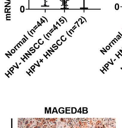
Wang et al.                                                                                                                           DNA Vaccines Enhance CPI Responses

TABLE 2 | Information of target antigen expression, HLA type, and detection of antigen-specific T cells in UK and Malaysia patient cohorts.

Patient ID                      Antigen expression (IHC)                          HLA-A2                 MAGED4B Tetramer+                              ELISpot

                             MAGED4B                       FJX1                                                                                 MAGED4B             FJX1

UoSno.1                   Pos                            Pos                      Pos                    Neg                                    Pos                 Pos
UoSno.2                                                                           Pos                    Pos
UoSno.3                                                                           Neg                    Neg                                    Pos                 Neg
UoSno.4                   Pos                            Pos                      Pos                    Pos                                    Pos                 Pos
UoSno.5                   Pos                            Pos                      Neg                    Neg                                    Pos                 Neg
UoSno.6                   Pos                            Pos                      Pos                    Pos
UoSno.7                   Pos                            Pos                      Pos                    Pos
UoSno.8                   Pos                            Pos                      Pos                    Pos
UoSno.9                   Pos                            Pos                      Neg
UoSno.10                  Pos                            Pos                      Pos                    Pos
UoSno.11                  Pos                            Pos                      Pos                    Pos
UoSno.12                  Pos                            Pos                      Neg                    Neg
UoSno.13                                                                          Pos                    Pos                                    Pos                 Pos
UoSno.14                  Pos                            Pos                      Pos                    Neg                                    Neg                 Neg
UoSno.15                                                                          Pos                    Neg
UoSno.16                                                                          Neg                    Neg                                    Neg                 Neg
UoSno.17                  Pos                            Pos
UoSno.18                  Pos                            Pos
UoSno.19                  Pos                            Pos
UoSno.20                  Pos                            Pos
UoSno.21                  Pos                            Pos
04-0017-17                Pos                            Pos                      Pos                    Pos
06-0008-19                Pos                            Neg                      Pos                    Pos
06-0011-19                Pos                            Pos                      Pos                    Pos
06-0012-19                Pos                            Pos                      Pos                    Pos
06-0005-19                Pos                            Pos                      Pos
06-0037-17                Pos                            Pos                      Neg                    Neg
06-0019-17                Pos                            Pos                      Neg
06-0024-19                Pos                            Pos                      Neg
01-0010-17                Pos                            Pos
01-0002-18                Pos                            Pos
04-0018-17                Pos                            Pos
04-0019-17                Pos                            Pos
04-0020-17                Pos                            Pos
04-0001-18                Pos                            Pos
04-0003-18                Pos                            Pos
04-0005-18                Pos                            Pos
06-0014-11                Pos                            Pos
06-0047-17                Pos                            Pos
06-0048-17                Pos                            Pos
06-0053-17                Pos                            Pos
06-0008-18                Pos                            Pos
06-0016-18                Pos                            Pos
06-0021-18                Pos                            Pos
06-0025-18                Pos                            Pos
06-0033-18                Pos                            Pos
06-0038-18                Pos                            Pos
06-0003-19                Pos                            Pos
06-0014-19                Pos                            Pos

Blank space indicating assay was not processed. FFPE blocks for samples UoSno.2, 3, 13,15 and 16 were not available at the time of the study.

pDom (p=0.018 and p=0.029 respectively; Figure 3F). The                                     Combination Therapy of pDom-M/F and
increase in PD-1 expression level amongst CD4+ and CD8+ T                                   Anti-PD-1 Inhibited Tumour Growth
cells indicated a combination with an anti-PD-1 antibody would                              We next investigated if vaccination with pDom-M/F in
likely to boost the T cell response.                                                        combination with anti-PD1 antibody would lead to an
    Further, there was no significant difference in body weight                              improved tumor control. Anti-PD1 group served as a
between pDom and pDom-M/F treated animals and                                               comparator while isotype control IgG plus pDom served as a
histopathology analysis of 5 major organs (kidney, lung, heart,                             negative control. When compared to IgG + pDom control group,
spleen and liver) revealed no pDOM-M/F induced toxicity (data                               growth inhibition was clearly demonstrated in pDom-M/F,
not shown).                                                                                 anti-PD-1 and the combined treatment group (at day 35,

Frontiers in Immunology | www.frontiersin.org                                           8                                        October 2021 | Volume 12 | Article 763086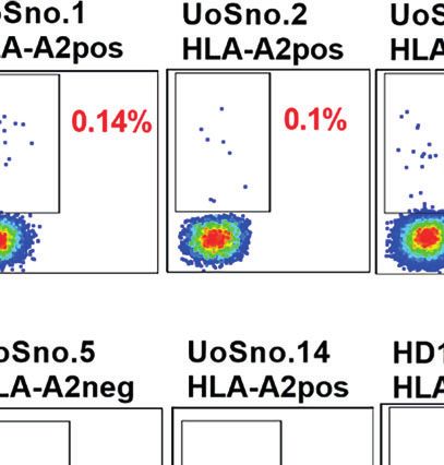
Wang et al.                                                                                                                DNA Vaccines Enhance CPI Responses

                                   A

                                   B

 FIGURE 2 | pDom-M and pDom-F vaccines are immunogenic in wildtype C57BL/6 and transgenic HLA-A2 mice. Augmentation of antigen-specific immune
 responses was observed in all mice that received pDom-M or pDom-F in comparison to mice which received pDom control. (A) C57BL/6 mice were vaccinated at
 day 1 and day 8 with pDom, pDom-M, or pDom-F. Splenocytes were harvested at day 22 to study antigen-specific immune responses by IFNg ELISpot. (B) HLA-A2
 tg mice were vaccinated at day 1 and day 22 with electroporation. Splenocytes were harvested at day 36 for IFNg ELISpot. Splenocytes were seeded at 2.5 x10⁵ to
 ELISpot plates and stimulated overnight with 1 µM of MAGED4B OPP or FJX1 OPP. All data are expressed as mean ± SD. (*p-value < 0.05).

TGI pDom-M/F: 49.16%, anti-PD-1: 60.11% and combination:                            The experiment was performed in BAF-bearing animals using the
96.7%; Figures 4B, C). Significantly, 6 of 8 mice (75%) that                         same protocol as for the BAM model. The growth inhibition in
received a combination of pDom-M/F and anti-PD-1 antibody                           the combination group was also prominent (at day 40, TGI
had complete tumor growth inhibition.                                               pDom-M/F: 37.78%, anti-PD-1: 71.59% and combination:
   We next used the BAF cells (engineered to overexpress human                      95.56%; Figures 4D, E). Fifty percent of animals (5 of 10) in
FJX1 only, Figure S6C), to evaluate the efficacy of pDom-M/F.                        the combination group had completely inhibited tumor growth

Frontiers in Immunology | www.frontiersin.org                                   9                                     October 2021 | Volume 12 | Article 763086
Wang et al.                                                                                                                 DNA Vaccines Enhance CPI Responses

                            A                                                        B

                            C

                                                                                            D

                            E

                            F

 FIGURE 3 | pDom-M/F vaccines enhance anti-tumour immunity by increasing infiltration of immune cells into the tumour. (A) Schematic of vaccination schedule,
 mice were inoculated with the BAM cell line and randomized to either received vaccination of pDom-M/F (pDom-M and pDom-F; 50 mg each vaccine per mouse) or
 pDom (50 mg per mouse) as vector control. Upon termination at endpoint, tumours from vaccinated animals were harvested for analysis of TILs. (B) Tumour growth
 inhibition was significant in mice that received pDom-M/F in comparison to mice which received pDom. Data is expressed as mean ± SEM. (C) Increased immune
 cells infiltration was observed in pDom-M/F group as opposed to pDom (left). IHC staining demonstrated CD8+ T cells were induced by pDom-M/F vaccination
 (right). Images shown were at 100X and 200X magnifications. (D) Analysis of IHC data demonstrated higher proportion of tumour from pDom-M/F group harbouring
 CD8+ T cells as compared to pDom vector control group. (E) pDom-M/F group significantly induced more CD4+ T cells. (F) Both PD1+CD4+ and PD1+CD8+ T
 cells were significantly upregulated in pDom-M/F vaccinated animals when compared to pDom group. Unless otherwise stated, all data are expressed as mean ± SD
 and pooled data from 2 independent experiments are shown for (B–F). Symbols *, **, ***, ns denote p < 0.05, p < 0.01, p < 0.001 and not significant respectively.

Frontiers in Immunology | www.frontiersin.org                                  10                                      October 2021 | Volume 12 | Article 763086
Wang et al. DNA Vaccines Enhance CPI Responses
A
B
C
D
E
FIGURE 4 | Combination therapy of pDom-M/F and anti-PD-1 inhibited tumour growth in vivo. (A) Schematic of vaccination schedule, mice were inoculated with the
BAM or BAF cells and randomized to either received 2 doses of pDom (50 mg per mouse) as vector control combined with 13 doses of isotype control (100 mg per
mouse, given every 3 days), 13 doses of anti-PD-1 (100 mg per mouse, given every 3 days), pDom-M/F (pDom-M and pDom-F; 50 mg each vaccine per mouse)
and the combination of pDom-M/F with anti-PD-1. Upon termination at end-point, mouse spleens were harvested for ELISpot. (B, D) The graphs indicated mean
tumour volumes of treatment group for BAM or BAF model respectively. Statistical analyses were conducted using with two-way ANOVA. (*p < 0.05; **p < 0.01;
ns, not significant). Statistics represented the comparison on terminated date (day 35 for BAM, day 40 for BAF). (C, E) Graphs of individual animal tumour volumes
for BAM and BAF models respectively comparing experimental groups including anti-PD-1 (green line), pDOM-M/F (blue line) and the combination of pDom-M/F with
anti-PD-1 (red line) to IgG/pDOM control (grey line).
Frontiers in Immunology | www.frontiersin.org 11 October 2021 | Volume 12 | Article 763086Wang et al. DNA Vaccines Enhance CPI Responses
throughout the experiment. We therefore were able to confirm We probed the immunogenicity of FJX1 using IVS ELISpot
that pDom-M/F DNA vaccines were able to significantly reduce designed to expand memory T responses in PBMCs. Three out
the tumor burden and this was enhanced by combination with of eight patients generated responses to FJX1, confirming pre-
anti-PD1 antibody achieving a complete clearance of tumor in existing memory T cell responses to FJX1. Notably, the same
50%-75% of mice in BAF and BAM model respectively. patients also demonstrated responses to MAGED4B.
T cell infiltration into tumor is an important criterion for a
successful immunotherapy and is linked to better patient
DISCUSSION prognosis (13, 14). Several approaches including targeted
therapy, radiotherapy and chemotherapy have also shown the
Therapeutic vaccine targeting tumor antigens is a promising ability to convert immunologically cold tumors into hot to
strategy to activate the immune system to eradicate cancer. This increase response to checkpoint inhibitors (55–57), however
strategy confers advantage over other non-specific therapies, as it these approaches are associated with treatment-associated side
does not only induce tumor-specific immune responses, but also effects. We demonstrated in this study that the pDom-M/F
promotes establishment of immunological memory. Early trials vaccination were able to inhibit the growth of MAGED4B and
in HNSCC had focused on targeting cancer driving mutations FJX1-expressing tumors in HLA-A2 transgenic mice by
such as p53 and K-Ras, however no significant clinical outcomes approximately 50%. Importantly, pDom-M/F vaccination were
were reported (45). Vaccines targeting MUC1 (NCT02544880) able to convert cold B16 tumors into hot tumors with an increase
and CEA (NCT00924092; NCT00027534) which are in phase I/II of CD8+ T cells. This is encouraging as an effective vaccination in
clinical trials have yet to report results while the phase I study cancer therapy is frequently associated with high degree of
targeting Survivin-2B demonstrated low efficacy (46). cytotoxic T cells infiltration into the tumor. We were not able
Several recent trials in solid tumors demonstrated significant to determine if these are antigen specific CD8+ T cells as our
immunogenicity (20–22, 25) with achieving partial or clinical HLA-A2 tetramer or the corresponding peptide was not
remission. Remarkably, the antigens which have been targeted presented in the HLA-A2 transgenic mice. The work is
are not only mutated antigens for which the central tolerance is currently underway to define T-cell epitopes to allow further
not expected but widely expressed antigens, tissue-specific characterization of antigen-specific responses in murine models.
antigens as well as cancer testis antigens (CTA) (25, 47, 48). High expression of PD-1 on TILs has been shown to impair the
The latter has long been thought of as “good” antigens due to anti-tumour immune responses in humans by engaging the PD-L1
their safety and immunogenicity; targeting NY-ESO1 and and to inhibit TCR-mediated proliferation and cytokine
MAGE-A3 recently have demonstrated clinical benefits (25). production (58). The upregulation of PD-1 levels in vaccinated
However, most well-characterized CTAs are not expressed in a mice supports our strategy to combine the pDom-M/F vaccine
large proportion of head and neck cancer (49). Previously, the with an anti-PD-1 antibody and this can potentially be applied in
expression of MAGED4B and FJX1 CTAs has been described in the clinic as anti-PD-1 antibodies have now been approved for the
HNSCC patients in Malaysia (26, 50, 51). Here we were able to treatment of recurrent/metastatic HNSCC. Promisingly, we
confirm their expression at the RNA levels using 522 HNSCC observed a complete tumour elimination or static tumour
samples deposited at TCGA. Through the collaborative effort of growth in the majority of mice that received both vaccine and
our Malaysian and UK teams, these findings have been further anti-PD-1 antibody treatment. Our results are one of the few
validated in two independent cohorts of patients in Malaysia TAA-based DNA vaccines that show near complete tumour
(n=28) and the UK (n=16), confirming 43/44 patients are co- clearance in preclinical models when combined with the anti-
expressed FJX1 and MAGED4B in both primary and recurrent PD-1 antibody. Overall, our study provides novel findings
tumors. This is a remarkably high frequency not observed for in which a DNA vaccine targeting TAAs frequently expressed in
other CTAs in HNSCC [reviewed in (52)]; our data also HNSCC is able to enhance the efficacy of anti-PD-1 therapy in
demonstrate consistency between several ethnic groups from preclinical settings. Clinical trials using selected epitopes including
the UK and Malaysia. Alcohol consumption and smoking are our own has focused on patients’ cohort expressing a particular
common etiological factors for HNSCC in both Malaysia and HLA allele (most frequently A2 and A24) (47, 48, 59). Although
UK, while betel quid chewing is specific to the Malaysia cohort we tested our vaccines in the HLA-A2 model, for clinical
(53, 54). Since the causes of the disease are different, the translation a full-length antigen vaccine is preferable to provide
discovery of antigens that are expressed in over 90% of a population-wide coverage irrespective of HLA genotype (31).
patients is unexpected and confers a rare chance for the This vaccine was designed with HPVneg patients in mind,
development of ‘off-the shelf’ cancer vaccine applicable HPVpos HNSCC patients may also benefit from this vaccine as
worldwide. These antigens are relatively unexplored and the antigens are also expressed in these cancers. Both antigens
therefore a very few defined epitopes are available (39, 50). are additionally co-expressed in non-small cell lung cancer
For MAGED4B, we were able to detect the tetramer specific (Figure S4), suggesting relevance of our approach for this
response in HLA-A2 patients (approx. 80%) with the levels that common cancer. A phase I/II clinical trial testing our DNA
are similar to those reported for mutated antigens reassuring vaccine delivered as doggybone DNA vaccine (33) in
that the antigen is remarkably immunogenic in this patient combination with anti-PD-1 antibody is due to begin
cohort. We were not able to generate a tetramer using the only recruitment of patients with recurrent HNSCC in the first half
HLA-A2 epitope (11mer) from FJX1 described previously (50). of 2022.
Frontiers in Immunology | www.frontiersin.org 12 October 2021 | Volume 12 | Article 763086Wang et al. DNA Vaccines Enhance CPI Responses
DATA AVAILABILITY STATEMENT MTA, SI, SL, GT, KM, RZ and ZAR. Analysis and interpretation
of data: CW, NSZ, SJC, CG, KL, SCC, and NS. Writing, review,
The original contributions presented in the study are included in and/or revision of the manuscript: All authors.
the article/Supplementary Material. Further inquiries can be
directed to the corresponding authors.
FUNDING
ETHICS STATEMENT This study is funded jointly by the Ungku Omar Fund and the
Medical Research Council UK Newton fund (MR/PO13414/1),
The studies involving human participants were reviewed and
and internal funding from Cancer Research Malaysia.
approved by 1. The Institutional Review Board of the Faculty of
Dentistry, University of Malaya; 2. Medical Research and Ethics
Committee, Ministry of Health, Malaysia; 3. UK Medical
Research and Ethics Committee; 4. Institutional approval at ACKNOWLEDGMENTS
Southampton University Hospitals NHS Foundation Trust,
Southampton, UK. The patients/participants provided their The authors would like to thank the Director General of Health
written informed consent to participate in this study. The Malaysia for the permission to publish this paper and Oral
animal study was reviewed and approved by 1. The Animal Cancer Research & Coordinating Centre (OCRCC) for
Ethics Committee of Universiti Kebangsaan Malaysia; 2. Animal providing information on patients follow up. The authors also
Ethics Committee of University of Southampton. thank all consented patients who participated in this study.
AUTHOR CONTRIBUTIONS SUPPLEMENTARY MATERIAL
Conception and design: KL, SCC, NS, CO, and GT. Development The Supplementary Material for this article can be found online
of methodology: CW, NSZ, SJC, CG, KL, SCC, and NS. at: https://www.frontiersin.org/articles/10.3389/fimmu.2021.
Acquisition of data: CW, NSZ, SJC, JD, CG, NZ, BL, RS, EK, 763086/full#supplementary-material
9. Riaz N, Havel JJ, Makarov V, Desrichard A, Urba WJ, Sims JS, et al. Tumor
REFERENCES and Microenvironment Evolution During Immunotherapy With Nivolumab.
1. Bray F, Ferlay J, Soerjomataram I, Siegel RL, Torre LA, Jemal A. Global Cell (2017) 171(4):934–49 e16. doi: 10.1016/j.cell.2017.09.028
Cancer Statistics 2018: GLOBOCAN Estimates of Incidence and Mortality 10. Ayers M, Lunceford J, Nebozhyn M, Murphy E, Loboda A, Kaufman DR, et al.
Worldwide for 36 Cancers in 185 Countries. CA: Cancer J Clin (2018) 68 IFN-Gamma-Related mRNA Profile Predicts Clinical Response to PD-1
(6):394–424. doi: 10.3322/caac.21492 Blockade. J Clin Invest (2017) 127(8):2930–40. doi: 10.1172/JCI91190
2. Wang C, Dickie J, Sutavani RV, Pointer C, Thomas GJ, Savelyeva N. Targeting 11. Havel JJ, Chowell D, Chan TA. The Evolving Landscape of Biomarkers for
Head and Neck Cancer by Vaccination. Front Immunol (2018) 9:830. Checkpoint Inhibitor Immunotherapy. Nat Rev Cancer (2019) 19(3):133–50.
doi: 10.3389/fimmu.2018.00830 doi: 10.1038/s41568-019-0116-x
3. Warnakulasuriya S. Living With Oral Cancer: Epidemiology With Particular 12. Jerby-Arnon L, Shah P, Cuoco MS, Rodman C, Su M-J, Melms JC, et al. A
Reference to Prevalence and Life-Style Changes That Influence Survival. Oral Cancer Cell Program Promotes T Cell Exclusion and Resistance to
Oncol (2010) 46(6):407–10. doi: 10.1016/j.oraloncology.2010.02.015 Checkpoint Blockade. Cell (2018) 175(4):984–97. e24. doi: 10.1016/
4. Burtness B, Harrington KJ, Greil R, Soulieres D, Tahara M, de Castro GJr., j.cell.2018.09.006
et al. Pembrolizumab Alone or With Chemotherapy Versus Cetuximab With 13. Wood O, Woo J, Seumois G, Savelyeva N, McCann KJ, Singh D, et al. Gene
Chemotherapy for Recurrent or Metastatic Squamous Cell Carcinoma of the Expression Analysis of TIL Rich HPV-Driven Head and Neck Tumors Reveals
Head and Neck (KEYNOTE-048): A Randomised, Open-Label, Phase 3 a Distinct B-Cell Signature When Compared to HPV Independent Tumors.
Study. Lancet (2019) 394(10212):1915–28. doi: 10.1016/S0140-6736(19) Oncotarget (2016) 7(35):56781–97. doi: 10.18632/oncotarget.10788
32591-7 14. Ganesan AP, Clarke J, Wood O, Garrido-Martin EM, Chee SJ, Mellows T,
5. Seiwert TY, Burtness B, Mehra R, Weiss J, Berger R, Eder JP, et al. Safety and et al. Tissue-Resident Memory Features are Linked to the Magnitude of
Clinical Activity of Pembrolizumab for Treatment of Recurrent or Metastatic Cytotoxic T Cell Responses in Human Lung Cancer. Nat Immunol (2017) 18
Squamous Cell Carcinoma of the Head and Neck (KEYNOTE-012): An (8):940–50. doi: 10.1038/ni.3775
Open-Label, Multicentre, Phase 1b Trial. Lancet Oncol (2016) 17(7):956–65. 15. Ward MJ, Thirdborough SM, Mellows T, Riley C, Harris S, Suchak K, et al.
doi: 10.1016/S1470-2045(16)30066-3 Tumour-Infiltrating Lymphocytes Predict for Outcome in HPV-Positive
6. Ferris RL, Blumenschein GJr., Fayette J, Guigay J, Colevas AD, Licitra L, et al. Oropharyngeal Cancer. Br J Cancer (2014) 110(2):489–500. doi: 10.1038/
Nivolumab for Recurrent Squamous-Cell Carcinoma of the Head and Neck. bjc.2013.639
N Engl J Med (2016) 375(19):1856–67. doi: 10.1056/NEJMoa1602252 16. Chen DS, Mellman I. Elements of Cancer Immunity and the Cancer–Immune
7. Patel SP, Kurzrock R. PD-L1 Expression as a Predictive Biomarker in Cancer Set Point. Nature (2017) 541(7637):321–30. doi: 10.1038/nature21349
Immunotherapy. Mol Cancer Ther (2015) 14(4):847–56. doi: 10.1158/1535- 17. Ochoa de Olza M, Navarro Rodrigo B, Zimmermann S, Coukos G. Turning
7163.MCT-14-0983 Up the Heat on non-Immunoreactive Tumours: Opportunities for Clinical
8. Davis AA, Patel VG. The Role of PD-L1 Expression as a Predictive Biomarker: Development. Lancet Oncol (2020) 21(9):e419–e30. doi: 10.1016/s1470-2045
An Analysis of All US Food and Drug Administration (FDA) Approvals of (20)30234-5
Immune Checkpoint Inhibitors. J Immunother Cancer (2019) 7(1):278. doi: 18. Aggarwal C, Cohen RB, Morrow MP, Kraynyak KA, Sylvester AJ, Knoblock
10.1186/s40425-019-0768-9 DM, et al. Immunotherapy Targeting HPV16/18 Generates Potent Immune
Frontiers in Immunology | www.frontiersin.org 13 October 2021 | Volume 12 | Article 763086Wang et al. DNA Vaccines Enhance CPI Responses
Responses in HPV-Associated Head and Neck Cancer. Clin Cancer Res (2019) 35. Koelzer VH, Lugli A, Dawson H, Hadrich M, Berger MD, Borner M, et al.
25(1):110–24. doi: 10.1158/1078-0432.Ccr-18-1763 CD8/CD45RO T-Cell Infiltration in Endoscopic Biopsies of Colorectal Cancer
19. Massarelli E, William W, Johnson F, Kies M, Ferrarotto R, Guo M, et al. Predicts Nodal Metastasis and Survival. J Transl Med (2014) 12:81.
Combining Immune Checkpoint Blockade and Tumor-Specific Vaccine for doi: 10.1186/1479-5876-12-81
Patients With Incurable Human Papillomavirus 16-Related Cancer: A Phase 2 36. Miksch RC, Schoenberg MB, Weniger M, Bosch F, Ormanns S, Mayer B, et al.
Clinical Trial. JAMA Oncol (2019) 5(1):67–73. doi: 10.1001/jamaoncol. Prognostic Impact of Tumor-Infiltrating Lymphocytes and Neutrophils on
2018.4051 Survival of Patients With Upfront Resection of Pancreatic Cancer. Cancers
20. Keskin DB, Anandappa AJ, Sun J, Tirosh I, Mathewson ND, Li S, et al. (Basel) (2019) 11(1). doi: 10.3390/cancers11010039
Neoantigen Vaccine Generates Intratumoral T Cell Responses in Phase Ib 37. Bratman SV, Bruce JP, O’Sullivan B, Pugh TJ, Xu W, Yip KW, et al. Human
Glioblastoma Trial. Nature (2019) 565(7738):234–9. doi: 10.1038/s41586-018- Papillomavirus Genotype Association With Survival in Head and Neck
0792-9 Squamous Cell Carcinoma. JAMA Oncol (2016) 2(6):823–6. doi: 10.1001/
21. Sahin U, Derhovanessian E, Miller M, Kloke BP, Simon P, Lower M, et al. jamaoncol.2015.6587
Personalized RNA Mutanome Vaccines Mobilize Poly-Specific Therapeutic 38. Yang W, Lee KW, Srivastava RM, Kuo F, Krishna C, Chowell D, et al.
Immunity Against Cancer. Nature (2017) 547(7662):222–6. doi: 10.1038/ Immunogenic Neoantigens Derived From Gene Fusions Stimulate T Cell
nature23003 Responses. Nat Med (2019) 25(5):767–75. doi: 10.1038/s41591-019-0434-2
22. Ott PA, Hu Z, Keskin DB, Shukla SA, Sun J, Bozym DJ, et al. An 39. Lim KP, Chun NA, Gan CP, Teo SH, Rahman ZA, Abraham MT, et al.
Immunogenic Personal Neoantigen Vaccine for Patients With Melanoma. Identification of Immunogenic MAGED4B Peptides for Vaccine Development
Nature (2017) 547(7662):217–21. doi: 10.1038/nature22991 in Oral Cancer Immunotherapy. Hum Vaccines Immunotherapeut (2014) 10
23. Hilf N, Kuttruff-Coqui S, Frenzel K, Bukur V, Stevanovic S, Gouttefangeas C, (11):3214–23. doi: 10.4161/hv.29226
et al. Actively Personalized Vaccination Trial for Newly Diagnosed 40. Gros A, Parkhurst MR, Tran E, Pasetto A, Robbins PF, Ilyas S, et al.
Glioblastoma. Nature (2019) 565(7738):240–5. doi: 10.1038/s41586-018-0810-y Prospective Identification of Neoantigen-Specific Lymphocytes in the
24. Krauss J, Krackhardt A, Jager E, Williams A, Wold H, Gerner L, et al. Abstract Peripheral Blood of Melanoma Patients. Nat Med (2016) 22(4):433–8.
CT217: An Open-Label, Phase I/IIa Study of VB10.NEO (DIRECT-01) in doi: 10.1038/nm.4051
Combination With Checkpoint Blockade in Patients With Locally Advanced 41. Firat H, Cochet M, Rohrlich PS, Garcia-Pons F, Darche S, Danos O, et al.
or Metastatic Solid Tumors Including Melanoma, NSCLC, Renal Cell Comparative Analysis of the CD8(+) T Cell Repertoires of H-2 Class I Wild-
Carcinoma, Urothelial Cancer or SSCHN. Cancer Res (2019) 79(13 Type/HLA-A2.1 and H-2 Class I Knockout/HLA-A2.1 Transgenic Mice. Int
Supplement):CT217–CT. doi: 10.1158/1538-7445.Am2019-ct217 Immunol (2002) 14(8):925–34. doi: 10.1093/intimm/dxf056
25. Sahin U, Oehm P, Derhovanessian E, Jabulowsky RA, Vormehr M, Gold M, 42. Pere H, Montier Y, Bayry J, Quintin-Colonna F, Merillon N, Dransart E,
et al. An RNA Vaccine Drives Immunity in Checkpoint-Inhibitor-Treated et al. A CCR4 Antagonist Combined With Vaccines Induces Antigen-Specific
Melanoma. Nature (2020) 585(7823):107–12. doi: 10.1038/s41586-020-2537-9 CD8+ T Cells and Tumor Immunity Against Self Antigens. Blood (2011) 118
26. Chong CE, Lim KP, Gan CP, Marsh CA, Zain RB, Abraham MT, et al. Over- (18):4853–62. doi: 10.1182/blood-2011-01-329656
Expression of MAGED4B Increases Cell Migration and Growth in Oral 43. Jong WY, Bhattacharya S, Yanamandra N, Kilian D, Shi H, Yadavilli S, et al.
Squamous Cell Carcinoma and is Associated With Poor Disease Outcome. Tumor-Immune Profiling of Murine Syngeneic Tumor Models as a
Cancer Lett (2012) 321(1):18–26. doi: 10.1016/j.canlet.2012.03.025 Framework to Guide Mechanistic Studies and Predict Therapy Response in
27. Chai SJ, Fong SCY, Gan CP, Pua KC, Lim PVH, Lau SH, et al. In Vitro Distinct Tumor Microenvironments. PloS One (2018) 13(11):e0206223.
Evaluation of Dual-Antigenic PV1 Peptide Vaccine in Head and Neck Cancer doi: 10.1371/journal.pone.0206223
Patients. Hum Vaccin Immunother (2019) 15(1):167–78. doi: 10.1080/ 44. Stevenson FK, Ottensmeier CH, Johnson P, Zhu D, Buchan SL, McCann KJ,
21645515.2018.1520584 et al. DNA Vaccines to Attack Cancer. Proc Natl Acad Sci USA (2004) 101
28. Goot-Heah K, Kwai-Lin T, Froemming GR, Abraham MT, Nik Mohd Rosdy Suppl 2:14646–52. doi: 10.1073/pnas.0404896101
NM, Zain RB. Human Papilloma Virus 18 Detection in Oral Squamous Cell 45. Carbone DP, Ciernik IF, Kelley MJ, Smith MC, Nadaf S, Kavanaugh D, et al.
Carcinoma and Potentially Malignant Lesions Using Saliva Samples. Asian Immunization With Mutant P53- and K-Ras-Derived Peptides in Cancer
Pac J Cancer Prevent: APJCP (2012) 13(12):6109–13. doi: 10.7314/ Patients: Immune Response and Clinical Outcome. J Clin Oncol (2005) 23
apjcp.2012.13.12.6109 (22):5099–107. doi: 10.1200/JCO.2005.03.158
29. Kreiter S, Selmi A, Diken M, Sebastian M, Osterloh P, Schild H, et al. 46. Miyazaki A, Kobayashi J, Torigoe T, Hirohashi Y, Yamamoto T,
Increased Antigen Presentation Efficiency by Coupling Antigens to MHC Yamaguchi A, et al. Phase I Clinical Trial of Survivin-Derived Peptide
Class I Trafficking Signals. J Immunol (2008) 180(1):309–18. doi: 10.4049/ Vaccine Therapy for Patients With Advanced or Recurrent Oral Cancer.
jimmunol.180.1.309 Cancer Sci (2011) 102(2):324–9. doi: 10.1111/j.1349-7006.2010.01789.x
30. Rice J, Elliott T, Buchan S, Stevenson FK. DNA Fusion Vaccine Designed to 47. Chudley L, McCann K, Mander A, Tjelle T, Campos-Perez J, Godeseth R, et al.
Induce Cytotoxic T Cell Responses Against Defined Peptide Motifs: DNA Fusion-Gene Vaccination in Patients With Prostate Cancer Induces
Implications for Cancer Vaccines. J Immunol (2001) 167(3):1558–65. High-Frequency CD8(+) T-Cell Responses and Increases PSA Doubling Time.
doi: 10.4049/jimmunol.167.3.1558 Cancer Immunol Immunother: CII (2012) 61(11):2161–70. doi: 10.1007/
31. Joseph-Pietras D, Gao Y, Zojer N, Ait-Tahar K, Banham AH, Pulford K, et al. s00262-012-1270-0
DNA Vaccines to Target the Cancer Testis Antigen PASD1 in Human Multiple 48. McCann KJ, Mander A, Cazaly A, Chudley L, Stasakova J, Thirdborough S,
Myeloma. Leukemia (2010) 24(11):1951–9. doi: 10.1038/leu.2010.196 et al. Targeting Carcinoembryonic Antigen With DNA Vaccination: On-
32. Pascolo S, Bervas N, Ure JM, Smith AG, Lemonnier FA, Perarnau B. HLA- Target Adverse Events Link With Immunologic and Clinical Outcomes. Clin
A2.1-Restricted Education and Cytolytic Activity of CD8(+) T Lymphocytes Cancer Res (2016) 22(19):4827–36. doi: 10.1158/1078-0432.CCR-15-2507
From Beta2 Microglobulin (Beta2m) HLA-A2.1 Monochain Transgenic H- 49. Laban S, Atanackovic D, Luetkens T, Knecht R, Busch CJ, Freytag M, et al.
2Db Beta2m Double Knockout Mice. J Exp Med (1997) 185(12):2043–51. Simultaneous Cytoplasmic and Nuclear Protein Expression of Melanoma
doi: 10.1084/jem.185.12.2043 Antigen-A Family and NY-ESO-1 Cancer-Testis Antigens Represents an
33. Allen A, Wang C, Caproni LJ, Sugiyarto G, Harden E, Douglas LR, et al. Linear Independent Marker for Poor Survival in Head and Neck Cancer. Int J
Doggybone DNA Vaccine Induces Similar Immunological Responses to Cancer (2014) 135(5):1142–52. doi: 10.1002/ijc.28752
Conventional Plasmid DNA Independently of Immune Recognition by 50. Chai SJ, Yap YY, Foo YC, Yap LF, Ponniah S, Teo SH, et al. Identification of
TLR9 in a Pre-Clinical Model. Cancer Immunol Immunother: CII (2018) 67 Four-Jointed Box 1 (FJX1)-Specific Peptides for Immunotherapy of
(4):627–38. doi: 10.1007/s00262-017-2111-y Nasopharyngeal Carcinoma. PloS One (2015) 10(11):e0130464.
34. Wang Z, Wu VH, Allevato MM, Gilardi M, He Y, Luis Callejas-Valera J, et al. doi: 10.1371/journal.pone.0130464
Syngeneic Animal Models of Tobacco-Associated Oral Cancer Reveal the 51. Chai SJ, Ahmad Zabidi MM, Gan SP, Rajadurai P, Lim PVH, Ng CC, et al. An
Activity of In Situ Anti-CTLA-4. Nat Commun (2019) 10(1):5546. Oncogenic Role for Four-Jointed Box 1 (FJX1) in Nasopharyngeal Carcinoma.
doi: 10.1038/s41467-019-13471-0 Dis Markers (2019) 2019:3857853. doi: 10.1155/2019/3857853
Frontiers in Immunology | www.frontiersin.org 14 October 2021 | Volume 12 | Article 763086You can also read