The role of RIPK1 mediated cell death in acute on chronic liver failure
←
→
Page content transcription
If your browser does not render page correctly, please read the page content below
www.nature.com/cddis

                  ARTICLE                  OPEN

                  The role of RIPK1 mediated cell death in acute on chronic liver
                  failure
                  Takayuki Kondo1,2,10, Stewart Macdonald1,10, Cornelius Engelmann1,3,4,10, Abeba Habtesion1, Jane Macnaughtan1, Gautam Mehta1,
                  Rajeshwar P. Mookerjee1, Nathan Davies1, Marco Pavesi5, Richard Moreau5,6,7,8, Paolo Angeli5,9, Vicente Arroyo5,
                                                           ✉
                  Fausto Andreola1,11 and Rajiv Jalan 1,11

                  © The Author(s) 2021

                      Acute-on-chronic liver failure (ACLF) is characterized predominantly by non-apoptotic forms of hepatocyte cell death. Necroptosis is
                      a form of programmed lytic cell death in which receptor interacting protein kinase (RIPK) 1, RIPK3 and phosphorylated mixed
                      lineage kinase domain-like (pMLKL) are key components. This study was performed to determine the role of RIPK1 mediated cell
                      death in ACLF. RIPK3 plasma levels and hepatic expression of RIPK1, RIPK3, and pMLKL were measured in healthy volunteers, stable
                      patients with cirrhosis, and in hospitalized cirrhotic patients with acutely decompensated cirrhosis, with and without ACLF (AD). The
                      role of necroptosis in ACLF was studied in two animal models of ACLF using inhibitors of RIPK1, necrostatin-1 (NEC-1) and SML2100
                      (RIPA56). Plasma RIPK3 levels predicted the risk of 28- and 90-day mortality (AUROC, 0.653 (95%CI 0.530–0.776), 0.696 (95%CI
1234567890();,:

                      0.593–0.799)] and also the progression of patients from no ACLF to ACLF [0.744 (95%CI 0.593–0.895)] and the results were validated
                      in a 2nd patient cohort. This pattern was replicated in a rodent model of ACLF that was induced by administration of
                      lipopolysaccharide (LPS) to bile-duct ligated rats and carbon tetrachloride-induced fibrosis mice administered galactosamine (CCL4/
                      GalN). Suppression of caspase-8 activity in ACLF rodent model was observed suggesting a switch from caspase-dependent cell
                      death to necroptosis. NEC-1 treatment prior to administration of LPS significantly reduced the severity of ACLF manifested by
                      reduced liver, kidney, and brain injury mirrored by reduced hepatic and renal cell death. Similar hepato-protective effects were
                      observed with RIPA56 in a murine model of ACLF induced by CCL4/GalN. These data demonstrate for the first time the importance
                      of RIPK1 mediated cell death in human and rodent ACLF. Inhibition of RIPK1 is a potential novel therapeutic approach to prevent
                      progression of susceptible patients from no ACLF to ACLF.

                      Cell Death and Disease (2022)13:5 ; https://doi.org/10.1038/s41419-021-04442-9

                  INTRODUCTION                                                                             death evolves from predominantly apoptosis to other non-
                  The most common hospital presentation of patients with cirrhosis                         apoptotic forms of cell death in ACLF, which are known to be
                  is an acute decompensation (AD) with jaundice, gastrointestinal                          more immunogenic [9–12].
                  bleeding, bacterial infection, acute onset of ascites or hepatic                            Necroptosis is a form of programmed necrosis that shares the
                  encephalopathy (HE), alone or in combination [1, 2]. Acute on                            same upstream receptors as apoptosis [tumor necrosis factor
                  chronic liver failure (ACLF) occurs in about 30% of these patients                       receptor 1 (TNFR1), TNFR2, TNF-related apoptosis including ligand
                  and is defined by the occurrence of hepatic and extra-hepatic                             (TRAIL) 1-2 and Fas]. However, under low ATP conditions or
                  organ failures associated with a 28-day mortality of about 30%.                          caspase inhibition, stimulation of these death receptors leads not
                  There are currently no clinically available therapies [2–7]. The PIRO                    to apoptosis but to cellular swelling and leakage of cellular
                  concept (Predisposition, Injury, Response and Organ Failures) has                        contents, which morphologically resembles necrosis. The pathway
                  been proposed to categorize ACLF patients into distinct patho-                           leading to necroptosis is an area of intense research but it is clear
                  physiological and prognostic groups [8]. We have previously                              that receptor-interacting protein kinase 1 (RIPK1) and RIPK3 are
                  shown that AD is associated with marked elevation in circulating                         central to the process [13]. Upon death receptor stimulation,
                  markers of hepatic cell death and this increases further as ACLF                         activation of caspase-8 results in RIPK1 and RIPK3 cleavage,
                  develops. Importantly, the data showed that the mode of cell                             leading to apoptosis; however, inhibition of caspase-8 preserves

                  1
                   Liver Failure Group, Institute for Liver and Digestive Health, University College London, London, UK. 2Department of Gastroenterology, Graduate School of Medicine, Chiba
                  University, Chiba, Japan. 3Section Hepatology, Clinic for Gastroenterology and Rheumatology, University Hospital Leipzig, Leipzig, Germany. 4Department of Hepatology and
                  Gastroenterology, Campus Virchow-Klinikum and Charité Campus Mitte, Charité - Universitaetsmedizin Berlin, Berlin, Germany. 5European Foundation of the study of Chronic
                  Liver Failure (EF-CLIF), Barcelona, Spain. 6Inserm, U1149, Centre de Recherche sur l’Inflammation (CRI), Clichy, Paris, France. 7UMRS1149, Université de Paris, Paris, France.
                  8
                   Assistance Publique-Hôpitaux de Paris, Service d’Hépatologie, Hôpital Beaujon, Clichy, France. 9Unit of Internal Medicine and Hepatology (UIMH), Department of Medicine -
                  DIMED University of Padova, Padova, Italy. 10These authors contributed equally: Takayuki Kondo, Stewart Macdonald, Cornelius Engelmann. 11These authors jointly supervised
                  this work: Fausto Andreola, Rajiv Jalan. ✉email: r.jalan@ucl.ac.uk
                  Edited by Professor Massimiliano Agostini

                  Received: 21 July 2021 Revised: 13 August 2021 Accepted: 29 November 2021

                  Official journal of CDDpress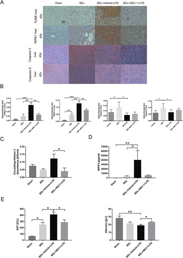
T. Kondo et al.
2
    the integrity of RIPK1 and RIPK3 allowing the formation of a
                                                                                        Table 1. Baseline characteristics stratified by the presence or absence
    multimolecular complex, known as the necrosome. Classically, the
                                                                                        of ACLF at enrollment (derivation cohort).
    necrosome complex formation depends on the interaction
    between RIPK1 and RIPK3 through the RHIM domain upon                                                               No ACLF             ACLF            p values
    phosphorylation at their kinase domains.                                                                           (n = 187)           (n = 124)
       RIPK3 phosphorylation is known to be dependent on both the                      Age (years)                     58 (50–67)          56 (49–64)        0.167
    kinase activity of RIPK1 and its own kinase activity [14]. The final
                                                                                       Male (n, %)                     129 (69.4)          85 (68)           0.804
    step of necrosome formation is RIPK3-mediated phosphorylation
    of the mixed lineage kinase like protein (MLKL); once phosphory-                   Etiology (n, %)
    lated, MLKL undergoes oligomerisation and translocation to the                        Alcohol                      86 (45.9)           70 (56.5)         0.011
    cell membrane, triggering not only the NLRP3 inflammasome [15]                         Alcohol + HCV                64 (34.2)           55 (44.4)         0.339
    but also resulting in cell swelling, rupture and release of damage-
                                                                                          HCV                          50 (26.7)           25 (20.2)         0.316
    associated molecular patterns (DAMPs) such as fragmented
    chromatin (mono- and poly-nucleosomes, core histones). These                          Non-alcoholic                11 (5.9)            6 (4.8)           0.803
    can drive further inflammasome activation leading to death of the                      steatohepatitis
    neighboring cells and can potentially modulate extra hepatic                          Other                        14 (7.5)            7 (5.6)           0.647
    organ failure [16–21].                                                             Active alcoholism at            17 (9.1)            29 (23.4)         0.003
       A number of studies have addressed the role of necroptosis in                   enrollment (n, %)
    various models of acute and chronic liver diseases [22]. Recent                    Ascites (n, %)                  61 (32.6)           83 (66.9)         0.066
    studies suggest a multifaceted role of RIPK3 in multiple causes of
                                                                                       Gastrointestinal                22 (11.8)           14 (14.3)       >0.999
    liver injury [23–25] whereas in the context of high-fat diet induced               bleeding (n, %)
    liver injury, RIPK3 seems to exert a protective role [16]. The role of
    RIPK3-dependent necroptosis is not known in the pathogenesis                       Bacterial infection (n, %)      19 (32.2)           15 (31.9)       >0.999
    of ACLF.                                                                           Organ failure (n, %)
       The aims of this study were to assess whether RIPK1 mediated                       Liver                        14 (7.5)            47 (37.9)
T. Kondo et al.
                                                                                                                                                               3
of ACLF. Inclusions: Cirrhotic patients admitted to the hospital with acute   Control group. Sham operated rats (n = 4) were also included in the
deterioration associated with a precipitating illness. Exclusions: Patients   analyses.
were excluded if they were admitted for reasons other than acute
decompensation of cirrhosis, had evidence of extra-hepatic malignancy or
hepatocellular carcinoma), patients who had undergone major surgery or
                                                                              Carbon tetrachloride (CCl4) plus Galactosamine (GalN) mouse
have unsolved surgical problems and pregnancy. Baseline characteristics       model of ACLF
are displayed in Tables S2 and S3.                                            Animals. For this model C57BL/6 male mice (~30 g b.w; 8–10 weeks-old)
                                                                              were randomly assigned to four groups,
Cohort 3. Samples from healthy volunteers and those with stable cirrhosis
(UCL Biobank Ethical Review Committee approval number NC.2017.16)               a. Vehicle (n = 8): gavaged with olive oil twice weekly for 6 weeks.
were obtained from archived bio-banked material at Royal Free Hospital          b. CCl4 (n = 8): gavaged with carbon tetrachloride (CCl4 0.5 mg/ml in
(London, UK).                                                                      olive oil—dose 0.5 ml/kg) twice weekly for 6 weeks, to induce a
                                                                                   chronic liver injury as described previously.
Cohort 4. Liver sections from patients with alcoholic hepatitis with or         c. CCl4 + GalN (n = 8): Three days after the last dose of CCl4 mice
without ACLF were obtained from the histology department of the Royal              were intraperitoneally injected with GalN 1000 mg/kg (Sigma, UK) in
                                                                                   saline. All animals were sacrificed 48 h after GalN injection under
Free Hospital in London (UCL Biobank Ethical Review Committee approval
                                                                                   general anesthesia.
number NC.2017.10) (baseline characteristics Table S4).
  Definitions for acute decompensation and ACLF are provided in the              d. CCl4 + GalN + RIPA56 (n = 8): RIPA56 (SML2100, Sigma UK) was
supplementary information.                                                         prepared as described for Nec-1. RIPA 56 (3 mg/kg, i.p) was injected
                                                                                   twice daily with the first injection 1 h after GalN injection. All animals
                                                                                   survived the 48 h observational period.
Measurement of human plasma RIPK3
Plasma samples from cohort 1 and 2 were obtained by centrifugation of
human blood at 2000 rpm for 10 min, and storage at −80 °C within 4 h of
collection. RIPK3 levels were measured in duplicate in 1:40 diluted plasma
                                                                              Animal blood and tissue handling, brain water determination
                                                                              Plasma was separated from arterial blood collected in heparin or EDTA
samples using a commercially available enzyme-linked immunosorbent
assay (ELISA) as per manufacturer’s protocol (Human RIPK3 ELISA,              tubes by refrigerated centrifugation (3500 rpm, 10 min) and stored at
CUSABIO, Wuhan, China). After addition of chromogenic substrate,              −80 °C until assessed for biochemistry, and other endopoints. Liver, brain,
absorbance was measured at 450 nm using a plate reader (FLUOstar              and kidney tissues were snap frozen and formalin-fixed for further analysis
Omega, BMG Labtech, UK).                                                      and histological assessment, respectively.
                                                                                 Immediately after termination, the whole brain was removed, and
                                                                              frontal cortex specimens (50 mm2) collected. The brain water content was
Animal studies                                                                calculated using the formula: Brain water (%) = (1−dry weight / wet
Two animal models were used for this study. The sample size was               weight) × 100.
estimated based on previous experiments [10, 11]. Investigators for
analyses were not blinded.
                                                                              Biochemistry and determination of plasma markers
                                                                              Clinical biochemistry was assessed using the COBAS system (COBAS
Bile duct ligation plus Lipopolysaccharide (LPS) rat model of                 Integra 400 plus, Roche Diagnostics).
ACLF                                                                             Plasma RIPK3 and Histone 3 levels were quantified using ELISAs [Rat
Animals. Male Sprague-Dawley rats (Charles River UK, Margate, UK)             RIPK3 ELISA kit (Wuhan Fine Biotech, China), EpiQuick total histone H3
(~300 g b.w., 8–10 weeks-old) underwent either sham operation                 quantification kit (Epigenetek, USA) respectively], according to manufac-
or bile duct ligation (BDL) to induce advanced fibrosis as described           turer protocols. Absorbance was determined using a plate-reader
previously [27]; ACLF was induced by intraperitoneal injection of             (FLUOstar Omega, BMG Labtech, UK).
Klebsiella pneumoniae LPS (Sigma, UK) (0.03 mg/ml in saline) 28 days
after BDL.                                                                    Caspase assays
                                                                              Caspase 3/7 and 8 activities were measured in liver extracts using Caspase-
Necrostatin 1. Nec1 (Calbiochem, Sigma Aldrich, UK) was prepared as           Glo assay kits (Promega) according to a modified protocol [28]. Diluted
follows. Briefly, Nec-1 was redissolved in N-2 methyl-pyrrolidone (NMP) to     lysates (10 μg/ml) were mixed with an equal volume of Caspase-Glo
make a 200 mg/ml stock solution further diluted to an intermediate            reagents (100 μl each) in white 96-well plate and incubated at room
concentration of 40 mg/ml in NMP. The 40 mg/ml solution was finally            temperature for 1 h. Luminescence was measured using a luminometer
diluted to a working concentration of 1 mg/ml in a solution of 30% (2-        (FLUOstar Omega, BMG Labtech, UK).
hydroxypropyl) β-cyclodextrin (Sigma, Cat No H107)/0.5% Citric acid.
Vehicle controls consisted of NMP and 30%(2-hydroxypropyl)
β-cyclodextrin/0.5% Citric acid (1:40 ratio).                                 Immunohistochemistry
                                                                              Expression of RIPK1, RIPK3, and pMLKL were evaluated by immunohis-
Study design. Animals underwent BDL surgery and were given 4 weeks to         tochemistry in liver sections of patients with alcoholic hepatitis with and
develop advanced fibrosis and portal hypertension [27]. The number per         without ACLF (Table S4). Histology of rodent model was performed using
treatment group was determined based on liver function test with two-         liver and kidney sections. The paraffin-embedded tissue sections were
tailed tests with type I and II errors set at 0.05 and 0.20, respectively.    deparaffinized using xylene and ethanol and endogenous peroxidase
                                                                              activity blocked in 0.3% H2O2 in water. After antigen retrieval by
Bile-duct ligation group. The animals were assigned randomly into 3           microwave, slides were blocked in 2.5% normal horse serum and then
groups 4-weeks after bile-duct ligation.                                      stained with following primary antibody (complete list of primary
                                                                              antibodies Table S5). Immunoperoxidase staining was completed using
  a. BDL + Vehicle (n = 4): BDL rats received an injection of intraper-       RTU Vectastatin universal Elite ABC kit (Vector Laboratories, UK), and 3.3′-
     itoneal (i.p.) vehicle control and were euthanised 6 h later under       diaminobenzidine was used to detect the target proteins. For negative
     general anesthesia.                                                      controls primary antibody was replaced with PBS and a rabbit IgG antibody
                                                                              was used as isotype controls (Table S5). Respective images are shown in
  b. BDL + Vehicle + LPS (n = 9): BDL rats received an i.p. injection of
                                                                              Fig. S2. RIPK3 antibody validation was performed by using a blocking
     vehicle control followed an i.p. injection of LPS (0.025 mg/kg), 3 h
     later. Animals were euthanised 3 h after LPS injection (or whenever      peptide as shown in Fig. S3.
     comatose).
  c. BDL + Nec-1 + LPS (n = 8): This group of BDL rats received an i.p.       In situ detection of cell death by the TUNEL assay
     injection of 3.3 mg/kg Nec-1 followed, by an i.p injection of LPS        Terminal deoxynucleotidyl transferase biotin-dUTP nick end labeling
     (0.025 mg/kg), 3 h later. Animals were euthanised 3 h after LPS.         (TUNEL) staining of deparaffinised, proteinase K-treated liver and kidney

Cell Death and Disease (2022)13:5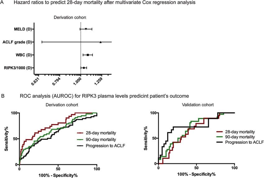
T. Kondo et al.
4
    sections was performed using the In-Situ Cell Death Detection kit, POD
    (Roche, UK) as per manufacturer’s protocol.                                   Table 2. Biomarkers stratified by patient group according to the PIRO
      Specimens were imaged with a Carl Zeiss Axio Scope. A1 microscope           concept (baseline—derivation cohort).
    equipment with N-Achroplan 10x/0.25 Ph1 and 40x/0.65 Ph2 objectives                                                RIPK3 (pg/ml)                p value
    and Axio Cam Mrc5 (Carl Zeiss Inc., Germany). Quantification of positive                                            Median (IQR)
    areas was assessed by measuring optical density or relative DAB positive
                                                                                 Disease severity
    areas (%) as appropriate with FIJI Image J software [29, 30].
                                                                                   Healthy controls (n = 21)           322 (136–493)
                                                                                   Stable cirrhosis (n = 42)           445 (320–565)
    Statistical analysis
    Human data are expressed as the mean ± standard deviation for normally         All decompensated (n = 311)         7816 (4500–140,905)
T. Kondo et al.
5
Fig. 1 RIPK3 plasma levels derivation cohort. A RIPK3 levels in patients with cirrhosis and acute decompensation from the derivation cohort
according the severity of liver disease, survival status, inflammation, type of organ failure, and other predisposition factors. Data were analyzed
using Student’s t test, Mann–Whitney U test or Kruskal–Wallis test followed by Fisher’s Least Significant Difference test as appropriate. *p <
0.05, **p < 0.01, ***p < 0.001. B Circulating RIPK3 levels showed a good correlation with markers of cell death such as K18 and cK18 as well as
markers of inflammation such as sCD163, IL6, IL8, IL1ra Markers were taken from previous analyses [K18 and cK18—MacDonald S et al. [9];
sCD163, IL6, IL8, IL1ra—Claria J et al. [5]] on the CANONIC cohort. Correlations were calculated as Pearson correlation coefficient.
associated with elevated levels of plasma markers of RIPK3 in Additional markers of cell death were also increased in
comparison to no ACLF (Tables S8 and S9, Fig. S4). The patients with ACLF. In particular, levels of cK18, K18 and
discrimination power (AUROC) of RIPK3 to predict survival was circulating nucleosomes were significantly raised in a step-wise
0.653 (95% CI, 0.530–0.776) and 0.696 (95% CI, 0.593–0.799) at 28 manner with increasing ACLF grade (Tables S8 and S9).
and 90 days, respectively (Table S6, Fig. 2B). Interestingly, as K18 increased more steeply than the cK18 with
Cell Death and Disease (2022)13:5T. Kondo et al.
6
Fig. 2 Multivariate COX-regression and ROC analysis. These figures delineate the prognostic relevance of RIPK3 levels. A Multivariate Cox-
regression analysis in 311 patients with acute decompensation (derivation cohort) considered age, MELD score, WBC, ACLF grade and RIPK3
levels as confounders for 28-day mortality. RIPK3 levels (HR 1.035; 95% CI 1.002–1.068; p = 0.037) and WBC (HR 1.081; 95% CI 1-026
1.026–1.138; p = 0.003) remained as independent predictors. B Thereafter, the predictive power (AUROC) of RIPK3 to predict 28-day and 90-
day mortality and progression to ACLF was calculated by ROC analysis in the derivation and validation cohorts. The AUROC of RIPK3 to predict
progression to ACLF was 0.650 (95% CI 0.562–0.738) in the derivation cohort and 0.744 (95% CI 0.595–0.892) in the validation cohort. The
discrimination power (AUROC) of RIPK3 to predict survival was 0.768 (95% CI, 0.679–0.858) and 0.676 (95% CI, 0.595–0.758) at 28 and 90 days
in the derivation cohort and 0.653 (95% CI, 0.530–0.776) and 0.696 (95% CI, 0.593–0.799) in the validation cohort, respectively.
Fig. 3 Immunohistochemistry human liver tissue. Representative images of liver biopsies of patients with alcoholic hepatitis with and
without ACLF stained for RIPK1, RIPK3, and pMLKL showing increased staining in the ACLF patients. Group comparisons were performed by
Mann–Whitney U test. *p < 0.05, **p < 0.01, ***p < 0.001.
increasing clinical severity, the cK18:K18 ratio also dropped [from Immunohistochemistry: RIPK1, RIPK3, and pMLKL
0.89 (0.58–2.50) in no ACLF, to 0.71 (0.50–2.44) in ACLF 1, and to In order to determine whether the circulating RIPK3 was
0.53 (0.34–1.91) in ACLF 2/3 (p = 0.081), respectively] suggesting, originating from the liver and reflected hepatic necroptosis,
as previously reported, that non-apoptotic rather than apoptotic biopsies from patients with AD with and without ACLF were
modes of cell death are relatively more important in the stained for RIPK1, RIPK3, and phosphorylated MLKL (pMLKL) (Table
pathophysiology of ACLF (Tables S8 and S9). S4, Fig. 3). Although RIPK1, RIPK3, and pMLKL were expressed in
Cell Death and Disease (2022)13:5T. Kondo et al.
7
biopsies of AD patients, this was considerably upregulated in
patients with ACLF and additional immuno-staining confirmed low
expression of cleaved caspase 3 and 8 (Fig. S5). The presence of
positive staining of all key components of the necroptotic
pathway and in particular the presence of the necrosome-
phosphorylated form of MLKL responsible for cellular death,
suggests that hepatic necroptosis is an important feature of
human ACLF.
The rodent model of ACLF recapitulates RIPK1 associated cell
death in human ACLF
In keeping with our previous studies [9], rodents who had
undergone bile-duct ligation (BDL, model of advanced fibrosis)
demonstrated significant alterations in liver function tests compared
with sham-operated controls (Table S10) but did not demonstrate
significant elevation in circulating nucleosomes or RIPK3 when
compared to sham-operated animals (Fig. S6). Immunohistochem-
istry of liver and kidney tissues from BDL animals demonstrated Fig. 4 Caspase activity liver tissue. LPS induces a switch from
positive staining for RIPK1 and RIPK3 in comparison to sham animals Caspase-dependent cell death to necroptosis. Caspase 8 and 3/7
(Fig. S6). Induction of ACLF in BDL animals by administration of LPS activities in liver tissue of sham, BDL, BDL + LPS and BDL + NEC-1 +
led to significant worsening of liver function tests [alanine LPS. Data are presented as box and whisker plots. Data were
analyzed using Student’s t test or Mann–Whitney U test. *p < 0.05,
aminotransferase (ALT), 79.4 ± 12.8 IU/L (mean ± SD); aspartate **p < 0.01, ***p < 0.001.
transaminase (AST), 412.9 ± 39.4 IU/L; p = 0.031 compared to BDL]
mirrored by increased circulating nucleosomes. In the BDL + LPS is causally related to the pathogenesis of ACLF, we explored whether
animals, a dramatic significant increase in plasma RIPK3 levels was treatment with NEC-1would prevent ACLF development.
also observed compared to BDL (p = 0.042) and this was NEC-1 treatment significantly decreased the expression of RIPK1
accompanied by increased protein expression of both RIPK1 and in NEC-1-treated “BDL + LPS” group compared to the “BDL +
RIPK3 in liver and kidney tissues (Fig. S6). These data strongly vehicle+LPS” group (p = 0.009) (Fig. 5A). Moreover, TUNEL
demonstrate that the onset of ACLF triggered by LPS administration staining showed a significant reduction in total cell death in the
to the BDL animals is associated with initiation of necroptosis within livers of the BDL + NEC-1+LPS compared to the BDL + Vehicle+LPS
3 h, confirming the findings in the ACLF patients. group (p = 0.001) (Fig. 5B), mirrored by a significant reduction in
circulating histone 3 levels in the NEC-1 treated group, compared
LPS induces a switch from caspase-dependent cell death to to BDL + Vehicle+LPS animals (p = 0.048) (Fig. 5C). Immunohis-
RIPK1 mediated cell death tochemistry on liver tissue after BDL + Vehicle+LPS showed a
To further explore the switch from predominantly apoptotic cell- reduction of cleaved caspase 3 and 8 after LPS injection (p = 0.05
death in cirrhosis to necroptosis in ACLF, we evaluated the relative for both), consistent with the reduced activities shown above, and
liver Caspases-3/7 and 8 activities in the animals described above. tended to increase after NEC-1 treatment (p < 0.001 for both)
Caspase-8 is a key regulator of the two forms of cell death (Fig. 5A, B) which mirrors the enzyme activity shown on Fig. 4. In
(apoptosis and necroptosis). Activation of caspase-8 (initiator order to further evaluate whether inhibition of RIPK1 could
caspase) promotes apoptosis through caspases-3/7 (executioner account for the observed attenuation of total cell death,
caspases). On the other hand, suppression of caspase-8 activity we measured circulating levels of RIPK3. Plasma RIPK3 levels in
shifts the balance towards necroptosis. Therefore, we investigated the BDL + NEC-1+LPS group reduced markedly compared to the
the activation pattern of both caspase-8 and caspase-3/7 in liver BDL + Vehicle+LPS (p = 0.073) (Fig. 5D). Furthermore, liver
homogenates. BDL livers showed a significant increase in caspase- preservation by NEC-1 was also demonstrated by significantly
3/7 activation compared to control, with a median activation of lower values of AST (p = 0.037) and a significantly higher albumin
2492 RLU (IQR, 1273–2988) and 396 RLU (IQR, 279–630), levels (p = 0.017) (Fig. 5E).
respectively (p = 0.0087). In contrast, caspase-8 activation did We then evaluated whether the preservation of liver function
not differ between the two groups with a median activation of induced by NEC-1 pre-treatment was accompanied by ameliora-
234.5 (IQR, 187.5–255.3) and 202 (IQR, 163.8–239.5), respectively tion of kidney and brain dysfunction. TUNEL staining showed a
(p = 0.425). In the “BDL + Vehicle+LPS” group a significant significant reduction in cell death in kidney sections of BDL + NEC-
reduction of all caspase activities was observed compared to 1+LPS group compared to BDL + vehicle+LPS (p = 0.002) (Fig.
BDL (caspase-3/7 [RLU] median 968 RLU (IQR 571-1495), p = 0.035 S7), which was associated with a significant reduction in creatinine
compared to BDL; caspase-8, median 164 RLU (IQR 128-182.5), p = (p = 0.020) and urea levels (p = 0.012) in BDL + NEC-1+LPS
0.021 compared to BDL]. NEC-1, inhibiting RIPK1-dependent compared to BDL + Vehicle+LPS (Fig. S7), demonstrating that
necroptosis, restored an apoptotic phenotype as demonstrated prevention of necroptosis by NEC-1 treatment protects also from
by caspases-3/7 and caspase-8 activation, similar to that observed kidney injury. Furthermore, a reduction in brain water content,
in the BDL group without LPS [caspase-3/7, median 2857 RLU (IQR although not statistically significant, was observed in the BDL +
1640-3890); caspase-8, median 326.5 (IQR 313.5-414.3), p = 0.626 NEC-1+LPS treated group compared to BDL + Vehicle+LPS (p =
and p = 0.020, compared to BDL, respectively] (Fig. 4). 0.100) (Fig. S7).
NEC-1 treatment prevents the occurrence of ACLF in the RIPK1 inhibition by RIPA56 prevents RIPK1/RIPK3 mediated
rodent ACLF model necroptosis in a CCl4-GalN mouse model
As increased liver expression of both RIPK1 and RIPK3 and elevated In order to validate the results of the experiments in the BDL
levels of TNFα [31–33] are associated with onset of the ACLF animals, we used a second model of ACLF and a more specific
phenotype in both humans and rodents, a canonical TNF-mediated inhibitor of RIPK1, RIPA56. In this mouse model, the chronic liver
RIPK1-RIPK3-dependent necroptosis might account for increased cell injury was induced by 6-weeks carbon tetrachloride (CCl4) gavage
death and organ failure in ACLF. To determine whether necroptosis followed by Galactosamine (GalN) injection as the second hit. This
Cell Death and Disease (2022)13:5T. Kondo et al.
8
Fig. 5 Effect of RIPK1 inhibition by Necrostatin 1. Histological assessment and biomarkers show the protective effect of pharmacological
RIPK1 inhibition against ACLF-related liver injury. A Representative images of liver tissue of sham, BDL, BDL + LPS and BDL + NEC-1 + LPS
stained for TUNEL, RIPK1, cleaved-Caspase 3 and 8. B The optical density of immunostaining intensity (mean ± SEM) was assessed by Image J
(mean ± SEM). D Plasma levels of AST and albumin of sham, BDL, BDL + LPS, and BDL + NEC-1 + LPS (mean ± SEM). C–E Plasma levels of ALT,
albumin, RIPK3 and histone 3 of sham, BDL, BDL + LPS and BDL + NEC-1 + LPS (mean ± SEM). Data were analyzed using Student’s t test or
Mann–Whitney U test. *p < 0.05, **p < 0.01, ***p < 0.001.
Cell Death and Disease (2022)13:5T. Kondo et al.
9
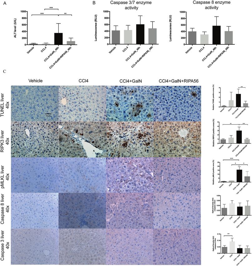
Fig. 6 Effect of RIPK1 inhibition by RIPA56 in a murine model of ACLF. C57BL/6 mice were treated with CCl4 for 6 weeks and subsequently
injected with GalN. All animals were sacrificed 48 h after GalN injection. A GalN injection was associated with a significant increase of ALT
levels. B Caspase 3/7 and 8 expression showed a mixed response without proving significance. C TUNEL staining of liver tissue showed that
GalN injection induced hepatocyte cell death with high expression of RIPK3 (magnification, 10x and 40x). RIPK1 inhibition by RIPA
56 significantly reduced ALT levels, abrogated tissue injury (TUNEL staining) and reduced the liver RIPK3, pMLK, cleaved-Caspase 3 and 8
expression. The optical density of immunostaining intensity (mean ± SEM) was measured. Data were analyzed using Student’s t test or
Mann–Whitney U test. Only significant p values were displayed *p < 0.05, **p < 0.01, ***p < 0.001.
model was designed to simulate ACLF that occurs due to a direct this model. RIPK1 inhibition by RIPA56 (selectivity shown by Ren Y
liver injury (unlike LPS) and produces predominant liver failure. ALT et al. [26]) significantly abrogated the tissue injury (ALT level p =
levels increased from 64.8 ± 54.2 U/L in CCl4 only treated animals to 0.006, TUNEL stain p < 0.001) and liver RIPK3 (p < 0.001) and pMLK
353.5 ± 284.2 U/L in the CCl4-GalN animals (p < 0.001) (Fig. 6A) and expression (p < 0.01) suggesting that ACLF is associated with RIPK1/
TUNEL staining of liver tissue demonstrated a significant hepatocyte RIPK3 mediated necroptotic cell death in the liver (Fig. 6B).
cell death after injection of GalN (p < 0.001) (Fig. 6B). RIPK3
immunohistochemistry showed that hepatocyte cell death was
associated with TUNEL positivity, increased RIPK3 (p < 0.01) and DISCUSSION
pMLK expression (p < 0.01) and decreased caspase 3 and 8 This study describes for the first time that plasma RIPK3 levels are
expression (Fig. 6B) principally defining a necroptotic cell death in associated with the progression of patients with no ACLF to ACLF,
Cell Death and Disease (2022)13:5T. Kondo et al.
10
severity of ACLF and mortality in two separate clinical cohorts on the potential role of necroptosis in ACLF can be translated into
suggesting that RIPK1 mediated cell death is an important clinical application as a therapeutic, needs further evaluation as
mechanism of cell death in the patients with ACLF. This was targeting RIPK3-mediated necroptosis was effective in preventing
associated with markedly increased expression of RIPK1, RIPK3, APAP induced acute liver failure in some studies [23, 39, 40] but
and pMLKL in liver tissues confirming the importance of this not in others [41]. Of concern, was the observation that in
pathway in mediating hepatocyte injury in these patients. The concanavalin A (ConA)-induced autoimmune hepatitis, RIPK3
important role of RIPK1 mediated cell death in ACLF was deletion was protective, whereas RIPK1 inhibition exacerbated
demonstrated in two animal models of ACLF. Administration of disease, accelerated animal death, and was associated with
two separate inhibitors of RIPK1, NEC-1 or RIPA56, resulted in the increased hepatocyte cell death [40]. However, other data
prevention of development of ACLF in the animal models. Taken suggested that pre-treatment with NEC-1 had protective effect
together these data point to the importance of necroptosis as against ConA mediated injury [42–44]. The mechanism underlying
being important in the pathogenesis of ACLF. these variable effects are unclear but may be related to the stage
We have previously reported the importance of onset of non- of the disease and potential activation of alternative pathways,
apoptotic cell death in patients with ACLF [9]. The significant particularly in the animals with RIPK3 deletion.
increase in the circulating levels of RIPK3 in patients with ACLF Circulating nucleosomes, a DAMP [45], also reflected the
supports the notion that RIPK1 mediated cell death is likely to be severity of ACLF. In addition, our study showed that circulating
the operative mechanism. RIPK3 is a key component in the nucleosomes were associated with brain, heart, and respiratory
necroptotic pathway alongside RIPK1 and pMLKL. It is also failure. Histones, components of nucleosomes released into the
associated with mitochondrial energy metabolism and production circulation from damaged cells, are known to promote inflamma-
of excess reactive oxygen species [20]. Circulating RIPK3 levels not tion [46]. Our results are in concert with the finding that
only reflected the severity of ACLF in the present study but also circulating nucleosomes are also elevated in other clinical
identified patients likely to progress to develop ACLF and patients conditions associated with necrosis such as severe acute
likely to die at 28-day and 90-day. These data were validated in a pancreatitis [47] and circulating histones are elevated in acute
second cohort although the AUROC for 28-day mortality was liver failure [46]. Histones released after tissue trauma may lead to
lower possibly due to the small sample size and different timing of multiple organ injury and death [48]. Several studies suggest that
sampling. Importantly RIPK3 levels correlated closely with markers histones are both biomarkers of disease progression and also
of systemic inflammation, which have been shown to be the possible therapeutic targets in sepsis, acute liver failure, and other
central in the mechanism underlying the development of ACLF inflammatory diseases [46–49]. In the rodent model of ACLF, NEC-
[31]. In order to determine whether this was simply an association 1 also prevented the LPS-induced liver damage and the
or whether RIPK1 mediated cell death was pathophysiologically consequent release of histone-3, which may have partially
important in the development of ACLF, we designed a study to contributed to its protective effect.
determine whether inhibition of the key upstream necroptosis Acute kidney injury is an important feature of ACLF [50] and
component RIPK1 using NEC-1 and RIPA56 would prevent the tubular cell death is an important pathophysiological character-
development of ACLF in rodent models. istic of this syndrome [51]. In the present study, we also showed
The most important observation in this study was that NEC-1, that renal dysfunction was associated with tubular injury.
an inhibitor of RIPK1, prevented hepatic and renal cell death in a Interestingly, the expression of RIPK1 and RIPK3 in the kidney of
BDL model of ACLF induced by injection of LPS, providing the first the animal model of ACLF was significantly increased. Previous
proof of the importance of this pathway in the pathogenesis of studies have also reported that plasma RIPK3 levels are elevated in
ACLF from AD. This model, extensively validated previously, patients with AKI following trauma [52]. Pharmacological inhibi-
mimics the clinical situation where ACLF is precipitated by tion of RIPK1 with NEC-1 protected against renal tubular cell death
bacterial infection and exhibits cerebral, circulatory and renal in our BDL model of ACLF suggesting that AKI in ACLF is due to
failure, all important extrahepatic diagnostic features of ACLF. The renal tubular necroptosis. Alternatively, or in addition, AKI in ACLF
observed over-expression of RIPK1 in the liver and kidney in the may be consequent on release of DAMPs from liver injury. The
animal model of ACLF was associated with evidence of cell death. study also showed that the brain water of the BDL animals with
It is intriguing to note that renal tubular injury was also prevented ACLF was markedly reduced in the NEC-1 treated group and this
with NEC-1 in this model suggesting that necroptosis may also be was independent of changes in the ammonia levels. The
the operative mechanism of kidney injury. In order to determine mechanism of protection of the brain by NEC-1 is not clear but
whether the RIPK1 mediated cell death was also important in a may be consequent on protection of liver cell death and DAMP
‘hepatic’ form of ACLF where liver injury predominates, a new release.
model of CCL4-GalN was developed. Interestingly, this model There are some limitations of our study. Firstly, the mechanism
develops severe liver injury but no extrahepatic organ failures, and source of the circulating RIPK3 is unclear. However, when
such as kidney and brain failure. In this second model RIPA56, comparing no organ failure and single organ failure, we observed
another inhibitor of RIPK1, was used as recently described to be that the elevated plasma RIPK3 levels were associated with single
more selective, potent and with no off-target effect compared to liver failure. In addition, histological assessment showed high
NEC-1, and its administration also abrogated severity of liver injury expression of RIPK3 in the liver of ACLF patients and also in the
significantly [14]. Taken together, the data suggest that necrop- animal model. The elevation of plasma RIPK3 is therefore likely to
tosis is likely to be pathophysiologically important in the be predominantly derived from the liver but could also be
pathogenesis of ACLF and RIPK1 is a potential therapeutic target. originating from the kidney as evidence of necroptosis was also
Normalizing RIPK1 is known to modulate the NF-κβ signaling observed in the rodent kidneys and the levels were also found to
pathway [34], which can promote cell survival thereby preventing be elevated in the patients with single kidney failure in the
development of ACLF [35]. An alternative mechanism of the patients in the CANONIC study. Secondly, NEC-1 has a short half-
protective effect of inhibition of RIPK1 may by through modula- life of about 1 h [53] and is known to have off-target effects, such
tion of necroptosis that is triggered in BDL animals by induction of as inhibition of indoleamne-2,3-dioxygenase (IDO), which can
senescence [36–38]. In addition, we showed that Caspase 8 modulate immune function [54] and this additional feature may
activity in ACLF rodent model (BDL + LPS) was suppressed have partially contributed to its observed effects. However,
suggesting a possible switch from apoptosis to necroptosis. administration of RIPA56, a specific inhibitor of RIPK1 with no
Interestingly, NEC-1 prevented loss of Caspase 8 activity and onset anti-IDO activity, had a similar protective effect suggesting that
of LPS-induced necroptosis. However, whether these observations necroptosis is indeed important in the development of ACLF and a
Cell Death and Disease (2022)13:5T. Kondo et al.
11
potential therapeutic target. We considered using a RIPK3 deletion 16. Roychowdhury S, McCullough RL, Sanz-Garcia C, Saikia P, Alkhouri N, Matloob A,
mouse but the fact that these animals develop variable degrees of et al. Receptor interacting protein 3 protects mice from high-fat diet-induced
liver injury and fibrosis and makes it an unsuitable model of liver injury. Hepatology. 2016;64:1518–33. https://doi.org/10.1002/hep.28676
cirrhosis and ACLF [16]. 17. Wu W, Liu P, Li J. Necroptosis: an emerging form of programmed cell death. Crit Rev
Oncol Hematol. 2012;82:249–58. https://doi.org/10.1016/j.critrevonc.2011.08.004
In conclusion, the novel observation that inhibition of RIPK1
18. Zhang DW, Shao J, Lin J, Zhang N, Lu BJ, Lin SC, et al. RIP3, an energy metabolism
with NEC-1 and RIPA56 resulted in prevention of occurrence of regulator that switches TNF-induced cell death from apoptosis to necrosis. Sci-
ACLF in the two animal models suggests that necroptosis is likely ence. 2009;325:332–6. https://doi.org/10.1126/science.1172308
to be of pathogenic importance and a potential therapeutic target 19. Linkermann A, Green DR. Necroptosis. N. Engl J Med. 2014;370:455–65. https://
to prevent the development of ACLF. This observation is doi.org/10.1056/NEJMra1310050
supported by data from two patient cohorts in whom elevated 20. Silke J, Rickard JA, Gerlic M. The diverse role of RIP kinases in necroptosis and
circulating levels of RIPK3 was associated with progression of inflammation. Nat Immunol. 2015;16:689–97. https://doi.org/10.1038/ni.3206
patients from AD to ACLF, severity of ACLF, and short-term 21. Wang H, Sun L, Su L, Rizo J, Liu L, Wang LF, et al. Mixed lineage kinase
mortality. Taken together, the study suggests that circulating domain-like protein MLKL causes necrotic membrane disruption upon
phosphorylation by RIP3. Mol Cell. 2014;54:133–46. https://doi.org/10.1016/j.
RIPK3 may serve as a potential biomarker to select patients for
molcel.2014.03.003
therapy with drugs targeting RIPK1 mediated cell death. 22. Dara L, Liu ZX, Kaplowitz N. Questions and controversies: the role of necroptosis
in liver disease. Cell Death Disco. 2016;2:16089 https://doi.org/10.1038/
cddiscovery.2016.89
DATA AVAILABILITY 23. Ramachandran A, McGill MR, Xie Y, Ni HM, Ding WX, Jaeschke H. Receptor
All data is available on request from the corresponding author. interacting protein kinase 3 is a critical early mediator of acetaminophen-induced
hepatocyte necrosis in mice. Hepatology. 2013;58:2099–108. https://doi.org/
10.1002/hep.26547
REFERENCES 24. Roychowdhury S, McMullen MR, Pisano SG, Liu X, Nagy LE. Absence of receptor
1. Jalan R, Williams R. Acute-on-chronic liver failure: pathophysiological basis of interacting protein kinase 3 prevents ethanol-induced liver injury. Hepatology.
therapeutic options. Blood Purif. 2002;20:252–61. https://doi.org/10.1159/ 2013;57:1773–83. https://doi.org/10.1002/hep.26200
000047017 25. Afonso MB, Rodrigues PM, Carvalho T, Caridade M, Borralho P, Cortez-Pinto H,
2. Jalan R, Gines P, Olson JC, Mookerjee RP, Moreau R, Garcia-Tsao G, et al. Acute-on et al. Necroptosis is a key pathogenic event in human and experimental murine
chronic liver failure. J Hepatol. 2012;57:1336–48. https://doi.org/10.1016/j. models of non-alcoholic steatohepatitis. Clin Sci (Lond). 2015;129:721–39.
jhep.2012.06.026 26. Ren Y, Su Y, Sun L, He S, Meng L, Liao D, et al. Discovery of a highly potent,
3. Bernardi M, Moreau R, Angeli P, Schnabl B, Arroyo V. Mechanisms of decom- selective, and metabolically stable inhibitor of Receptor-Interacting Protein 1
pensation and organ failure in cirrhosis: From peripheral arterial vasodilation to (RIP1) for the treatment of systemic inflammatory response syndrome. J Med
systemic inflammation hypothesis. J Hepatol. 2015;63:1272–84. https://doi.org/ Chem. 2017;60:972–86.
10.1016/j.jhep.2015.07.004 27. Harry D, Anand R, Holt S, Davies S, Marley R, Fernando B, et al. Increased sensi-
4. Moreau R. The pathogenesis of ACLF: the inflammatory response and immune tivity to endotoxemia in the bile duct-ligated cirrhotic rat. Hepatology.
function. Semin Liver Dis. 2016;36:133–40. https://doi.org/10.1055/s-0036-1583199 1999;30:1198–205.
5. Clària J, Stauber RE, Coenraad MJ, Moreau R, Jalan R, Pavesi M, et al. Systemic 28. Liu D, Li C, Chen Y, Burnett C, Liu XY, Downs S, et al. Nuclear import of proin-
inflammation in decompensated cirrhosis: characterization and role in acute-on- flammatory transcription factors is required for massive liver apoptosis induced
chronic liver failure. Hepatology. 2016;64:1249–64. https://doi.org/10.1002/ by bacterial lipopolysaccharide. J Biol Chem. 2004;279:48434–42.
hep.28740 29. Schneider CA, Rasband WS, Eliceiri KW. NIH image to ImageJ: 25 years of image
6. Moreau R, Jalan R, Gines P, Pavesi M, Angeli P, Cordoba J, et al. Acute-on-chronic analysis. Nat Methods. 2012;9:671–5. https://doi.org/10.1038/nmeth.2089
liver failure is a distinct syndrome that develops in patients with acute decom- 30. Schindelin J, Arganda-Carreras I, Frise E, Kaynig V, Longair M, Pietzsch T, et al.
pensation of cirrhosis. Gastroenterology. 2013;144:1426–37. https://doi.org/ Fiji: an open-source platform for biological-image analysis. Nat Methods.
10.1053/j.gastro.2013.02.042. 1437 e1421-1429 2012;9:676–82. https://doi.org/10.1038/nmeth.2019
7. Angeli P, Ginès P, Wong F, Bernardi M, Boyer TD, Gerbes A, et al. Diagnosis and 31. Claria J, Arroyo V, Moreau R. The acute-on-chronic liver failure syndrome, or when
management of acute kidney injury in patients with cirrhosis: revised consensus the innate immune system goes astray. J Immunol. 2016;197:3755–61. https://doi.
recommendations of the International Club of Ascites. J Hepatol. 2015;62:968–74. org/10.4049/jimmunol.1600818
https://doi.org/10.1016/j.jhep.2014.12.029 32. Balasubramaniyan V, Kumar Dhar D, Warner AE, Li W-YV, Amiri AF, Bright B,
8. Jalan R, Stadlbauer V, Sen S, Cheshire L, Chang YM, Mookerjee RP. Role of pre- et al. Importance of Connexin-43 based gap junction in cirrhosis and acute-on-
disposition, injury, response and organ failure in the prognosis of patients with chronic liver failure. J Hepatol. 2013;58:1194–1200. https://doi.org/10.1016/j.
acute-on-chronic liver failure: a prospective cohort study. Crit Care. 2012;16:R227 jhep.2013.01.023
https://doi.org/10.1186/cc11882 33. Li F, Miao L, Sun H, Zhang Y, Bao X, Zhang D. Establishment of a new acute-on-
9. Macdonald S, Andreola F, Bachtiger P, Amoros A, Pavesi M, Mookerjee R, et al. chronic liver failure model. Acta Pharm Sin B. 2017;7:326–33. https://doi.org/
Cell death markers in patients with cirrhosis and acute decompensation. Hepa- 10.1016/j.apsb.2016.09.003
tology. 2018;67:989–1002. https://doi.org/10.1002/hep.29581 34. Christofferson DE, Yuan J. Necroptosis as an alternative form of programmed cell
10. Engelmann C, Adebayo D, Oria M, De Chiara F, Novelli S, Habtesion A, et al. death. Curr Opin Cell Biol. 2010;22:263–8. https://doi.org/10.1016/j.
Recombinant alkaline phosphatase prevents acute on chronic liver failure. Sci ceb.2009.12.003
Rep. 2020;10:389 https://doi.org/10.1038/s41598-019-57284-z 35. Chen S, Lv X, Hu B, Shao Z, Wang B, Ma K, et al. RIPK1/RIPK3/MLKL-mediated
11. Engelmann C, Sheikh M, Sharma S, Kondo T, Loeffler-Wirth H, Zheng YB, et al. necroptosis contributes to compression-induced rat nucleus pulposus cells
Toll-like receptor 4 is a therapeutic target for prevention and treatment of liver death. Apoptosis. 2017;22:626–38. https://doi.org/10.1007/s10495-017-1358-2
failure. J Hepatol. 2020;73:102–12. 36. Aravinthan A, Pietrosi G, Hoare M, Jupp J, Marshall A, Verrill C, et al. Hepatocyte
12. Engelmann C, Claria J, Szabo G, Bosch J, Bernardi M. Pathophysiology of expression of the senescence marker p21 is linked to fibrosis and an adverse
decompensated cirrhosis: portal hypertension, circulatory dysfunction, inflam- liver-related outcome in alcohol-related liver disease. PLoS ONE. 2013;8:e72904
mation, metabolism and mitochondrial dysfunction. J Hepatol. 2021;75:S49–S66. https://doi.org/10.1371/journal.pone.0072904
https://doi.org/10.1016/j.jhep.2021.01.002. Suppl 1 37. Aravinthan A, Scarpini C, Tachtatzis P, Verma S, Penrhyn-Lowe S, Harvey R,
13. Kaczmarek A, Vandenabeele P, Krysko DV. Necroptosis: the release of damage- et al. Hepatocyte senescence predicts progression in non-alcohol-related
associated molecular patterns and its physiological relevance. Immunity. fatty liver disease. J Hepatol. 2013;58:549–56. https://doi.org/10.1016/j.
2013;38:209–23. https://doi.org/10.1016/j.immuni.2013.02.003 jhep.2012.10.031
14. Majdi A, Aoudjehane L, Ratziu V, Islam T, Afonso MB, Conti F, et al. Inhibition of 38. Wan J, Benkdane M, Alons E, Lotersztajn S, Pavoine C. M2 kupffer cells promote
receptor-interacting protein kinase 1 improves experimental non-alcoholic hepatocyte senescence: an IL-6-dependent protective mechanism against alco-
fatty liver disease. J Hepatol. 2020;72:627–35. https://doi.org/10.1016/j. holic liver disease. Am J Pathol. 2014;184:1763–72. https://doi.org/10.1016/j.
jhep.2019.11.008 ajpath.2014.02.014
15. Kang TB, Yang SH, Toth B, Kovalenko A, Wallach D. Caspase-8 blocks kinase 39. An J, Mehrhof F, Harms C, Lättig-Tünnemann G, Lee SLL, Endres M, et al. ARC is a
RIPK3-mediated activation of the NLRP3 inflammasome. Immunity. novel therapeutic approach against acetaminophen-induced hepatocellular
2013;38:27–40. https://doi.org/10.1016/j.immuni.2012.09.015 necrosis. J Hepatol. 2013;58:297–305. https://doi.org/10.1016/j.jhep.2012.10.002
Cell Death and Disease (2022)13:5T. Kondo et al.
12
40. Deutsch M, Graffeo CS, Rokosh R, Pansari M, Ochi A, Levie EM, et al. Divergent The German Research Foundation (DFG) funded C.E. (EN 1100/2-1). We would like to
effects of RIP1 or RIP3 blockade in murine models of acute liver injury. Cell Death thank Andrew Hall for helping with the additional experiments.
Dis. 2015;6:e1759 https://doi.org/10.1038/cddis.2015.126
41. Dara L, Johnson H, Suda J, Win S, Gaarde W, Han D, et al. Receptor interacting
protein kinase 1 mediates murine acetaminophen toxicity independent of the AUTHOR CONTRIBUTIONS
necrosome and not through necroptosis. Hepatology. 2015;62:1847–57. https:// The corresponding author certifies that all listed authors participated meaningfully in
doi.org/10.1002/hep.27939 the study and that they have seen and approved the final manuscript. T.K.
42. Jouan-Lanhouet S, Arshad MI, Piquet-Pellorce C, Martin-Chouly C, Moigne-Muller contributed to study design, data collection and analysis, experiments, and drafted
GL, Van Herreweghe F, et al. TRAIL induces necroptosis involving RIPK1/RIPK3- the paper. S.M. contributed to study design, data collection, analysis, and
dependent PARP-1 activation. Cell Death Differ. 2012;19:2003–14. https://doi.org/ experiments. A.H. contributed to data collection, and experiments. C.E. contributed
10.1038/cdd.2012.90 to data collection and analysis, experiments, and drafted the paper. J.M., G.M., and R.
43. Arshad MI, Piquet-Pellorce C, Filliol A, L’Helgoualc’h A, Lucas-Clerc C, Jouan- P.M. contributed to data collection. N.D. contributed to experiments. R.J. contributed
Lanhouet S, et al. The chemical inhibitors of cellular death, PJ34 and Necrostatin- to study design, data analysis, and drafted the paper. F.A. contributed to study
1, down-regulate IL-33 expression in liver. J Mol Med. 2015;93:867–78. https://doi. design, data collection and analysis, experiments, and drafted the paper.
org/10.1007/s00109-015-1270-6
44. Zhou Y, Dai W, Lin C, Wang F, He L, Shen M, et al. Protective effects of necrostatin-
1 against concanavalin A-induced acute hepatic injury in mice. Mediators
Inflamm. 2013;2013:706156–15. https://doi.org/10.1155/2013/706156 COMPETING INTERESTS
45. Marsman G, Zeerleder S, Luken BM. Extracellular histones, cell-free DNA, or Rajiv Jalan has research collaborations with Yaqrit and Takeda. Rajiv Jalan is the
nucleosomes: differences in immunostimulation. Cell Death Dis. 2016;7:e2518 inventor of OPA, which has been patented by UCL and licensed to Mallinckrodt
https://doi.org/10.1038/cddis.2016.410 Pharma. He is also the founder of Yaqrit Ltd. (a spin out company from University
46. Wen Z, Lei Z, Yao L, Jiang P, Gu T, Ren F, et al. Circulating histones are major College London), Cyberliver Ltd. and Hepyx Ltd. C.E. has on-going research
mediators of systemic inflammation and cellular injury in patients with acute liver collaboration with Merz Pharmaceutical and Novartis. The other authors do not
failure. Cell Death Dis. 2016;7:e2391 https://doi.org/10.1038/cddis.2016.303 have any CoI to declare.
47. Penttila AK, Rouhiainen A, Kylänpää L, Mustonen H, Puolakkainen P, Rauvala H,
et al. Circulating nucleosomes as predictive markers of severe acute pancreatitis.
J Intensive Care. 2016;4:14 https://doi.org/10.1186/s40560-016-0135-6 ADDITIONAL INFORMATION
48. Kawai C, Kotani H, Miyao M, Ishida T, Jemail L, Abiru H, et al. Circulating extra- Supplementary information The online version contains supplementary material
cellular histones are clinically relevant mediators of multiple organ injury. Am J available at https://doi.org/10.1038/s41419-021-04442-9.
Pathol. 2016;186:829–43. https://doi.org/10.1016/j.ajpath.2015.11.025
49. Xu J, Zhang X, Pelayo R, Monestier M, Ammollo CT, Semeraro F, et al. Extracellular Correspondence and requests for materials should be addressed to Rajiv Jalan.
histones are major mediators of death in sepsis. Nat Med. 2009;15:1318–21.
https://doi.org/10.1038/nm.2053 Reprints and permission information is available at http://www.nature.com/
50. Gines P, Schrier RW. Renal failure in cirrhosis. N. Engl J Med. 2009;361:1279–90. reprints
https://doi.org/10.1056/NEJMra0809139
51. Shah N, Dhar D, Mohammed FEZ, Habtesion A, Davies NA, Jover-Cobos M, et al. Publisher’s note Springer Nature remains neutral with regard to jurisdictional claims
Prevention of acute kidney injury in a rodent model of cirrhosis following in published maps and institutional affiliations.
selective gut decontamination is associated with reduced renal TLR4 expression.
J Hepatol. 2012;56:1047–53. https://doi.org/10.1016/j.jhep.2011.11.024
52. Shashaty MG, Reilly JP, Sims CA, Holena DN, Qing D, Forker CM, et al. Plasma
levels of Receptor Interacting Protein Kinase-3 (RIP3), an essential mediator of
necroptosis, are associated with acute kidney injury in critically ill trauma Open Access This article is licensed under a Creative Commons
patients. Shock. 2016;46:139–43. https://doi.org/10.1097/SHK.0000000000000596 Attribution 4.0 International License, which permits use, sharing,
53. Jagtap PG, Degterev A, Choi S, Keys H, Yuan J, Cuny GD, et al. Structure-activity adaptation, distribution and reproduction in any medium or format, as long as you give
relationship study of tricyclic necroptosis inhibitors. J Med Chem. appropriate credit to the original author(s) and the source, provide a link to the Creative
2007;50:1886–95. https://doi.org/10.1021/jm061016o Commons license, and indicate if changes were made. The images or other third party
54. Muller AJ, DuHadaway JB, Donover PS, Sutanto-Ward E, Prendergast GC. Inhibi- material in this article are included in the article’s Creative Commons license, unless
tion of indoleamine 2,3-dioxygenase, an immunoregulatory target of the cancer indicated otherwise in a credit line to the material. If material is not included in the
suppression gene Bin1, potentiates cancer chemotherapy. Nat Med. article’s Creative Commons license and your intended use is not permitted by statutory
2005;11:312–9. https://doi.org/10.1038/nm1196 regulation or exceeds the permitted use, you will need to obtain permission directly
from the copyright holder. To view a copy of this license, visit http://creativecommons.
org/licenses/by/4.0/.
ACKNOWLEDGEMENTS
This work was supported by the funds of the Liver Failure Group, ILDH, Division of
© The Author(s) 2021
Medicine, UCL. T.K. was supported by a fellowship from Uehara Memorial Foundation.
Cell Death and Disease (2022)13:5You can also read