Presentation of cerebral and cervical arterial dissections in Botucatu, Brazil: case series
←
→
Page content transcription
If your browser does not render page correctly, please read the page content below
ISSN 1677-7301 (Online)
Brief Communications
Presentation of cerebral and cervical arterial dissections in
Botucatu, Brazil: case series
Apresentação de dissecções arteriais cerebrais e cervicais em Botucatu, Brasil: uma série
de casos
Gabriel Pinheiro Modolo1 , Elaine Keiko Fujisao1 , Niura Aparecida de Moura Ribeiro Padula2 ,
Felipe Aranibar Soares da Silva1 , Gustavo José Luvizutto1 , Marcone Lima Sobreira1 , Rodrigo Bazan3 ,
Carlos Clayton Macedo de Freitas1
Abstract
Spontaneous dissection of the cervical and cerebral arteries is an important cause of stroke and disability in young
patients. In this report, the authors present a case series of patients with spontaneous carotid, vertebral, or cerebral artery
dissection who underwent digital angiography. A review of the published literature on this subject is also presented.
Keywords: artery dissection; stroke; endovascular treatment.
Resumo
A dissecção espontânea das artérias cervicais e cerebrais é uma causa importante de acidente vascular cerebral
e incapacidade em pacientes jovens. Neste relato, é apresentada uma série de casos de pacientes com dissecção
espontânea da artéria carótida, vertebral ou cerebral submetidos à angiografia digital. Além disso, é fornecida uma
revisão da literatura sobre esse assunto.
Palavras-chave: dissecção da artéria; acidente vascular cerebral; tratamento endovascular.
How to cite: Modolo, G. P., Fujisao, E. K., Padula, N. A. M. R., et al. Presentation of cerebral and cervical arterial dissections
in Botucatu, Brazil: case series. J Vasc Bras. 2021;20:e20200242. https://doi.org/10.1590/1677-5449.200242
1
Universidade Estadual Paulista – UNESP, São Paulo, SP, Brasil.
2
Universidade Estadual de Campinas – UNICAMP, Faculdade de Ciências Médicas – FCM, Campinas, SP, Brasil.
3
Faculdade de Medicina de Ribeirão Preto – USP, Ribeirão Preto, SP, Brasil.
Financial support: None.
Conflicts of interest: No conflicts of interest declared concerning the publication of this article.
Submitted: December 24, 2020. Accepted: April 05, 2021.
The study was carried out at Hospital das Clínicas da Faculdade de Medicina de Botucatu (UNESP), Botucatu, SP, Brazil.
Copyright© 2021 The authors. This is an Open Access article distributed under the terms of the Creative Commons Attribution License, which
permits unrestricted use, distribution, and reproduction in any medium, provided the original work is properly cited.
Modolo et al. J Vasc Bras. 2021;20:e20200242. https://doi.org/10.1590/1677-5449.200242 1/6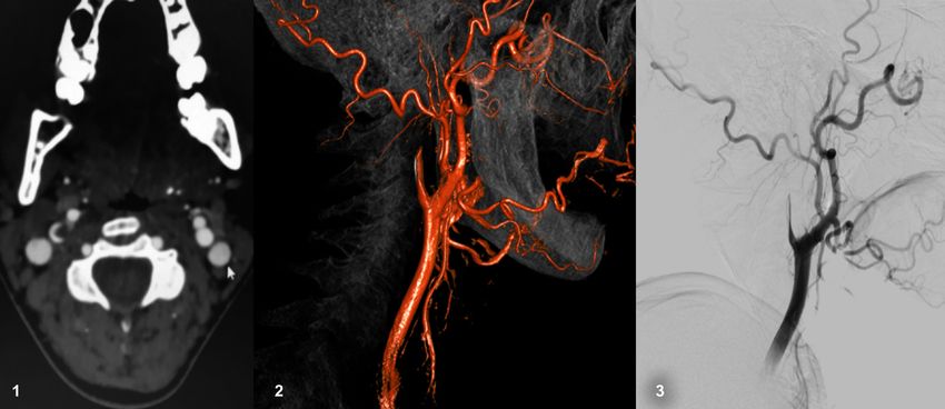
Case series of cervical and cerebral artery dissections

INTRODUCTION                                                             risk factors such as systemic arterial hypertension,
                                                                         smoking, diabetes, and clinical and/or endovascular
   Cerebrovascular disease is the leading cause of                       therapy were also analyzed.
severe disability and mortality in Brazil.1,2 Spontaneous
dissection of the carotid and vertebral arteries is
                                                                         RESULTS
responsible for 2% of all ischemic strokes, with
higher occurrence in young people,3,4 affecting their                        Of the 49 patients with carotid or vertebral/basilar
most productive years.5-8                                                artery (extra or intracranial) dissection, 42 presented
   The most common risk factors include arterial                         with an ischemic event due to vessel stenosis or arterial
hypertension, smoking, dyslipidemia, fibromuscular                       thrombus embolism, and 6 presented with SAH due
dysplasia, collagen disorders, trauma, and migraine                      to rupture of the vessel and formation of a dissecting
with aura.3,7,9-11 Sudden onset neck pain associated with                aneurysm. The mean patient age was 55 years, and
nausea, vomiting, and signs of neurological deficits                     51% of the patients were female. Dissection occurred
are strong indicators of cervical artery dissection.                     at the carotid level in 31 patients and in the vertebral
Dissection leads to formation of a mural hematoma,                       and basilar arteries or their branches in 18 patients.
resulting in vascular stenosis, pseudoaneurysm, and                          Headache and cervical pain were the most
eventual rupture of the vessel.10 Although the clinical                  common symptoms. In up to one-third of all cases,
presentation may indicate a serious condition such as                    nausea, vomiting, and coordination deficits were
stroke or subarachnoid hemorrhage (SAH), prognosis                       found principally in vertebrobasilar territory events.
and recovery are typically positive and most patients                    Additionally, sensory/motor and visual deficits were
achieve functional independence.5-7 In addition, the                     common. In cases of dissection with pseudoaneurysm,
incidence of recurrent stroke in these patients is                       the signs and symptoms were associated with SAH
usually low.12                                                           and impaired consciousness.
   Diagnosis of cervical or vertebral dissection is                          The most common risk factors included systemic
based on a combination of clinical and radiological                      arterial hypertension, smoking, diabetes, and
findings. Typically, the vessel is analyzed with                         dyslipidemia. Almost 10% of all cases had a prior
computed angiography tomography (CTA) or magnetic                        history of migraine. Hyperhomocysteinemia and
resonance angiography (MRA). However, the gold                           hypothyroidism were rare.
standard is digital subtraction angiography (DSA).                           Treatment followed the stroke unit protocols.
This method, although more invasive, allows possible                     Antiplatelet therapy combined with statin was the most
treatment of stenosis and local complications such as                    frequent treatment (63.2%). Dual antiplatelet therapy
pseudoaneurysms. Clinical management of this disease                     was indicated for 22.4%, anticoagulant combined with
is still a matter of debate. Therefore, the main aim                     statin was administered in 4%, and 46.9% underwent
of this study was to trace the epidemiological profile                   endovascular treatment. Mechanical thrombectomy
of this disease in our region within the last 5 years.                   associated with application of intracranial or extracranial
                                                                         stents in cases of dissection with tandem occlusion
PATIENTS AND METHODS                                                     was indicated for 5 patients (10.2%) with intracranial
    The protocol was approved by the institution Ethics                  occlusion, only 1 of whom received recombinant tissue
Committee Botucatu Medical School (number 535/2012)                      plasminogen activator. Pure intracranial thrombectomy
and informed consent was obtained. We created a                          was performed in 2 patients (4%).
database with information extracted from the Botucatu                        Patients with SAH and pseudoaneurysm formation
Stroke Unit electronic medical record. We identified                     were treated in the acute phase predominantly with
60 patients with cerebral or cervical arterial dissection                platinum coils and intracranial stents, excluding brain
who underwent DSA. Eleven patients were excluded                         circulation injury, and it was necessary to occlude the
due to a lack of information in their medical records                    vessel as treatment for the pseudoaneurysm in only
or to a different final diagnosis after undergoing DSA.                  1 patient. Most cases had an mRS
Case series of cervical and cerebral artery dissections
The cervical and intracranial carotid arteries were DISCUSSION
the most often compromised (60% of all cases).
The lesion pattern was stenosis in all cases of cervical Cerebral and cervical artery dissection are important
carotid, and pseudoaneurysm in most intracranial causes of stroke, especially for people under 45 years,
carotid cases (18 of 19 patients). Cervical lesions where they may be responsible for approximately one-
had ischemic presentation, and intracranial lesions fifth of all cases.5-7 The mean patient age in our case
presented as SAH. Vertebral arteries were affected series was 55 years (range, 16–85), and these findings
in almost one-third of all cases. The angiographic are probably related due to the selection criteria used
findings are shown in Table 2. for this study. All patients in our institution underwent
CTA in the acute stroke phase and were only sent to
the angiography suite when there were complications
Table 1. Demographic characteristics of entire sample (n = 49). or the diagnosis was dubious. This may also explain
Variables n % the large number of SAH cases in our sample.
Age (years) 55 (16-85) The pathophysiology of the disease is related to
Patients < 45 years old 12 24.48 tears in the arterial endothelium and an intraluminal
Sex (Female) 25 51.02 hematoma that causes delamination of the vessel wall
NIHSS at admission (median) 2 (0-9) layers, resulting in a false lumen. When cerebral or
NIHSS at discharge (median) 0 (0-3.5) cervical vessels are affected, a stroke is caused by
mRS at admission (median) 0 (0-1) local growth of the false lumen with occlusion of
mRS at discharge (median) 0 (0-2) the vessel, thrombus formation with focal or distal
Arterial hypertension 32 65.30 occlusion, compromise of the perforated artery ostium,
Smoking 27 55.10 and hemorrhage due to vessel wall rupture, aneurysm,
Dyslipidemia 15 30.61 or pseudoaneurysm formation.
Diabetes 10 20.40 Our study results were consistent with the main
Migraine 5 10.20 common risk factors,3,5,11,13-16 showing that most patients
Obesity 5 10.20 had hypertension, dyslipidemia, and were smokers.
Previous arterial disease (MI, stroke, 10 20.40 We found that only 10% of patients had migraine and
or PAD)
no patients had hyperhomocysteinemia, but this result
Clinical Presentation
may be due to the older patient ages in our study.
Pain (Headache or cervical) 15 30.61
Based on clinical presentation, many SAH patients
Hemiparesis 12 24.48
were selected for DSA. When the cervical carotid or
Aphasia 2 4.08
vertebral territories were compromised, we observed
Impaired consciousness 8 16.32
widely described clinical symptoms such as headache,
Visual impairment 5 10.20
cervicalgia, hemiparesis, visual deficits, vertigo, and
Ataxia 6 12.45
ataxia. Ischemic stroke was more common, although
Subarachnoid hemorrhage 6 12.45
this was probably due to the requirement for DSA
NIHSS: National Institute of Health Stroke Scale; mRS: modified Rankin Scale;
MI: myocardial infarction; PAD: peripheral artery disease. treatment in our patient sample. Notably, we found
many patients presenting with tandem occlusions, as
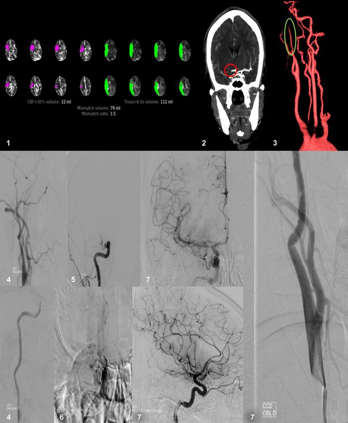
shown in Figure 1.
Table 2. Angiographic findings and treatment modality (n = 49). A diagnosis of cerebral and cervical arterial dissection
Compromised artery n % is typically based on clinical and radiological results.
Cervical carotid 12 24.48 Usually, findings include crescent-shaped thickenings,
Intracranial carotid 19 38.77 eccentric narrowed lumens, pencil, rat-tail, or candle-
Vertebral 14 28.57 flame shaped occlusions, pseudoaneurysms, and intimal
Basilar 1 2.04 flaps with false lumens (Figure 2). Most patients present
Posterior cerebral 2 4.08 with a single vessel lesion. However, patients with
Superior cerebellar 1 2.04 vessel wall disease, such as fibrodysplasia, may show
Angiography pattern multiple lesions and compromised vessels (Figure 3).
Pseudoaneurysm 18 36.73 Lesions are more common in the V3 segment of
Stenosis 31 63.26 vertebral arteries, and the cervical segment of the
Treatment carotid artery. In these cases, the vessel tear tends
Anticoagulation 2 4.08 not to affect the carotid bulb. The disease is typically
Antiaggregation 31 63.26 more frequent in the cervical carotid artery, as we
Endovascular 23 46.93 observed in our patient sample.
Modolo et al. J Vasc Bras. 2021;20:e20200242. https://doi.org/10.1590/1677-5449.200242 3/6Case series of cervical and cerebral artery dissections Figure 1. Patient that was admitted with acute stroke due to tandem occlusion and underwent mechanical thrombectomy. 1. Perfusion CT showing an acute stroke with large penumbra. 2. Median cerebral artery occlusion on CTA. 3. Internal carotid dissection with “pencil sign” on 3D CTA. 4. Angiographic image of carotid dissection and retarded contrast flow. 5. Median cerebral artery occlusion on DSA. 6. Position of stent retriever. 7. Post thrombectomy intracranial and cervical carotid images. The treatment approach for cerebral and cervical findings, many authors prefer to use antiplatelet therapy, arterial dissection is currently a matter of debate. since major hemorrhagic complications tend to be less A Cochrane systematic review comparing antiaggregants frequent with this treatment.12 No consensus exists and anticoagulants found no differences in death, stroke on dual or single antiaggregant use. In the CADISS recurrence, or functional outcomes.17-19 Based on these trial, administration of single or dual agents was a Modolo et al. J Vasc Bras. 2021;20:e20200242. https://doi.org/10.1590/1677-5449.200242 4/6
Case series of cervical and cerebral artery dissections

Figure 2. Radiology findings in arterial dissection case. 1. Crescent sign. 2 and 3. Flame shaped carotid occlusion.

Figure 3. Bilateral spontaneous vertebral artery dissection in a patient with suspected fibrodysplasia.

decision made by the investigator, and consequently                      wall compromise. Stroke recurrence is low, and very
the results could not be analyzed separately.20                          few patients present with new strokes, regardless of
    Thrombolysis and thrombectomy may safely be                          the treatment approach.3,6,12,15,19
performed in cases of ischemic stroke, and no patient should
be denied reperfusion therapies.21,22 Figure 3 illustrates               CONCLUSIONS
a successful case of mechanical thrombectomy
                                                                           Cerebral and cervical artery dissections are a
in a patient with acute stroke and cervical carotid
                                                                         common cause of stroke for which potential treatment
dissection. Endovascular treatment of arterial
                                                                         options exists to prevent poor outcomes. Although
dissection is thought to be safe and may be an option
                                                                         sometimes neglected, this disease should be recognized
in selected cases, especially when complications such
                                                                         and understood by specialists including neurologists,
as pseudoaneurysms are found.23
                                                                         neuroradiologists, and vascular surgeons.
    The functional prognosis for cerebral and cervical
arterial dissections tends to be positive, with most patients
                                                                         ACKNOWLEDGEMENTS
achieving functional independence.5,15,17,24 However,
cases with extensive infarct areas and SAH may have                         We would like to thank the hemodynamic and
poor recovery, and the functional outcome is more                        stroke unit at the Botucatu Medical School for
dependent on cerebral damage than it is on vessel                        supporting our research.

Modolo et al. J Vasc Bras. 2021;20:e20200242. https://doi.org/10.1590/1677-5449.200242                                                      5/6
Case series of cervical and cerebral artery dissections
REFERENCES 19. Lyrer P, Engelter S. Antithrombotic drugs for carotid artery
dissection. Cochrane Database Syst Rev. 2010;(10):CD000255.
1. Brazilian Cerebrovascular Disease Society. Primeiro consenso PMid:20927720.
brasileiro do tratamento da fase aguda do acidente vascular 20. Limaye K, Abla AA. We will use antiplatelets as our first choice for
cerebral. Arq Neuropsiquiatr. 2001;59(4):972-80. http://dx.doi. prevention of stroke recurrence in Cervical Arterial Dissection After
org/10.1590/S0004-282X2001000600026. PMid:11733849. Reading CADISS--will you? World Neurosurg. 2015;84(5):1182-4.
2. Saposnik G, Del Brutto OH, Iberoamerican Society of Cerebrovascular http://dx.doi.org/10.1016/j.wneu.2015.09.012. PMid:26376201.
Diseases. Stroke in South America: a systematic review of incidence, 21. Traenka C, Jung S, Gralla J, et al. Endovascular therapy versus
prevalence, and stroke subtypes. Stroke. 2003;34(9):2103-7. http:// intravenous thrombolysis in cervical artery dissection ischemic
dx.doi.org/10.1161/01.STR.0000088063.74250.DB. PMid:12907823. stroke – Results from the SWISS registry. Eur Stroke J. 2018;3(1):47-
3. Pieri A, Spitz M, Valiente RA, Avelar WM, Silva GS, Massaro AR. 56. http://dx.doi.org/10.1177/2396987317748545. PMid:31008337.
Dissecção espontânea das artérias carótidas e vertebrais em uma 22. Bernardo F, Nannoni S, Strambo D, Bartolini B, Michel P, Sirimarco
população multiétnica. Arq Neuropsiquiatr. 2007;65(4-A):1050-5. G. Intravenous thrombolysis in acute ischemic stroke due to
http://dx.doi.org/10.1590/S0004-282X2007000600029. intracranial artery dissection: a single-center case series and a
4. Griffiths D, Sturm J. Epidemiology and etiology of young stroke. Stroke review of literature. J Thromb Thrombolysis. 2019;48(4):679-84.
Res Treat. 2011;2011:209370. http://dx.doi.org/10.4061/2011/209370. http://dx.doi.org/10.1007/s11239-019-01918-6. PMid:31302824.
PMid:21789269. 23. Spanos K, Karathanos C, Stamoulis K, Giannoukas AD. Endovascular
5. Reges DS, Mazzeo M, Rosalino R, Gagliardi VDB, Cerqueira LG, treatment of traumatic internal carotid artery pseudoaneurysm. Injury.
Gagliardi RJ. Cervical arterial dissection: Clinical characteristics 2016;47(2):307-12. http://dx.doi.org/10.1016/j.injury.2015.09.015.
in a neurology service in São Paulo, Brazil. Arq Neuropsiquiatr. PMid:26453153.
2019;77(9):632-7. http://dx.doi.org/10.1590/0004-282x20190108. 24. Engelter ST, Traenka C, Lyrer P. Dissection of cervical and cerebral
PMid:31553393. arteries. Curr Neurol Neurosci Rep. 2017;17(8):59. http://dx.doi.
6. Dziewas R, Konrad C, Dräger B, et al. Cervical artery dissection - org/10.1007/s11910-017-0769-3. PMid:28667505.
Clinical features, risk factors, therapy and outcome in 126 patients.
J Neurol. 2003;250(10):1179-84. http://dx.doi.org/10.1007/s00415-
003-0174-5. PMid:14586598. *Correspondence
7. Mergeani A, Popescu D, Antochi F. Spontaneous intracranial Gustavo José Luvizutto
internal carotid artery dissection. Rom J Neurol Rev Rom Neurol. Universidade Federal do Triângulo Mineiro – UFTM, Centro de
2010;9(4):200-2. Pesquisas Professor Aluízio Rosa Prata, Departamento de Fisioterapia
8. Wang G, Zhang Z, Ayala C, Dunet DO, Fang J, George MG. Costs of Aplicada
hospitalization for stroke patients aged 18-64 years in the United Rua Vigário Carlos, 100, Sala 410, 4º andar - Bairro Abadia
States. J Stroke Cerebrovasc Dis. 2014;23(5):861-8. http://dx.doi. CEP 38025-350 - Uberaba (MG), Brasil
org/10.1016/j.jstrokecerebrovasdis.2013.07.017. PMid:23954598. Tel.: (34) 99915-1611
E-mail: gluvizutto@gmail.com
9. Shin DH, Hong JM, Lee JS, et al. Comparison of potential risks
between intracranial and extracranial vertebral artery dissections. Eur
Neurol. 2014;71(5-6):305-12. http://dx.doi.org/10.1159/000357867. Author information
PMid:24662973. GPM – MSc in Clínica Médica, Universidade Estadual Paulista
10. Pezzini A, Grond-Ginsbach C, Debette S, et al. Genetics of cervical (UNESP).
artery dissection. Riv Ital di Neurobiol. 2008;5(1):22-31. EKF – PhD, Programa de Pós-graduação Fisiopatologia em Clínica
Médica, Universidade Estadual Paulista (UNESP) and full member,
11. Guidetti D, Rota E, Morelli N, Immovilli P. Migraine and stroke:
Academia Brasileira de Neurologia (ABN), Sociedade Brasileira de
“vascular” comorbidity. Front Neurol. 2014;5:193. http://dx.doi.
Neurofisiologia Clínica (SBNC), Liga Brasileira de Epilepsia (LBE) and
org/10.3389/fneur.2014.00193. PMid:25339937.
Young Epilepsy Section (YES - ILAE).
12. CADISS trial investigators, Markus HS, Hayter E, Levi C, Feldman NAMR - PhD in Ciências Médicas, Faculdade de Ciências Médicas,
A, Venables G, Norris J. Antiplatelet treatment compared with Universidade Estadual de Campinas (FCM/UNICAMP).
anticoagulation treatment for cervical artery dissection (CADISS): FASS - Medical student, Universidade Estadual Paulista (UNESP).
a randomised trial. Lancet Neurol. 2015;14(4):361-7. http://dx.doi. GJL - MSc in Fisioterapia e Doutor em Neurologia, Universidade
org/10.1016/S1474-4422(15)70018-9. PMid:25684164. Estadual Paulista (UNESP).
13. Wolfe CDA, Giroud M, Kolominsky-Rabas P, et al. Variations MLS – PhD in Bases Gerais da Cirurgia and tenured professor,
in stroke incidence and survival in 3 areas of Europe. Stroke. Universidade Estadual Paulista (UNESP).
2000;31(9):2074-9. http://dx.doi.org/10.1161/01.STR.31.9.2074. RB - MSc and PhD in Neurologia, Faculdade de Medicina de Ribeirão
PMid:10978032. Preto (USP), and Departamento Científico (DC) coordinator in
14. Touzé E, Gauvrit JY, Moulin T, Meder JF, Bracard S, Mas JL, Multicenter Reabilitação Neurológica at Academia Brasileira de Neurologia (ABN).
Survey on Natural History of Cervical Artery Dissection. Risk of CCMF - MSc and PhD in Bases Gerais da Cirurgia, Universidade
stroke and recurrent dissection after a cervical artery dissection: a Estadual Paulista, (UNESP).
multicenter study. Neurology. 2003;61(10):1347-51. http://dx.doi.
org/10.1212/01.WNL.0000094325.95097.86. PMid:14638953. Author contributions
15. Taylor FR. Incidence and outcome of cervical artery dissection: a Conception and design: GPM, EKF, RB, CCMF
population-based study - Commentary. Headache. 2007;47(3):460. Analysis and interpretation: NAMR
16. Debette S. Pathophysiology and risk factors of cervical artery Data collection: EKF, FASS, CCMF
dissection: what have we learnt from large hospital-based cohorts? Writing the article: GJL, MLS
Curr Opin Neurol. 2014;27(1):20-8. http://dx.doi.org/10.1097/ Critical revision of the article: RB, CCMF
WCO.0000000000000056. PMid:24300790. Final approval of the article*: GPM, EKF, NAMR, FASS, GJL,
17. Arnold M, Kappeler L, Georgiadis D, et al. Gender differences in MLS, RB, CCMF
spontaneous cervical artery dissection. Neurology. 2006;67(6):1050- Statistical analysis: GJL, RG, MLS, CCMF
2. http://dx.doi.org/10.1212/01.wnl.0000237341.30854.6a. Overall responsibility: RB, CCMF
PMid:17000975.
18. Perry BC, Al-Ali F. Spontaneous cervical artery dissection: the *All authors have read and approved of the final version of the
Borgess classification. Front Neurol. 2013;4:133. PMid:24062720. article submitted to J Vasc Bras.
Modolo et al. J Vasc Bras. 2021;20:e20200242. https://doi.org/10.1590/1677-5449.200242 6/6You can also read