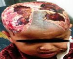
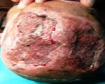
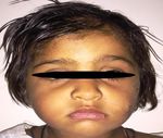
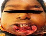
Diverse spectrum of facial dog bite presentation and their management
←
→
Page content transcription
If your browser does not render page correctly, please read the page content below
International Surgery Journal
Jain RK et al. Int Surg J. 2018 Sep;5(9):3017-3022
http://www.ijsurgery.com                                                                   pISSN 2349-3305 | eISSN 2349-2902

                                                                DOI: http://dx.doi.org/10.18203/2349-2902.isj20183452
Original Research Article

 Diverse spectrum of facial dog bite presentation and their management
                 Rakesh Kumar Jain, Gautam Prakash*, Manojit Midya, Pankaj Sharma

  Department of Plastic Surgery, SMS Medical College and Hospital, Jaipur, Rajasthan, India

  Received: 03 August 2018
  Accepted: 10 August 2018

  *Correspondence:
  Dr. Gautam Prakash,
  E-mail: drgprakash86@gmail.com

  Copyright: © the author(s), publisher and licensee Medip Academy. This is an open-access article distributed under
  the terms of the Creative Commons Attribution Non-Commercial License, which permits unrestricted non-commercial
  use, distribution, and reproduction in any medium, provided the original work is properly cited.

   ABSTRACT

   Background: Dog bite patients are frequently encountered in our hospital seeking immediate as well as delayed
   reconstruction. More than two third of dog bite injuries involve head, neck and scalp region. Facial dog bites present a
   challenge for the surgeon, as they lead to cosmetic disfigurement and psychological trauma to the patient. Following
   thorough washout and debridement, we have used various reconstructive techniques for definitive management of
   wounds like- primary repair, V-Y advancement flap, nasolabial flap, SSG, FTG and Karapandzic flap. Purpose of the
   present study is to share our experiences in management of dog bite wounds on the face in both adult and paediatric
   patients with available reconstructive options to maximize the functional and cosmetic outcomes by using basic
   principles of surgery.
   Methods: Present study was a single centre retrospective study conducted in a tertiary care centre from February
   2013 to January 2018. Total 497 patients of dog bite who presented in the emergency department were enrolled. Out
   of them 310 patients had involvement of head, neck and scalp requiring surgical intervention in any form.
   Results: In last five years, we have encountered mid face predilection in face, head and neck cases. Out of 310 cases,
   lip (25.16%) and cheek (24.51%) were involved in majority of the patients. Flap cover surgery is required in majority
   of the scalp and nose group of patients, as there is less mobility of tissue present in surrounding region, while cheek
   and lip were managed with primary closure in most of the patients.
   Conclusions: Although most of the dog bites are preventable, but cases of dog bite are increasing continuously. Child
   should never be left alone with dogs and, if they are fear of dogs, it’s better not to obtain dogs. As far now, it’s a
   major concern for treating physician or surgeon to provide optimal cosmetic as well as functional outcome. Early
   surgical intervention for wound management gives better results with the use of basic principles of plastic surgery.

   Keywords: Dog bite, Dog bite management, Facial injury

INTRODUCTION                                                        challenge for the surgeon, as they lead to cosmetic
                                                                    disfigurement and psychological trauma to the patient.
A substantial association has been seen between dogs and
humans.1 Dog bite patients are frequently encountered in            Although dog bites have been reported on almost all
our hospital seeking immediate as well as delayed                   areas of the body, but there is predilection for head, neck
reconstruction. More than two third of dog bite injuries            and scalp in pediatric age group and for upper limbs in
involve head, neck and scalp region.2,3 Most of the dog             adults.5 Young children are more prone for head and neck
bite injuries are deep seated, as skin flaps may appear             involvement due to relatively large head size, short
viable but underlying tissue is devitalized requiring               stature and less fear of mishap. Also, young children are
proper assessment (Table 1).4 Facial dog bites present a            physically less able to defend themselves and escape.6

                                                        International Surgery Journal | September 2018 | Vol 5 | Issue 9   Page 3017
Jain RK et al. Int Surg J. 2018 Sep;5(9):3017-3022
Grossly dogs are categorized as: pets (restricted and Out of them 310 patients had involvement of head, neck
supervised); family dogs (partially restricted, wholly and scalp. Only the patients who needed surgical
dependent); community dogs (unrestricted, partially intervention (wounds more than 2 cm) were included in
dependent); and feral dogs (unrestricted, independent). In the study. Patients having wounds less than 2 cm, small
India maximum dogs belong to last three categories.7 laceration, immune-deficient, using immunosuppressive
agent, autoimmune disorder and diabetes were excluded.
Table 1: Lackmann classification of dog bite injury.
Our protocol for management of dog bite wound was to
Classification Definition debride the wound thoroughly, and wash with 20% liquid
Superficial lesion without muscle soap, 3% hydrogen peroxide and normal saline. Repeated
Type I
involvement wash of two to three times for total of approximately 15
Deep lesion with muscle minutes was done under running saline. Local anesthetic
Type II was used judiciously if needed for adequate debridement.
involvement
Deep lesion with muscle All the necrotic, slough tissue and dirt particles were
Type III debrided till healthy surrounding tissue.
involvement and tissue defect
Type III combined with vascular
Type IVa Simple lacerations were primarily repaired in layers, and
damage or nerve lesion
Type III combined with bone composite defects were managed with flap cover on
Type IVb emergency basis. Delayed cover was done only in cases
damage or organ involvement
who had concomitant life-threatening injuries.
Prophylactic antibiotics (combination of amoxicillin and
The purpose of the present study is to share our
clavulanic acid) was given in intraoperative and
experiences in management of dog bite wounds on the
postoperative period. We advised azithromycin to the
face in both adult and pediatric patients with available
patients who were found to be allergic to penicillin.8
reconstructive options to maximize the functional and
Infection was assessed on the basis of three major
cosmetic outcomes by using basic principles of surgery.
criteria: fever (body temperature ≥380C), abscess, and
We have used various reconstructive techniques for
lymphangitis; or four of five minor criteria: wound-
definitive management of wounds like: primary repair, V-
associated erythema that extended more than 3 cm from
Y advancement flap, nasolabial flap, SSG, FTG and
the edge of the wound, tenderness at the wound site,
Karapandzic flap. Regardless of surgical techniques used,
swelling at the site, purulent drainage, white cell count in
few patients develop complications or unsightly scars,
the peripheral blood >12,000/ml. In consultation with
which may require revision surgery like scar revision or
anti-rabies department, anti-rabies vaccination (ARV)
expander placement.
was done. Tetanus antitoxin was administered to all
patients who were not immunized previously or those
METHODS
who did not remember their last booster dose. Rabies
immunoglobulin was administered to all patients.
This is a single center retrospective study conducted in a
tertiary care center, SMS Hospital Jaipur from February
RESULTS
2013 to January 2018. Data of total 497 patients of dog
bite who presented in the emergency department was
In last five years, we have encountered mid face
collected (Table 2).
predilection in face, head and neck cases. Out of 310
cases, lip (25.16%) and cheek (24.51%) were involved in
Table 2: Patient demographic data.
majority of the patients. Flap cover surgery is required in
majority of the scalp and nose group of patients, as there
Demographic variables Number of patients (%)
is less mobility of tissue present in surrounding region,
Sex while cheek and lip were managed with primary closure
Male 208 (67.09%) in most of the patients. We are showing results of four
Female 102 (32.90%) different anatomic regions.
Age (years)
50 21 as per our protocol mentioned above. Immediate cover of
Yes 221 (71.29%) scalp wound with split skin graft and primary closure of
Patient familiar with dog facial laceration was done. In the postoperative period,
No 89 (28.71%)
facial and scalp wounds healed satisfactorily (Figure 2).
International Surgery Journal | September 2018 | Vol 5 | Issue 9 Page 3018Jain RK et al. Int Surg J. 2018 Sep;5(9):3017-3022
After 3 months, next surgery was planned and a 200ml
silicone expander was placed in scalp in the right
temporo-parietal region. Expander was inflated with
normal saline at one-week intervals.
Figure 4: Post-operative photograph after one year
following expander placement, flap advancement and
scar revision surgery.
Case 2
Figure 1: Post dog bite scalp avulsion in a two-year Three-year-old boy presented to our outpatient
girl. department with dog bite over lower lip. Patient had full
thickness tissue loss of two third of left side of lower lip.
(Figure 5).
Figure 2: Follow up photograph seven days after split
thickness skin grafting. Figure 5: Post dog bite 2/3rd full thickness central
lower lip defect with bilateral intact oral commissure.
After achieving adequate expansion which took three
months, the expander was removed under general Patient was thoroughly evaluated, and primary care was
anesthesia and flap advancement was done. Post done as per our protocol. Immediate reconstruction with
operatively patient had a good pattern of hair distribution left sided Karapandzic flap was done (Figure 6 and 7)
and a stable facial scar (Figure 3 and 4). Post-operative day 7 photograph (Figure 8).
Figure 3: Tissue expander was placed in occipital Figure 6: Intra-operative photograph after
region after 3 months. debridement and marking of incision line.
International Surgery Journal | September 2018 | Vol 5 | Issue 9 Page 3019Jain RK et al. Int Surg J. 2018 Sep;5(9):3017-3022
After initial management, immediate definitive
reconstruction with lip switch for upper lip defect and
paramedian forehead flap for nose defect was done
(Figure 10).
Figure 7: Intra-operative photograph after raising
and advancement of flaps.
Figure 10: Intra OP photograph showing lip switch
flap for upper lip defect and paramedian forehead
flap for nose defect.
Figure 8: Post op photograph after seven days
showing acceptable scar line and oral competence.
Figure 11: Post op photograph after two months
Case 3 follow up.
A thirty-five-year-old female came to us with dog bite After three weeks forehead and lip switch flap
over nose and upper lip with full thickness loss of tissue detachment and in-setting were done (Figure 11).
and defect of nose and upper central lip (Figure 9).
Figure 9: Post dog bite full thickness defect of central Figure 12: Post dog bite lower lip full thickness
upper lip, collumella and bilateral ala of nose in a 35- laceration in a 38-year male after seven days.
year female.
International Surgery Journal | September 2018 | Vol 5 | Issue 9 Page 3020Jain RK et al. Int Surg J. 2018 Sep;5(9):3017-3022
midface is the center of attraction in face, hence in view
of disfigurement, immediate primary repair is preferred.
Most of our patients (71.29%) were familiar to the dog
and were either owners of the pet, or some relatives were
the owner, or it was a common neighborhood pet, similar
to study by Kaye.14 Hence this shows familiarity of dog
with the victim is not an absolute surety against getting
bitten by it.15-17 There is no standard protocol for wound
closure surgery either primary repair or flap cover.
Previously, in view of risk of infection, usually wounds
were left for healing by secondary intension or delayed
repair.18,19 As per recent studies immediate primary repair
of facial dog bite wounds give the best cosmetic and
functional outcomes.2, 18
Figure 13: Post op photograph after seven days. Table 4: Various surgical procedure in dog bite of
head neck.
Case 4
Type of No. of
Site involved
Thirty-eight-year-old male presented to our outpatient operation patients
department 5 days after dog bite over lower lip (Figure Primary closure 14
Scalp
12). He had received three doses of ARV in local hospital SSG/flap cover 45
in his town. Re-suturing was done under general Primary closure 03
anesthesia, in which margins were freshened, slough and Nose
Flap cover 27
necrotic tissue was debrided. Primary closure of skin Primary closure 48
flaps was done. Post op result after seven days (Figure Cheek
Flap cover 28
13).
Primary closure 43
Lip
DISCUSSION Flap cover 35
Primary closure 16
Eyelid
Dog bite is a serious concern for parents as well as Flap cover 07
clinicians. Majority of the patients in our study are in Primary closure 12
younger age group with grade IV injury. Other studies Flap cover 18
Multiple site
also have similar data of dog bite in pediatric age Primary closure
14
group.9,10 Also, young boys are more commonly affected and flap cover
in comparison to girls. This is in sync with our society
and cultural scenario where boys are more commonly Scalp and nose have less tissue mobility; hence primary
involved in outdoor games/ activities while girls usually closure is not possible in large wounds and require flap
remain indoors. Various other studies have also shown cover surgery. In contrary to above cheek, lip and eyelid
male dominance in dog bite (Table 2).2,10,11 have sufficient laxity which allows primary closure of
wounds (Table 4).
Table 3: Distribution dog bite on face.
Table 5: complications and revision surgery.
Site involved Number of patients
Scalp 59 Complications Number of cases
Nose 30 Alopecia 38
Cheek 76 Ectropion 6
Lip 78 Hypertrophic scar 17
Eyelid 23 Microstomia 8
Multiple 44
Present study also points similar figures as previously
As a child plays with the dog, hugs, kisses or pulls the studied, showing low rate of infection n = 4 patients
tail, they might unintentionally provoke it for biting. (1.29%) in facial dog bite after primary repair.20
These activities place the face in closest position; making
it the most vulnerable area for dog bites in children. All four patients were managed with debridement and
Involvement of midface structures like lip and nose is injectable antibiotics. None of patient had rabies or
predominant.12,13 In the present study midface is more intracranial infection during treatment and follow up
frequently involved similar to other studies (Table 3). As period so far.
International Surgery Journal | September 2018 | Vol 5 | Issue 9 Page 3021Jain RK et al. Int Surg J. 2018 Sep;5(9):3017-3022
It is clear from the present study that primary repair of 5. Baker MD, Lanuti M. The management and
facial dog bite gives best outcome, as wound heal by outcome of lacerations in urban children. Ann
primary intention leading to less scarring and deformity Emerg Med. 1990;19:1001-5.
formation. In contrary, wounds will experience 6. Alizadeh K, Shayesteh A, Xu L Min. An
inflammation-hyperplasia of granulation tissue in algorithmic approach to operative management of
secondary intention healing, leads to broad and ugly scar complex pediatric dog bites: 3-year review of level I
formation. Wounds near angle of mouth, ala of nose, regional referral pediatric trauma hospital. Plast
eyelid leads to severe cosmetic as well as functional Reconstr Surg Glob Open. 2017;5:1431.
disfigurement after secondary intention healing. 7. Chaudhuri S. Rabies prevention and dog population
management. india’s official dog control policy in
Revision surgeries were performed in follow up period context of WHO guidelines. Ecollage. 2005.
for unavoidable complications like scar, alopecia and 8. Goldstein EJ. Current concepts on animal bites:
microstomia etc. (Table 5). Scar revision was the most bacteriology and therapy. Curr Clin Top Infect Dis.
frequently performed procedure in revision surgery 1999;19:99-111.
group. Majority of the patients were very much satisfied 9. Borud LJ, Friedman DW. Dog bites in New York
after revision surgeries for alopecia and scar. City. Plast Reconstr Surg. 2000;106:987-90.
10. Mitchell RB, Nanez G, Wagner JD, Kelly J. Dog
CONCLUSION bites of the scalp, face and neck in children.
Laryngoscope. 2003;113:492-5.
Although most of the dog bites are preventable, but cases 11. Savino F, Gallo E, Serraino P, Oggero R, Silvestro
of dog bite are increasing continuously. It is clear from L, Mussa GC. Dog bites in children less than
various studies that majority of the dog bites are in fourteen years old in Turin. Minerva Pediatricia.
pediatric age group. Most of the dog bites are 2002;54:237-42.
preventable, as children must be taught not to interact 12. Akhtar N, Smith MJ, McKirdy S, Page RE. Surgical
with unfamiliar dogs. Child should never be left alone delay in the management of dog bite injuries in
with dogs and, if they are fear of dogs, it’s better not to children, does it increases the risk of infection? J
obtain dogs. Before planning to obtain a dog, we must Plast Reconstr Aesthetic Surg. 2006;59:80-85.
enquire about aggressive nature or any abnormal 13. Palmer J, Rees M. Dog bites of the face: a 15-year
behavior of the dog. Proper vaccination of the obtained review. Br J Plast Surg. 1983;36:315-8.
dog should be insured. 14. Kaye AE, Belz JM, Kirschner RE. Pediatric dog bite
injuries: a 5-year review of the experience at the
Hence, pediatrician and health care worker must Children’s Hospital of Philadelphia. Plast Reconstr
participate in the dog bite awareness programs. As far Surg. 2009;124:551-8.
now, it’s a major concern for treating physician or 15. Weiss HB, Friedman D, Coben JH. Incidence of dog
surgeon to provide optimal cosmetic as well as functional bite injuries treated in emergency departments.
outcome. Early surgical intervention for wound JAMA. 1998;279:51-3.
management gives better results. 16. Bernardo LM, Gardner MJ, Rosenfield RL, Cohen
B, Pitetti R. A comparison of dog bite injuries in
Funding: No funding sources younger and older children treated in a pediatric
Conflict of interest: None declared emergency department. Pediatr Emerg Care.
Ethical approval: The study was approved by the 2002;18:247-49.
Institutional Ethics Committee 17. Gershman KA, Sacks JJ, Wright JC. Which dog
bites? A case control study of risk factors. Pediatr.
REFERENCES 1994;93:913-7.
18. Lackmann G, Wolfgang D, Isselstein G, Tollner U.
1. Davis SJM, Valla R Francois. Evidence for Surgical treatment of facial dog bites injuries in
domestication of the dog 12,000 years ago in the children. Craniomaxillofac Surg. 1992;20:81-6.
Natufian of Israel. Nature. 1978;276:608-10. 19. Jone RC, Shires GT. Bites and stings of animals and
2. Mcheik JN, Vergnes P, Bondonny JM. Treatment of insects. Principles of surgery. New York: McGraw-
facial dog bite injuries in children: a retrospective Hill; 1979.
study. J Pediatr Surg. 2000;35:580-3. 20. Barry EL. Facial dog bite injuries in children:
3. Calkins C, Bensard D, Patrick D, Karrer F. Life treatment and outcome assessment. J Craniofac
threatening dog attacks: a devastating combination Surg. 2013;24:384-6.
of penetrating and blunt injuries. J Pediatr Surg.
2001;36:1115-7. Cite this article as: Jain RK, Prakash G, Midya M,
4. Galloway RE. Mammalian bites. J Emerg Med. Sharma P. Diverse spectrum of facial dog bite
1988;6:325-31. presentation and their management. Int Surg J
2018;5:3017-22.
International Surgery Journal | September 2018 | Vol 5 | Issue 9 Page 3022You can also read


