Preproteins couple the intrinsic dynamics of SecA to its ATPase cycle to translocate via a catch and release mechanism
←
→
Page content transcription
If your browser does not render page correctly, please read the page content below
Article

Preproteins couple the intrinsic dynamics of SecA to
its ATPase cycle to translocate via a catch and
release mechanism
Graphical abstract                                                 Authors
                                                                   Srinath Krishnamurthy,
                                                                   Marios-Frantzeskos Sardis,
                                                                   Nikolaos Eleftheriadis, ...,
                                                                   Ana-Nicoleta Bondar,
                                                                   Spyridoula Karamanou,
                                                                   Anastassios Economou

                                                                   Correspondence
                                                                   tassos.economou@kuleuven.be

                                                                   In brief
                                                                   Combining biophysical and biochemical
                                                                   tools, Krishnamurthy et al. show how
                                                                   preproteins activate the intrinsically
                                                                   dynamic membrane-associated Sec
                                                                   translocase. Preprotein signal peptides
                                                                   close the clamp and mature domains
                                                                   increase motor dynamics. Nucleotides
                                                                   drive the translocase between
                                                                   conformational states that catch and
                                                                   release preproteins through frustrated
                                                                   prongs and lead to their translocation.
Highlights
d   Preproteins couple the dynamics of the SecA ATPase motor
    to its preprotein clamp

d   Preprotein binding causes increased motor dynamics leading
    to ADP release

d   Nucleotide states subtly alter the intrinsic dynamics of Sec
    translocase

d   Clients are translocated via a nucleotide-dependent ‘‘catch
    and release’’ mechanism

           Krishnamurthy et al., 2022, Cell Reports 38, 110346
           February 8, 2022 ª 2022 The Author(s).
           https://doi.org/10.1016/j.celrep.2022.110346                                            ll
ll
                                                                                                                          OPEN ACCESS

Article
Preproteins couple the intrinsic
dynamics of SecA to its ATPase cycle
to translocate via a catch and release mechanism
Srinath Krishnamurthy,1 Marios-Frantzeskos Sardis,1 Nikolaos Eleftheriadis,1 Katerina E. Chatzi,1 Jochem H. Smit,1
Konstantina Karathanou,2 Giorgos Gouridis,1,3,4 Athina G. Portaliou,1 Ana-Nicoleta Bondar,2,5,6 Spyridoula Karamanou,1
and Anastassios Economou1,7,*
1KU  Leuven, University of Leuven, Rega Institute, Department of Microbiology and Immunology, 3000 Leuven, Belgium
2Freie Universität Berlin, Department of Physics, Theoretical Molecular Biophysics Group, Arnimallee 14, 14195 Berlin, Germany
3Molecular Microscopy Research Group, Zernike Institute for Advanced Materials, University of Groningen, Nijenborgh 4,

9747 AG Groningen, the Netherlands
4Structural Biology Division, Institute of Molecular Biology and Biotechnology (IMBB-FORTH), Nikolaou Plastira 100, Heraklion, Crete, Greece
5University of Bucharest, Faculty of Physics, Atomiștilor 405, 077125 Ma  gurele, Romania
6Forschungszentrum Ju    €lich, Institute of Computational Biomedicine, IAS-5/INM-9, Wilhelm-Johnen Straße, 5428 Ju €lich, Germany
7Lead contact

*Correspondence: tassos.economou@kuleuven.be
 https://doi.org/10.1016/j.celrep.2022.110346

SUMMARY

Protein machines undergo conformational motions to interact with and manipulate polymeric substrates. The
Sec translocase promiscuously recognizes, becomes activated, and secretes >500 non-folded preprotein
clients across bacterial cytoplasmic membranes. Here, we reveal that the intrinsic dynamics of the translo-
case ATPase, SecA, and of preproteins combine to achieve translocation. SecA possesses an intrinsically
dynamic preprotein clamp attached to an equally dynamic ATPase motor. Alternating motor conformations
are finely controlled by the g-phosphate of ATP, while ADP causes motor stalling, independently of clamp
motions. Functional preproteins physically bridge these independent dynamics. Their signal peptides pro-
mote clamp closing; their mature domain overcomes the rate-limiting ADP release. While repeated ATP
cycles shift the motor between unique states, multiple conformationally frustrated prongs in the clamp
repeatedly ‘‘catch and release’’ trapped preprotein segments until translocation completion. This universal
mechanism allows any preprotein to promiscuously recognize the translocase, usurp its intrinsic dynamics,
and become secreted.

INTRODUCTION                                                              Protein intrinsic dynamics and disorder are multi-leveled (Hen-
                                                                       zler-Wildman et al., 2007; Yang et al., 2014) and essential for a
Protein machines modify, reshape, disaggregate, and transport          protein assembly and interactions (Dunker et al., 2002; Fuxreiter
nucleic acids and polypeptides (Avellaneda et al., 2017; Flechsig      et al., 2014): motions between subunits (quaternary), within a
and Mikhailov, 2019; Kurakin, 2006) by converting between              chain (global), of a domain (rigid body), and of segments (local).
auto-inhibited and active states commonly relying on intrinsic         It is unclear how intrinsic dynamics couple allostery to protein
structural dynamics (Nussinov et al., 2018). A fascinating para-       function (Loutchko and Flechsig, 2020; Zhang et al., 2019),
digm is the bacterial Sec translocase that secretes preprotein cli-    even less so in multi-liganded/partner enzymes that operate
ents across the inner membrane. Its SecA ATPase subunit, a four        hierarchically, like the Sec translocase.
domain Superfamily 2 helicase (Figure S1A), binds non-folded              Cytoplasmic SecA is dimeric, ADP-bound and quiescent, and
clients, nucleotides, lipids, chaperones, and the SecYEG chan-         chaperones clients (Sianidis et al., 2001). Its helicase motor
nel (De Geyter et al., 2020; Rapoport et al., 2017; Tsirigotaki        (comprising nucleotide binding domains [NBDs] 1/2) is fused to
et al., 2017a). A multi-tiered intrinsic dynamics nexus of sub-re-     an ATPase-suppressing C-domain and a preprotein binding
actions activates the translocase (Corey et al., 2019; Gouridis        domain (PBD; rooted via a stem in NBD1; Figure S1A). The
et al., 2013; Krishnamurthy et al., 2021; Sardis and Economou,         PBD intrinsically rotates from a distal ‘‘wide-open’’ position to-
2010), requiring minor energetic input from ligands. While part-       ward NBD2 (‘‘closed’’) (Ernst et al., 2018; Krishnamurthy et al.,
ners and nucleotides prime the dynamics landscape of the trans-        2021; Sardis and Economou, 2010; Vandenberk et al., 2019) to
locase, 500 loosely conserved clients activate it at the expense      clamp mature domains (Bauer and Rapoport, 2009). SecYEG
of energy via a universal mechanism (Tsirigotaki et al., 2017a).       binding (Figure 1AII, ‘‘primed’’) enhances local dynamics in

                                                                    Cell Reports 38, 110346, February 8, 2022 ª 2022 The Author(s). 1
                     This is an open access article under the CC BY-NC-ND license (http://creativecommons.org/licenses/by-nc-nd/4.0/).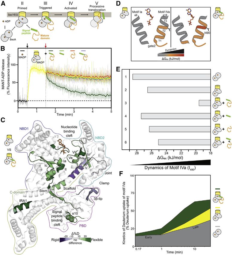
ll
OPEN ACCESS Article
Figure 1. Preproteins induce ADP release by
enhancing motor dynamics
(A) Cytoplasmic ADP-bound, quiescent SecA2 (I)
binds asymmetrically to SecYEG through the
active protomer (II, gray oval). The preprotein is
targeted to the translocase through bivalent signal
peptide or mature domain binding. Signal peptide
binding alone ‘‘triggers’’ (III), yet only preprotein
binding activates (IV), the translocase for ATP
hydrolysis cycles that result in processive
translocation (V).
(B) ADP release assay. The fluorescence intensity
of MANT-ADP increases upon binding to the
translocase (black arrow). Reactions were
chased (red arrow) with the indicated ligands (see
STAR Methods). The drop in fluorescence in-
tensity corresponds to the MANT-ADP release
from SecA. Transparent lines: raw fluorescence
traces (n = 3–4). solid lines: smoothened
data (LOWESS). Preprotein: proPhoA1-122, mature
domain: PhoA23-122.
(C) Effect of proPhoA1-122 binding on the local
dynamics (HDX-MS) of SecYEG:SecA2:ADP. D
uptake differences between SecYEG:SecA2:
ADP (top pictogram: ‘‘reference’’) and SecYEG:
SecA2:ADP:preprotein (bottom pictogram: ‘‘test’’)
shown. Purple: decreased; green: increased dy-
namics; no difference: transparent gray. ADP: or-
ange sticks. Domains are contoured.
(D) DGex values representing protein dynamics
were calculated by PyHDX from HDX-MS data
(Smit et al., 2021a) and mapped onto a cartoon of
the closed-gate2 state with its two helices
comprising helicase motifs Ia (a6) and IVa (a18;
three turns). Dynamics of SecYEG:SecA2:ADP in
preprotein free (left) and bound (right) state are
shown. I490 (between turns two and three) reports
on motif IVa dynamics. ADP: orange sticks. See
also Figure S1D.
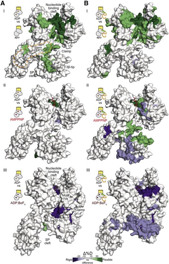
(E) DGex values (from D) for I490 (motif IVa) determined under the indicated conditions. Decreased DGex values: increased dynamics.
(F) D uptake kinetic plots of peptide aa488-501 (motif IVa), shown as a percentage of its full deuteration control (Table S1), for SecA2:ADP (gray), Se-
cYEG:SecA2:ADP (yellow), and SecYEG:SecA2:ADP:preprotein (green). Labeling timepoints (0.17, 0.5, 1, 2, 5, 10, and 30 m) and SD values (
ll
Article                                                                                                                    OPEN ACCESS

client-less translocase can no longer release ADP and becomes           Motif IVa of gate2 senses ligands via its intrinsic
quiescent.                                                              dynamics
                                                                        Motif IVagate2 is intriguing. It has elevated basal dynamics, is sen-
RESULTS                                                                 sitive to multiple interactants (Krishnamurthy et al., 2021), and is
                                                                        located between the ADP-binding motif Va and motifs IV and Vb
Preprotein-stimulated ADP release from the helicase                     on NBD2 b-strands (Figure S1E). It comprises a flexible linker fol-
motor                                                                   lowed by a three-turn helix with a constantly dynamic first half
To stimulate ATP turnover at the translocase clients destabilize        (Figure 1D, left, orange; Figure S1D, left) and a conditionally dy-
the SecA:ADP state. We tested this by monitoring the binding            namic second half (turns two–three). Preprotein binding
and release of MANT-ADP from SecYEG:SecA2 upon preprotein               increased dynamics specifically of I490 (Figures 1D and S1D,
addition (Figure 1B). The fluorescence intensity of MANT-ADP in-        right), yielding a quantitative assay. Channel binding marginally
creases upon binding to SecA (37 C; Figure 1B; black arrow)            affects dynamics of SecA2:ADP (Figure 1E, compare lane 2 to
(Galletto et al., 2005; Karamanou et al., 2005; Krishnamurthy           1). Preprotein binding to SecYEG:SecA2:ADP significantly
et al., 2021), and it remains high (Figure 1B, yellow line), indi-      further enhances dynamics (lane 3), while signal peptides (lane
cating tight ADP binding. Preprotein addition (proPhoA1-122;            4) or mature domains (lane 5) added alone, or together (lane 6),
Chatzi et al., 2017) (Figure 1B, orange arrow) causes a drop in         cause minor effects.
fluorescence intensity (green line), indicative of MANT-ADP                Motif IVa shows biphasic D uptake kinetics that suggest mod-
release. Release is not observed in soluble SecA2 (37 C; Fig-          ulation of its energy landscape in two differentially flexible
ure S1CI) nor at 10 C with channel-bound SecA2 (II). ATP excess        conformational steps (Figure 1F, gray), due to varying D uptake
on channel-bound SecA2 is as efficient in MANT-ADP release as           kinetics in different regions of the analyzed peptide or combined
proPhoA1-122 (2 mM; 37 C; III), while signal peptide (Figure 1B,       H bonding and solvent accessibility effects. Channel binding
green) or mature domain (orange) alone, or combined in trans            increased selectively the second-phase dynamics (yellow). Pre-
(purple), at concentrations several fold over their Kd, are not.        protein added on top increased both phases (green), suggesting
Since only physiological conditions induce ADP release, this re-        a major loosening of motif IVa’s conformational landscape.
action is on pathway.                                                   Neither step alone is sufficient; together they complete activation
                                                                        of the translocase, and preprotein could not be replaced either
Preprotein-enhanced motor dynamics underlie ADP                         by signal peptide (Figure S1FI) nor by mature domain (II) added
release                                                                 alone or together in trans (III).
To study if preprotein-stimulated ADP release correlates with              Therefore, motif IVa of gate2 senses clients via its intrinsic
changes in the local dynamics of SecYEG:SecA2:ADP, we                   dynamics.
used HDX-MS and derived Gibbs free energy of exchange per
residue (DGex), which maps flexible or rigid regions (Figure S1D)       ADP-antagonized, signal peptide-regulated motif IVa
(Krishnamurthy et al., 2021; Smit et al., 2021a).                       dynamics
  To quantify the effect of preprotein binding on translocase dy-       The contribution of each preprotein moiety to activating the
namics, we compared the deuterium (D) uptake of SecYEG:                 translocase was probed next. proPhoA’s signal peptide margin-
SecA2:ADP with (Figure S1D, right) or without (left; reference)         ally affected the dynamics of SecYEG:SecA2:ADP (Figures 1E
preprotein and derived a DD uptake map. Preprotein binding              and S1FI; Table S1). Presuming that bound ADP antagonized
enhanced dynamics of the motor (at helicase motifs [roman nu-           subtle dynamics effects, we tested signal peptide binding on
merals] and parallel b-sheets; Figure 1C; green hues; Figure S1E),      SecYEG:SecA2. This time we observed increased motif IVa
the mature domain (IRA1, stem) and signal peptide (PBDa10; Fig-         localized dynamics: at turn two of motif IVa and the middle of
ures S1A and S1D) binding sites (Keramisanou et al., 2006). In          the scaffold (Figure 2A). Time-dependent dynamics of motif IVa
parallel, it decreased dynamics in the joint/scaffold start and         revealed that signal peptide binding increased the dynamics of
the 3b-tipPBD (purple hues). The effects in the motor occuring          the early but not the late phase (Figure 2B, compare line to
far from preprotein binding sites (Figure S1B) are likely allosteric.   shaded yellow), albeit less than did the preprotein (compare
As preprotein did not significantly alter soluble SecA2 dynamics        line to shaded green; Figure S2A). This was corroborated by
(Table S1), these effects must be on pathway.                           I490 dynamics (Figure S2B).
  Motor motifs I, II, and Va directly bind ATP phosphates (Papa-           The measurable but minor effect the signal peptide had on
nikolau et al., 2007) (Figure S1E, left), while motifs IV and V are     SecA’s conformational landscape (compared to the preprotein
important parallel b-strands of NBD2. Motifs Ia and IVa form            effect), primarily at motif IVagate2 likely underlies triggering
the lateral gate2, which occupies closed or open states affecting       (Figure 1AIII).
NBD1 and 2 association (Figure S1E, right) and regulate access
to the nucleotide cleft (see below) and ATP hydrolysis (Fig-            Signal peptide-induced translocase triggering occurs
ure S1E, left) (Karamanou et al., 2007). Increased dynamics at          via motif IVa dynamics
these motifs indicates weakened nucleotide contacts in the mo-          To understand how signal peptides control translocase dy-
tor (Figure 1B).                                                        namics via motif IVa, we used Prl (protein localization) mutants
  In conclusion, preprotein binding alters motor dynamics at in-        in SecA(PrlD) or SecY(PrlA). These gain-of-function mutants
ternal and peripheral gate2 helicase motifs (Figures 1D–1F),            secrete clients devoid of signal peptides (Figure S2E) (Flower
drives ADP release (Figure 1B), and restarts the ATPase cycle.          et al., 1994; Huie and Silhavy, 1995) by structurally mimicking

                                                                                              Cell Reports 38, 110346, February 8, 2022 3
ll
     OPEN ACCESS                                                                                                                Article
                                                                                            Figure 2. Signal peptides trigger the trans-
                                                                                            locase by enhancing gate2 dynamics
                                                                                            (A) Long-range signal peptide effect (dashed ar-
                                                                                            row) on the local dynamics of channel-primed
                                                                                            SecA2. Regions showing differential D uptake
                                                                                            in SecYEG:SecA2:signal peptide compared to
                                                                                            SecYEG:SecA2 are mapped onto a single SecA
                                                                                            protomer, as indicated. Only increased dynamics
                                                                                            were observed (green).
                                                                                            (B) D uptake kinetic plots of a motif IVa peptide
                                                                                            (aa488-501, as in Figure 1E but without ADP) in
                                                                                            SecYEG:SecA2:signal peptide were compared
                                                                                            with the kinetics of the same peptide from SecA2
                                                                                            (gray), SecYEG:SecA2 (yellow), and SecYEG:
                                                                                            SecA2:preprotein (green). The ADP-bound (Fig-
                                                                                            ure 1E) and free (Figure S2A) states had minor
                                                                                            differences. Selected kinetic regime focuses on
                                                                                            timepoints (10 s, 30 s, 1 m, 2 m) that show the
                                                                                            maximum differences; SD values (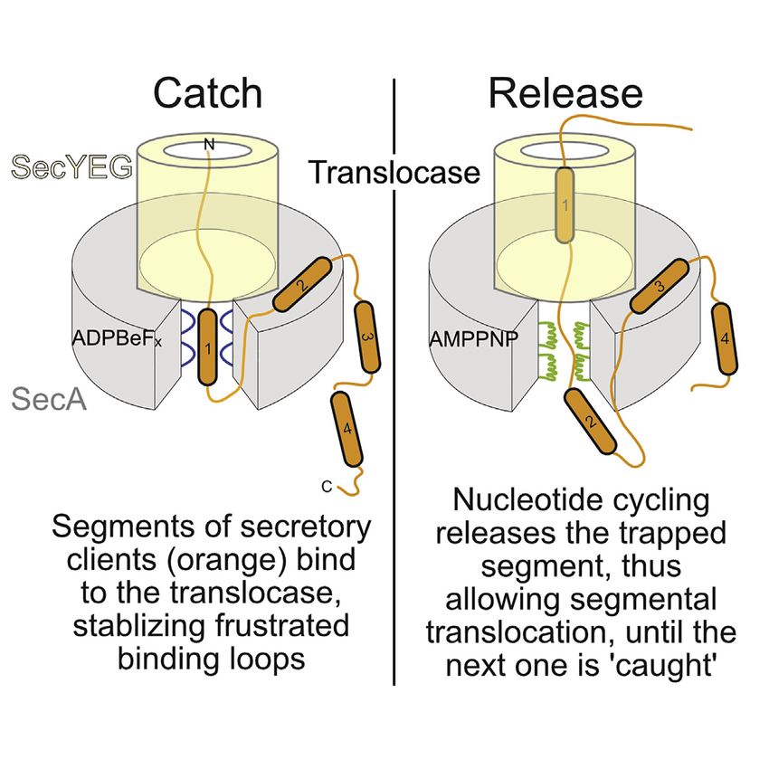
ll
Article                                                                                                                                OPEN ACCESS

                                                                                   (i.e., open plus closed states) in 98% of the active protomers
                                                                                   (lanes 4–6), irrespective of ADP (Figure S3BIII). Signal peptides
                                                                                   alone can replicate this in 85% of the active protomers (Figure 3A,
                                                                                   lanes 7–9). The already triggered SecYPrlA4EG:SecA2 exhibits
                                                                                   clamp closing in the absence of preprotein or signal peptide
                                                                                   (lanes 10–12). Signal peptide-driven clamp closing (Figure 3A,
                                                                                   lanes 7–9) is not accompanied by detectable secondary structure
                                                                                   or flexibility changes inside the PBD or nucleotide cleft (Fig-
                                                                                   ure 2A). This rigid body motion is rather uncoupled from nucleo-
                                                                                   tide turnovers in the helicase motor (see below). The freely
                                                                                   diffusing wild-type SecA2 maintains its clamp equilibrium at the
                                                                                   wide-open state (Figure 3B, lanes 1–3) and signal peptides
                                                                                   cannot close it (Figure S3BI). In contrast, in diffusing, spontane-
                                                                                   ously triggered SecAPrlDI, clamp equilibria shift toward closed
                                                                                   states in the absence of channel or signal peptides (Figure 3B,
                                                                                   lanes 4–6) but less so in SecAPrlDII that require the channel for trig-
                                                                                   gering (lanes 7–9; Figures S3BIVb and S3BIVc and S3C).
                                                                                      To test if NBD2-PBD interaction in the closed clamp is func-
                                                                                   tionally important, we mutated the conserved 3b-tipPBD that
                                                                                   binds NBD2 (Figure S3A). The generated SecA(TGR342AAA)
                                                                                   failed to become triggered by signal peptide (Figure 3C,
                                                                                   lane 4), SecYPrlA4 (lane 6), or their combination (lane 7).
                                                                                   SecA(TGR342AAA) binds to channel/preproteins (Figure S4A)
                                                                                   yet fails to stimulate its ATPase, secreted in vitro or in vivo (Fig-
                                                                                   ures S4B–S4D).
                                                                                      Furthermore, we locked the clamp in the closed state through
                                                                                   engineered disulfides (Chatzi et al., 2017; Sardis et al., 2017) and
                                                                                   tested functionality. The SecAlocked closed was permanently trig-
                                                                                   gered, independently of channel or preprotein (Figure 3D, lanes
                                                                                   1–3), akin to SecAPrlDI mutants (Figure S2C, lanes 4–5). Reduc-
                                                                                   tion of the disulfide reinstated a channel plus preprotein require-
                                                                                   ment for triggering (Figure 3D, lanes 4–6).
                                                                                      Signal peptide-driven clamp closing and increased motif IVa
                                                                                   dynamics underlie translocase triggering.

                                                                                   The signal-peptide cleft crosstalks to motif IVa via two
                                                                                   main H-bond pathways
                                                                                   To determine how clamp closing might allow the signal peptide
                                                                                   binding cleft to crosstalk with motif IVa, we determined the H
                                                                                   bond networks, including water-mediated bridging, between
                                                                                   the two allosterically connected sites. In all simulations motif
Figure 3. Clamp closing underlies translocase triggering                           IVa (Figures 3E and 3F, red spheres) interconnected within a
(A and B) The distribution of population percentages of the preprotein clamp in
                                                                                   local H bond network extending to most SecA’s residues (Krish-
channel-bound (A) and free (B) SecA2, determined by confocal smFRET
(Krishnamurthy et al., 2021). In channel-bound conditions, only data for the       namurthy et al., 2021). Graph analysis determined the most
active, channel-bound protomer (blue oval) are shown. n R 3; mean (±SEM).          frequently visited or shortest possible H bond pathways that
Also see Figure S3.                                                                could be potentially altered along the reaction coordinate of
(C and D) Activation energy (Ea) of wild-type SecA, SecA(TGR342AAA) (C) and a      SecA. Through these the signal peptide cleft in PBD (purple
double cysteine SecA derivative (D) under the indicated conditions; oxidized =     spheres) could communicate with motif IVa. Two main routes
locked closed; reduced = unlocked open clamp.
                                                                                   were proposed. One, via the NBD2Joint/PBDBulb interface of the
(E and F) H bond pathways connecting motif IVa (red) to the signal peptide cleft
(purple) through the stem (E; orange), or the PBD-NBD2 interface (F; cyan),
                                                                                   closed clamp (Figure 3F, cyan spheres), was experimentally
derived from graph analysis of MD simulations of ecSecA2VDA with an open           tested above. The other, via the PBDStem/a8 interface that binds
gate2. PDB:2VDA                                                                    mature domains and interconnects to the second half of gate2
                                                                                   (Y134; Figure 3E, orange spheres), was tested below.
or apart (high and low FRET, respectively; Figures S3A and
S3B).                                                                              The signal-peptide cleft crosstalks to motif IVa through
  In SecYEG:SecA2 the PBD of the active protomer samples all                       the stem/a8 interface
three states, with a preference for the wide open (Figure 3A,                      During open-closed state transitions the stem/a8 interface,
lanes 1–3; Figure S3BIIa). Preprotein binding closes the clamp                     which binds mature domains (Chatzi et al., 2017), is

                                                                                                         Cell Reports 38, 110346, February 8, 2022 5
ll
      OPEN ACCESS                                                                                                                       Article

                                                                                  conformationally altered (Figures 4A and S3A). As this interface
                                                                                  is pried open, local hydrophobic stem b strands/a8 interactions
                                                                                  likely change. The interface extends to a three strand b-sheet
                                                                                  with the highly dynamic b6Stem and b24C-tail and involves
                                                                                  L187a8 that packs against A373 of b12Stem (Figure 4A).
                                                                                     To alter hydrophobic packing at the stem/a8 interface, we
                                                                                  substituted A373Stem with large hydrophobic residues (V, I, F)
                                                                                  and L187a8 with V, I, A. All derivatives displayed Prl phenotypes
                                                                                  in vitro (SecAPrlDII; Figure 4B, lanes 3–8) or in vivo (Figure 4C,
                                                                                  lanes 6–8), with L187A as the weakest one. A373V has been
                                                                                  the only known Prl outside the nucleotide binding cleft (Flower
                                                                                  et al., 1994; Huie and Silhavy, 1995). M191A (a control), located
                                                                                  one turn after L187 at the end of a8 and of the stem/a8 interface
                                                                                  (Figure 4A), was not a Prl (Figure 4B, lane 9). All derivatives were
                                                                                  functional in vivo (Figure S4E).
                                                                                     Signal peptides may alter hydrophobic packing at the mature
                                                                                  domain binding patch on the stem/a8 interface.

                                                                                  Binding of mature domains drives ADP release and ATP
                                                                                  turnover
                                                                                  Mature domains bind to the stem/a8 interface (Chatzi et al.,
                                                                                  2017) (Figure S1B, orange surface). Yet, only preproteins stimu-
                                                                                  late ADP release (Figure 1B) and ATP turnover on the translocase
                                                                                  (Figure 4D, lane 3) (Karamanou et al., 2007). Alone (lane 4; in
                                                                                  excess: lane 7) or with signal peptide added in trans (lane 5),
                                                                                  mature domains can poorly stimulate the ATPase (1.5, 2, and
                                                                                  3-fold, respectively). In contrast, the signal peptide cannot
                                                                                  (lane 6; in excess: lane 8). Mature domains alone marginally in-
                                                                                  crease the local dynamics of the helicase motor in SecYEG:
                                                                                  SecA2:ADP (Figure S4F). Minor effects are also seen upon trans
                                                                                  addition of mature domain plus signal peptide (Figure S4G), thus
                                                                                  explaining their inability to fully stimulate the ATPase when the
                                                                                  two preprotein moieties are unconnected.
                                                                                     We presumed that signal peptides might optimize the stem/a8
                                                                                  interface for mature domains to bind and stimulate ATP turnover.
                                                                                  So, we screened for mutant derivatives at this interface with high
                                                                                  basal ATPase activity, mimicking the mature domain-bound
                                                                                  state. L187A and A373V (SecAPrlDI; Figure 4B) displayed
                                                                                  elevated ATPase compared to free SecA2 (Figure 4D, lanes
                                                                                  9–10), while derivatives at the back of a8 were non-functional
                                                                                  (Figures S4H–S4J).
                                                                                     The conformational effects on stem/a8 and the motor ATPase
                                                                                  seem coupled. The mature domain binding site residue M191
                                                                                  (Figure 4A) disentangled them. Freely diffusing SecA(M191A)
                                                                                  displayed elevated basal (Figure 4D, lane 11) and hyper-stimu-
Figure 4. Mature domain-driven ADP release and ATP turnovers                      lated translocation (Figure S4K, lane 5) ATPase but was neither
(A) Structure (aligned on NBD1) and residues at the stem/a8 region including
                                                                                  a Prl (Figure 4B, lane 9) nor had changed motif IVa dynamics (Ta-
b24C-tail. DGex values are shown for the SecYEG:SecA2:preprotein state in the
open (left; ecSecA2VDA; PDB:2VDA) or closed (right; ecSecA2VDA MD model)
                                                                                  ble S1). Stem/a8 interfacial residues appear critical for mature
clamp.                                                                            domains to control ADP release from the motor.
(B) Activation energy (Ea) of indicated stem SecAPrlDII mutants in channel-          The roles of signal peptides and mature domains in translo-
primed states, compared to wild-type translocase (as in Figure 3B).               case activation are inter-connected but divergent and converge
(C) In vivo translocation of proPhoA or pro(L8Q)PhoA (defective signal peptide)   at stem/a8. This hub couples conformational cues from signal
by the indicated translocases. n = 6; mean values (±SEM).                         peptide-driven clamp closing to promote mature domain bind-
(D) The ATPase activity of SecA in basal (B), membrane bound (M), and
                                                                                  ing, ADP release, and motif IVa dynamics in the motor.
translocating conditions (T). Signal peptide (30 or 60 [excess] mM; mature
domain (PhoAcys-; 20 or 40 [excess] mM). n = 3–6; mean values (±SEM).
                                                                                  Nucleotides finely control the intrinsic dynamics of SecA
                                                                                  Co-ordinated signal peptide/mature domain docking releases
                                                                                  ADP from SecA, allowing fresh ATP binding. Using analogs as

6 Cell Reports 38, 110346, February 8, 2022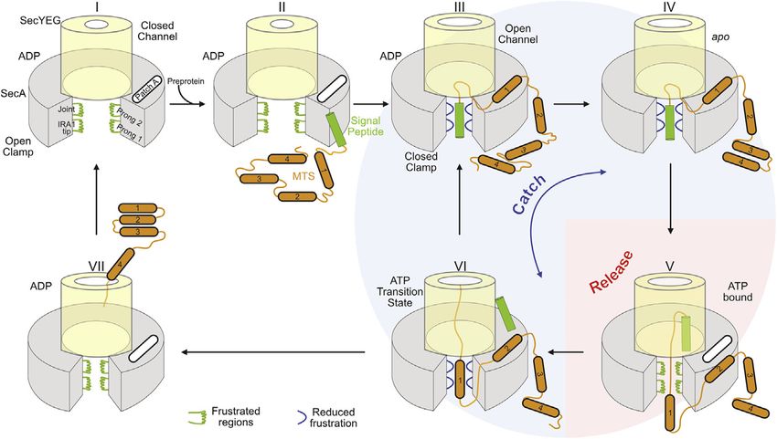
ll
Article                                                                                                                              OPEN ACCESS

                                                                                    ing releases bound ADP (Figure 1B) and causes widespread
                                                                                    elevated dynamics in the motor and localized ones in the stem,
                                                                                    scaffold, IRA1, and PBD (Figure 5AI). The dynamics of SecYEG:
                                                                                    SecA2:AMPPNP and SecYEG:SecA2:ADP differ marginally (Fig-
                                                                                    ures 5AII and S5CII). ADP:BeFX stabilizes additionally the motor
                                                                                    (motifs I, IVa, V/Va) and the clamp (stem, a13, and the 3b-tipPBD
                                                                                    that binds NBD2; Figure 5AIII). Motif Va harbors the R509 finger,
                                                                                    crucial in g-phosphate recognition and regulation of motor con-
                                                                                    formations (Keramisanou et al., 2006). Our results agree with the
                                                                                    crystal structure of SecYEG:SecA:ADP:BeFx (Figure S6A) (Zim-
                                                                                    mer et al., 2008) where PBDbulb and NBD2 home into each other
                                                                                    while PBD3b-tip flips toward NBD2motifIVa, salt-bridging R342 to
                                                                                    E487 (Figures S6B and S6C; Video S1). Conversion to the ADP
                                                                                    state reverses rigidification through minor local changes (Fig-
                                                                                    ure S6DVIII). Some of these intra-protomeric changes enhance
                                                                                    dynamics of the dimerization interface (Figure S5CI), suggesting
                                                                                    preparation for dissociation (Gouridis et al., 2013). These effects
                                                                                    are specific to channel-bound SecA2. ADP:BeFX contacts in the
                                                                                    soluble SecA2 motor are weak (Table S1) with higher dynamics
                                                                                    than ADP (Figure S6DIV).
                                                                                       The Q motif, that tightly binds the immutable adenine ring
                                                                                    (Figure S1E), showed negligible dynamics in the presence of
                                                                                    nucleotide consistent with similar motor affinities for ATP ana-
                                                                                    logs. It is the mutable g-phosphate in AMPPNP and ADP:BeFX
                                                                                    that bind to NBD2 and stabilize different motor conformations,
                                                                                    while ADP does not (Figures S1E and S5A). Missing g-phos-
                                                                                    phate contacts weaken NBD2-ADP association, allow higher
                                                                                    nucleotide mobility inside the pocket, and increase motif I dy-
                                                                                    namics (Figure S6DVIII).
                                                                                       Despite only minor chemical differences, nucleotides stabilize
                                                                                    unique conformational SecA states, largely via NBD2/g-phos-
                                                                                    phate, and promote multiple, minor local dynamics changes.
                                                                                    None of these significantly alter PBD motion (Figure S6E).

                                                                                    Preproteins regulate nucleotide-controlled dynamics in
Figure 5. Nucleotide states and preproteins drive distinct translo-                 channel-bound SecA
case conformations                                                                  How do preproteins exploit the nucleotide-regulated dynamics
(A and B) The local dynamics (differential D uptake) of the SecYEG:SecA2 at         of the primed translocase leading to translocation? For this, we
the indicated nucleotide state (I, II and III; ‘‘test’’) were compared to SecYEG:   followed SecYEG:SecA2:nucleotide dynamics (as in Figure 5A)
SecA2:ADP (‘‘reference’’), in the absence (A) or presence (B) of preprotein.
                                                                                    with a bound secretory client (15 mM proPhoA1-122; >50 fold
Orange dashed line in (A) I: PatchA. I and II mapped on surface representations
of a protomer of ecSecA2VDA (open clamp; PDB:2VDA) and ecSecA3DIN (III;
                                                                                    over Kd; Figures 5B and S5D).
closed-flipped clamp; PDB:3DIN).                                                       Motor dynamics with ADP were enhanced (Figures 5BI and
                                                                                    S6C), due to preprotein-induced ADP release (Figure 1B).
                                                                                    Compared to ADP, AMPPNP enhanced the dynamics in mo-
mimics of ATPase cycle stages we examined how they modulate                         tifs I, Ic, III, and VI (Figures 5BII and S1E) and inside the clamp
SecYEG:SecA2 dynamics using HDX-MS (Figure S5A). Nucleo-                            (3b-tip, b24C-tail) and decreased them at the signal peptide
tides bind in a positively charged cleft using residues from                        binding cleft (stem, a10, a13). The contrasting dynamics flank-
both NBD1 and 2, mostly NBD1; b and g-phosphates also con-                          ing PBD suggested that ATP binding divergently affects the
tact NBD2 (Figure S5B) (Krishnamurthy et al., 2021; Papanikolau                     signal peptide and mature domain binding sites. ADP:BeFX
et al., 2007). We monitored the dynamics of the ADP (2 mM; 104-                     reduced dynamics in all NBD2 helicase motifs without
fold excess over Kd), apoprotein (i.e., empty cleft due to prepro-                  affecting NBD1 and rigidified clamp areas (stem, a13, 3b-tip,
tein binding; Figure 1B), ATP (mimic: non-hydrolyzable AMP-                         joint, scaffold; Figure 5BIII). These changes coincide with
PNP), and ATP hydrolysis transition (mimic: ADP:BeFX) (Zimmer                       signal peptide-driven, nucleotide-independent clamp closing
et al., 2008) states. SecYEG:SecA2:ADP dynamics were                                (Figure S3BIII). The preprotein coupled the otherwise uncon-
compared to all other states (Figures 5 and S5C; ‘‘reference,’’                     nected nucleotide-regulated motor dynamics to clamp
top versus bottom).                                                                 motions.
   The suppressed SecA2:ADP dynamics are somewhat relieved                             Nucleotides intimately regulate motor dynamics with minor ef-
by channel priming (Krishnamurthy et al., 2021). Preprotein bind-                   fects on preprotein binding regions. However, clients not only

                                                                                                         Cell Reports 38, 110346, February 8, 2022 7
ll
OPEN ACCESS Article
Figure 6. Translocase binds and regulates
dynamics of preprotein islands
(A–D) Multi-parametric analysis of SecA flexibility
mapped on ecSecA2VDA (open clamp; PDB:2VDA).
C-tail from ecSecA1M6N (PDB:1M6N). (A) DGex
values for SecA2, red hues: high flexibility (i.e.,
DGex = 11–16 kJ/mol). (B) Frustrated regions
(Frustratometer; Parra et al., 2016). (C) Total
displacement of normal modes 7–12 (unweighted
sum) (details in Figure S7C). (D) Predicted intrinsic
disorder (MobiDB-lite aggregator; MobiDB data-
base). Predictions/consensus: high/extensive
multi tool (orange); moderate (Yellow).
(E) Flexibility map of free or translocase-bound
proPhoA1-122 (I) and PhoA23-122 (II) as indicated
(residue level absolute dynamics; percent D up-
take), aligned below a linear map with signal pep-
tide and MTS (mature domain targeting signals) 1
and 2 and known secondary structural features of
native PhoA. Percent D uptake: >90%: hyper-
flexible/disordered residues; 90–60%: increased
flexibility. Purple: dynamics of PhoA23-122 with
signal peptide added in trans. Dashed gray: signal
peptide flexibility in free proPhoA1-122 (from I).
Differences >10% are considered significant.
n = 3; SD values >1% are shown (vertical lines).
(F) Flexibility map of the indicated SecYEG:
SecA2:proPhoA1-122 regions (with percent D up-
take differences between states) as the translo-
case goes through the nucleotide cycle (right, as
colored): ADP-ground state; ADP release (apo;
light b), ATP bound (mimic: AMPPNP), and ATP
hydrolysis transition state (mimic: ADP:BeFX).
strengthen nucleotide effects at the motor, but they also cause We further probed intrinsic dynamics in SecA’s clamp using
nucleotide-modulated dynamics at multi-valent preprotein bind- normal mode analysis (NMA), a mathematical description of
ing sites for successful translocation. atomic vibrational motions and protein flexibility (Bahar
et al., 2010; Kovacs et al., 2004). The model generates a set
Locally frustrated prongs in the SecA clamp allow client of normal modes, where all Ca atoms are oscillating with the
promiscuity same frequency. The lowest frequency normal modes
The translocase handles hundreds of dissimilar clients presum- contribute the most to protein domain dynamics and the asso-
ably binding to the same SecA sites. For universal chaperone ciated Ca displacement is calculated (Figure S7C; Hinsen,
promiscuity, frustrated, dynamic elements in chaperones may 1998; Tiwari et al., 2014). Motif IVa, JointNBD2, 3b-tipPBD,
recognize frustrated regions on non-folded clients (He et al., and the signal peptide binding cleft show maximum displace-
2016; Hiller, 2019). Dynamic regions seen around the SecA ment during vibrational motions (Figure 6C; blue shades) and
clamp might exert similar mechanisms. To test this we compared practically coincide with the HDX-MS-determined dynamic
the dynamic islands determined by HDX-MS (Figure 6A, orange/ islands and the frustrated regions. Finally, we tested the
red) to frustrated predicted regions (Parra et al., 2016). Most frus- intrinsic disorder propensity of SecA using online predictors
trated inter-residue contacts (Figure 6B; green lines) closely such as the MobiDB database (Piovesan et al., 2021). Several
overlap with the experimentally determined dynamic islands of the flexible regions identified by HDX-MS yield high pre-
(Figure 6A). Clamp closing upon signal peptide binding (Fig- dicted disorder scores (Figure 6D), eight of which (including
ure 3A, lanes 7–12) would allow the 3b-tip, prong1, and joint to in the PBD, NBD2, IRA1-tip, and the C-tail; Orange) were
interact, forming a contiguous frustrated region (Figure S7AII) from a wide tool consensus.
that could trap clients through local favorable interactions. In Altered dynamics in the flexible prongs are subject to direct
the ATP hydrolysis transition state (ADP:BeFX; Figure S7B), nucleotide modulation and promiscuous, local, rigidifying inter-
two parallel regions of frustration and the closed clamp could actions with non-folded clients. These would couple client catch
enclose client chains during channel entry. and release to the ATPase cycle.
8 Cell Reports 38, 110346, February 8, 2022ll
Article OPEN ACCESS
Signal peptide-driven clamp closing enhances mature of the signal peptide throughout the ATPase cycle (Figure 6FI)
domain binding are consistent with the client remaining largely tethered to the
Nucleotide-regulated motor or clamp dynamics allow for multi- translocase via its signal peptide, while mature domain parts
valent localized, transient interactions of non-folded clients associate or dissociate more dynamically (Burmann et al.,
with SecA. To probe how dynamics translate to client transloca- 2013). During ATP hydrolysis transition, all client regions that
tion steps, we monitored the dynamics of proPhoA1-122 by HDX- bind SecA become rigidified (Figures 6FI–6FIII), likely reflecting
MS (Figure S7D). This client contains three necessary and tight trapping inside the ADP:BeFX-driven rigidified closed-flip-
sufficient elements for translocase binding and secretion: a ped clamp (Figure 5BIII). Upon ATP to ADP hydrolysis the trans-
signal peptide and two mature domain targeting signals locase becomes more dynamic (Figure 5BIV) and modestly
(MTS1-2; Figure 6E, top) (Chatzi et al., 2017). relaxes its grasp on the client (Figure 6F, brown to green). In
In solution, proPhoA1-122 is highly flexibile (Figure 6EI, gray), this recreated ATP cycle, every translocase state has distinct
consistent with extensive non-foldedness (>90% D uptake; Fig- ‘‘catch and release’’ consequences on each on the three client
ure 6EI, pink area) and lack of stable secondary structure (typi- islands.
cally 20%–40% D uptake; Tsirigotaki et al., 2017b). Only three
islands show some backbone stabilization signifying H DISCUSSION
bonding/transient acquisition of secondary structure (85% alter dynamics to ensure translocation.
D uptake). These rigidified islands reflect stabilized H bonds Evolution prevented a readily activated SecA2, favoring quies-
either within secondary structure elements or externally and cence (Krishnamurthy et al., 2021). The translocase conforma-
are a direct demonstration of binding to SecA. Mature domain tional ensemble becomes activated by regulating pre-existing
binding to SecYEG:SecA2 exhibited less rigidification (Fig- subunit dynamics (Ahdash et al., 2019; Corey et al., 2019). The
ure 6EII, compare black to orange and to Figure 6EI) that barely full compendium of pre-existing conformations arise from ther-
increased by trans addition of signal peptide (Figure 6EII, purple mal atomic vibrations (Figures 6C and S7C) (Bahar et al., 2010;
line) or use of SecYPrlA4:SecA2 (Figure S7E, purple), and it never Chen and Komives, 2021; Dobbins et al., 2008; Smit et al.,
reached that seen with preprotein. These data rationalize why 2021c). These are differentially sampled over minor energetic
preprotein moieties must be covalently connected for maximal barriers (Henzler-Wildman and Kern, 2007) tipped over by li-
translocase interaction. gands (e.g., ATP, preproteins) that bias overpopulation of certain
equilibrium states. These minor energetic requirements allow
The ATP cycle selectively alters SecA interaction with point mutations to mimic ligand binding effects (e.g., triggering,
preprotein islands ATPase stimulation, enhanced dynamics) (Figure 2C) (Gouridis
To dissect how the ATPase cycle influences the dynamics of the et al., 2009, 2013; Karamanou et al., 2007).
islands of proPhoA1-122 that bind SecA (Figures 6FI–6FIII), we SecA bears two distinct modules, an ATPase hardwired onto a
monitored them in the four translocase:nucleotide analog con- preprotein clamp (Figure 7I, gray), which assemble onto the
formations (Figure 6F, right). channel (yellow). Both modules display distinct local and domain
When transitioning from the ADP-bound to the apoprotein to dynamics, largely uncoupled from each other and each finely
the AMPPNP-bound translocase, the signal peptide region controlled by a ligand. Non-folded clients couple the dynamics
shows slight rigidification and a significant one on the ADP: of the two modules. By binding to multiple SecA sites, clients
BeFX-bound translocase (Figure 6FI, red to brown). MTS1 dy- physically bridge the modules, all the while tuning their dynamics
namics are unchanged when transitioning from the ADP-bound (Figure 7II). Gate2 and stem control this coupling and regulate
to the apo translocase but increase in the AMPPNP state (Fig- the transduction of conformational signals downstream, to effec-
ure 6FII, blue to red) and decrease in the ADP:BeFX state (red tuate enzymatic activation, first with ADP release (Figures 7III
to brown). For MTS2, as the ATPase cycle progresses from and 7IV), followed by fresh ATP binding (Figure 7V). The nucleo-
ADP-bound to apo to AMPPNP state, its dynamics increase tide state of the motor dictates the conformation of frustrated
incrementally (Figure 6FIII, green to blue to red) but decrease prongs in the clamp. As a result, the prongs catch and release
significantly in the ADP:BeFX state (red to brown). the client chain, at multiple locations, biasing its forward motion
Our results suggest that SecA binds all three client elements in (Figures 7III and 7VI).
the ADP and apo states of a translocation cycle. ATP binding Despite overall similarities between polypeptides, only secre-
(mimic: AMPPNP) enhances SecA dynamics (Figure 5BII) and tory preproteins are legitimate translocase clients. Alone, signal
loosens its grip on the mature domain. The decreased dynamics peptides and mature domains alter distinctly but partially the
Cell Reports 38, 110346, February 8, 2022 9ll
OPEN ACCESS Article
Figure 7. Catch and release model for pre-
protein translocation
See text for details. Green: enhanced and blue:
reduced frustrated regions. PatchA: MTS binding
site.
(Keramisanou et al., 2006; Papanikolau
et al., 2007; Sianidis et al., 2001).
An underappreciated feature of secre-
tory clients is their elevated intrinsic dy-
namics, which when reduced, abrogated
secretion (Sardis et al., 2017). HDX-MS
uniquely captured islands of dynamics in
the secretory chain (e.g., signal peptide,
early mature domain, and MTSs) (Figures
6EI and 7II) (Sardis et al., 2017) that
respond to the transient, nucleotide
dynamics of the switches, with inadequate functional effects states of the translocase (Figure 6E). How does SecA, a weak
(Figures 3 and 4). Signal peptides close the clamp (Figure 3A, cytoplasmic and strong membrane-associated holdase (Gouri-
lanes 10–12), partially elevate gate2 dynamics (Figures 2A and dis et al., 2009), promiscuously recognize its clients? Chaper-
2B), and loosen the channel (Figures 7II and 7III) (Fessl et al., ones may recognize frustrated regions in clients through their
2018; Knyazev et al., 2014). Mature domains partially increase own frustrated regions (He and Hiller, 2019; Hiller, 2019), as
the motor’s dynamics (Figure S4G) and drive inefficient ADP does SecA (Figure 6B). Client’s or chaperone’s frustrated re-
release (Figure 4D). It is the synergy between the two covalently gions can sample a wide conformational and sequence space,
connected moieties that secures adequate motor dynamics for until they interact tightly (Ferreiro et al., 2014; He and Hiller,
ADP release (Figures 1B, 1CII, and 1CIII), a key rate limiting 2018). Thus, a chaperone can promiscuously interact with hun-
step (Fak et al., 2004; Robson et al., 2009; Sianidis et al., dreds of non-folded clients without rigid lock-and-key recogni-
2001). This mechanism expels random cytoplasmic proteins tion. In SecA, the four highly dynamic, locally frustrated prongs
from secretion. around the clamp (Figures 6A and 6B and 7I–7III), the electroneg-
ATP binding to the motor initiates translocation (Figure 7V). ativity of the clamp (Figure S5B), and its adjustable width due to
As the translocase cycles through nucleotide states it manipu- PBD/NBD2 rigid body motions enhance plasticity and interac-
lates the client’s dynamics. The ATP-bound translocase binds tions, potentially accommodating partially folded structures
signal peptides, releasing mature domain segments (Figure 6E, (Tsirigotaki et al., 2018). Client-translocase interactions reduce
red). In the transition state it catches both signal peptide and dynamics both in the frustrated SecA prongs (Figures 5BIII and
mature domain targeting signals (MTS) regions (Figures 5BIII 7, green) and the corresponding frustrated elements of the client
and 6B and 6E). In the ADP-bound state, a succeeding region (Figure 6E). Such a mechanism permits high affinity yet transient
from the bound client will induce ADP release (Figure 1B) to interactions with the client and fast release, formulating an
restart an ATP hydrolysis cycle. Cycles repeat for as long as optimal solution for secretion. Tighter recognition of unfolded cli-
succeeding mature domain segments are available to bind to ents might have impeded SecA’s processivity.
the translocase and drive ADP release (Figures 7III–7VI). In their SecA biases vectorial forward motion, uncommon in soluble
absence, SecA:ADP remains quiescent, diffuses to the cyto- chaperones. Presumably, local interactions of frustrated
plasmic pool, and dimerizes (Gouridis et al., 2013). Secretion prongs are sufficient to stall backward sliding of the exported
is achieved through such repetitive client catch and release cy- chain yet loose enough to allow forward motion of untethered
cles regulated by nucleotide turnovers (Figure 7). The mecha- segments through the channel. This catch and release mech-
nism is generic for initial as well as subsequent processive anism is important for translocation. Release cycles allow
translocation steps (Figure 1AV). Later in translocation, cleaved chain segments to enter the channel by Brownian motion (Al-
signal peptides are replaced by hydrophobic MTSs (Chatzi len et al., 2016) catch cycles would bind a downstream
et al., 2017) (Figure 7VI). segment and prevent back-slippage (Figures 7V and 7VI),
SecA dynamics sense the slightest chemical change in nucle- like a ‘‘brake’’ (Vandenberk et al., 2019). This mechanism is
otides and invite a rethink of the role of ATP hydrolysis in trans- also compatible with a power stroke, if catching actively
location. Rather than driving major deterministic strokes, carries along chain segments into the channel (Catipovic
nucleotides subtly, stepwise, bias SecA’s intrinsic dynamics et al., 2019) or with a continuum ratchet, SecA moving sto-
(Figure 5) by affecting residues that line the nucleotide cleft. chastically along a periodic potential, coupled to ATP cycling,
The limited, transient interaction of the g-phosphate of ATP providing the required time correlation for net vectorial motion
and its transition states with NBD2 bias motor conformational (Magnasco, 1993). All models depend on catch signals like
cycles, which stop when it is absent (Figures 1B and 7I–7VI) MTSs (Figures 6E and 6F).
10 Cell Reports 38, 110346, February 8, 2022ll
Article OPEN ACCESS
A short preprotein with limited folding permitted dissection of SUPPLEMENTAL INFORMATION
translocase binding from folding propensities but the same
Supplemental information can be found online at https://doi.org/10.1016/j.
fundamental principles revealed likely apply to longer clients.
celrep.2022.110346.
Most secretory proteins are flexible with delayed folding but
some may rapidly fold (Arkowitz et al., 1993; Gupta et al.,
ACKNOWLEDGMENTS
2020; Tsirigotaki et al., 2018). Translocase dynamics may
counter such inherent folding forces alone, or in concert with We are grateful to: T. Cordes for sharing software for smFRET data analysis.
chaperones (De Geyter et al., 2020; Fekkes et al., 1997). Our research was funded by grants (to AE): MeNaGe (RUN #RUN/16/001;
KU Leuven); ProFlow (FWO/F.R.S. - FNRS "Excellence of Science - EOS" pro-
Limitations of the study gram grant #30550343); DIP-BiD (#AKUL/15/40 - G0H2116N; Hercules/FWO);
CARBS (#G0C6814N; FWO); Profound (WoG Research Training Network,
SecA2 binds asymmetrically to SecYEG, using one of its proto-
FWO, Protein folding/non-folding and dynamics; #W002421N) and (to AE
mers. SecA2 is essential to initiate translocation, but later mono- and SK): FOscil (ZKD4582-C16/18/008; KU Leuven) and (to A-NB): by the
merizes. The ensemble nature of HDX-MS cannot delineate the Excellence Initiative of the German Federal and State Governments via
differences in dynamics between the two protomers. This will the Freie Universität Berlin, and by allocations of computing time from the
require immobilized single-molecule techniques. North-German Supercomputing Alliance, HLRN. S.Kr. was an FWO
Here, we monitored client protein dynamics using ATP ana- [PEGASUS]2 MSC fellow; N.E. was an MSCA SoE FWO fellow; J.H.S. is a
PDM/KU Leuven fellow; G.G. was a Rega Foundation postdoctoral program
logs that are assumed to mimic distinct stages of the nucleotide
fellow. This project has received funding from the Research Foundation Flan-
cycle during translocation. These analogs may not accurately ders (FWO) and the European Union’s Horizon 2020 research and innovation
represent stages in the ATP hydrolysis cycle, therefore weak- program under the Marie Sk1odowska-Curie grant agreements No. 665501
ening our interpretation. Furthermore, to synchronize ATP- and 195872.
dependent translocation of all clients in the ensemble and follow
their dynamics by HDX-MS during processive translocation re- AUTHOR CONTRIBUTIONS
mains challenging and is not explored in this study.
This study monitored translocation steps using the minimal S.Kr. purified proteins and membranes, did biochemical and fluorescence as-
functional translocase SecYEG:SecA2 and a single preprotein. says, designed and performed HDX-MS work and data analysis. M.F.S. and
K.E.C. purified proteins, performed molecular biology, in vivo and in vitro
The model we present here may not fully explain in vivo delivery
biochemical and biophysical assays. N.E. purified and labeled proteins and
and secretion of multiple preproteins from the ribosome to the performed smFRET experiments and data analysis. J.H.S. developed PyHDX
translocase that involves chaperones and feedback mechanisms. software and analyzed HDX-MS data, adapted FRET burst analysis for Micro-
time200 output data, and performed NMA analysis. K.K. performed MD simu-
STAR+METHODS lations and graph analysis of H bond networks. G.G. performed biochemical,
molecular biology, and biophysical assays, analyzed data and advised on
smFRET. A.G.P. performed molecular cloning and mutagenesis. A.N.B. set
Detailed methods are provided in the online version of this paper up and supervised the MD simulations and graph analysis. S.K. designed
and include the following: and supervised molecular biology experiments, purified proteins, performed
biochemical and biophysical assays and data analysis. A.E. did structure
d KEY RESOURCES TABLE and data analysis and designed experiments. S.Kr. and A.E. wrote the first
d RESOURCE AVAILABILITY draft and finalized it with contributions from S.K., A.N.B., J.H.S., and N.E. All
B Lead contact authors reviewed and approved the final manuscript. A.E. and S.K. conceived
B Materials availability and managed the project.
B Data and code availability
d EXPERIMENTAL MODEL AND SUBJECT DETAILS DECLARATION OF INTERESTS
d METHOD DETAILS
The authors declare no competing interests.
B List of buffers
B Molecular cloning
Received: August 31, 2021
B Protein purification Revised: November 22, 2021
B MANT-ADP release assays Accepted: January 12, 2022
B Dynamics of the Sec translocase by HDX-MS Published: February 8, 2022
B HDX-MS data visualization
B Dynamics of client proteins by HDX-MS SUPPORTING CITATIONS
B Determination of DGex values
The following reference appears in the Supplemental information: Baker et al.,
B Single-molecule fluorescence microscopy and PIE
2001; Lacabanne et al., 2020.
B H-bonding graph analysis
B Normal mode analysis and intrinsic disorder prediction
REFERENCES
B Miscellaneous
d QUANTIFICATION AND STATISTICAL ANALYSIS Ahdash, Z., Pyle, E., Allen, W.J., Corey, R.A., Collinson, I., and Politis, A.
B MANT-ADP release assays (2019). HDX-MS reveals nucleotide-dependent, anti-correlated opening and
B HDX-MS data closure of SecA and SecY channels of the bacterial translocon. Elife 8, e47402.
B Biochemical assays Allen, W.J., Corey, R.A., Oatley, P., Sessions, R.B., Baldwin, S.A., Radford,
B smFRET data S.E., Tuma, R., and Collinson, I. (2016). Two-way communication between
Cell Reports 38, 110346, February 8, 2022 11ll
OPEN ACCESS Article
SecY and SecA suggests a Brownian ratchet mechanism for protein transloca- Ferreiro, D.U., Komives, E.A., and Wolynes, P.G. (2014). Frustration in biomol-
tion. Elife 5, e15598. ecules. Q. Rev. Biophys. 47, 285–363.
Arkowitz, R.A., Joly, J.C., and Wickner, W. (1993). Translocation can drive the Fessl, T., Watkins, D., Oatley, P., Allen, W.J., Corey, R.A., Horne, J., Baldwin,
unfolding of a preprotein domain. EMBO J. 12, 243–253. S.A., Radford, S.E., Collinson, I., and Tuma, R. (2018). Dynamic action of the
Avellaneda, M.J., Koers, E.J., Naqvi, M.M., and Tans, S.J. (2017). The chap- Sec machinery during initiation, protein translocation and termination. Elife
erone toolbox at the single-molecule level: from clamping to confining. Protein 7, e35112.
Sci. 26, 1291–1302. Flechsig, H., and Mikhailov, A.S. (2019). Simple mechanics of protein ma-
chines. J. R. Soc. Interf. 16, 20190244.
Bahar, I., Lezon, T.R., Bakan, A., and Shrivastava, I.H. (2010). Normal mode
analysis of biomolecular structures: functional mechanisms of membrane pro- Flower, A.M., Doebele, R.C., and Silhavy, T.J. (1994). PrlA and PrlG suppres-
teins. Chem. Rev. 110, 1463–1497. sors reduce the requirement for signal sequence recognition. J. Bacteriol. 176,
5607–5614.
Baker, N.A., Sept, D., Joseph, S., Holst, M.J., and McCammon, J.A. (2001).
Electrostatics of nanosystems: application to microtubules and the ribosome. Fuxreiter, M., Toth-Petroczy, A., Kraut, D.A., Matouschek, A., Lim, R.Y., Xue,
Proc. Natl. Acad. Sci. U.S.A 98, 10037–10041. B., Kurgan, L., and Uversky, V.N. (2014). Disordered proteinaceous machines.
Chem. Rev. 114, 6806–6843.
Bauer, B.W., and Rapoport, T.A. (2009). Mapping polypeptide interactions of
the SecA ATPase during translocation. Proc. Natl. Acad. Sci. U.S.A 106, Galletto, R., Jezewska, M.J., Maillard, R., and Bujalowski, W. (2005). The
20800–20805. nucleotide-binding site of the Escherichia coli DnaC protein: molecular topog-
raphy of DnaC protein-nucleotide cofactor complexes. Cell Biochem. Biophys.
Burmann, B.M., Wang, C., and Hiller, S. (2013). Conformation and dynamics of
43, 331–353.
the periplasmic membrane-protein-chaperone complexes OmpX-Skp and
tOmpA-Skp. Nat. Struct. Mol. Biol. 20, 1265–1272. Gelis, I., Bonvin, A.M., Keramisanou, D., Koukaki, M., Gouridis, G., Karama-
nou, S., Economou, A., and Kalodimos, C.G. (2007). Structural basis for
Catipovic, M.A., Bauer, B.W., Loparo, J.J., and Rapoport, T.A. (2019). Protein
signal-sequence recognition by the translocase motor SecA as determined
translocation by the SecA ATPase occurs by a power-stroke mechanism.
by NMR. Cell 131, 756–769.
EMBO J. 38, e101140.
Gouridis, G., Karamanou, S., Gelis, I., Kalodimos, C.G., and Economou, A.
Chatzi, K.I., Gouridis, G., Orfanoudaki, G., Koukaki, M., Tsamardinos, I., Kar-
(2009). Signal peptides are allosteric activators of the protein translocase. Na-
amanou, S., and Economou, A. (2011). The signal peptides and the early
ture 462, 363–367.
mature domain cooperate for efficient secretion. FEBS J. 278, 14.
Gouridis, G., Karamanou, S., Koukaki, M., and Economou, A. (2010). In vitro
Chatzi, K.E., Sardis, M.F., Tsirigotaki, A., Koukaki, M., Sostaric, N., Konijnen-
assays to analyze translocation of the model secretory preprotein alkaline
berg, A., Sobott, F., Kalodimos, C.G., Karamanou, S., and Economou, A.
phosphatase. Methods Mol. Biol. 619, 157–172.
(2017). Preprotein mature domains contain translocase targeting signals that
are essential for secretion. J. Cel. Biol. 216, 1357–1369. Gouridis, G., Karamanou, S., Sardis, M.F., Scharer, M.A., Capitani, G., and
Economou, A. (2013). Quaternary dynamics of the SecA motor drive translo-
Chen, W., and Komives, E.A. (2021). Open, engage, bind, translocate: the
case catalysis. Mol. Cell 52, 655–666.
multi-level dynamics of bacterial protein translocation. Structure 29, 781–782.
Gupta, R., Toptygin, D., and Kaiser, C.M. (2020). The SecA motor generates
Corey, R.A., Ahdash, Z., Shah, A., Pyle, E., Allen, W.J., Fessl, T., Lovett, J.E.,
mechanical force during protein translocation. Nat. Commun. 11, 3802.
Politis, A., and Collinson, I. (2019). ATP-induced asymmetric pre-protein
folding as a driver of protein translocation through the Sec machinery. Elife Hanson, J., Yang, Y., Paliwal, K., and Zhou, Y. (2017). Improving protein disor-
8, e41803. der prediction by deep bidirectional long short-term memory recurrent neural
networks. Bioinformatics 33, 685–692.
Cryar, A., Groves, K., and Quaglia, M. (2017). Online hydrogen-deuterium ex-
change traveling wave ion mobility mass spectrometry (HDX-IM-MS): a sys- Hartl, F.U., Lecker, S., Schiebel, E., Hendrick, J.P., and Wickner, W. (1990).
tematic evaluation. J. Am. Soc. Mass Spectrom. 28, 1192–1202. The binding cascade of SecB to SecA to SecY/E mediates preprotein targeting
to the E. coli plasma membrane. Cell 63, 269–279.
De Geyter, J., Portaliou, A.G., Srinivasu, B., Krishnamurthy, S., Economou, A.,
He, L., and Hiller, S. (2018). Common patterns in chaperone interactions with a
and Karamanou, S. (2020). Trigger factor is a bona fide secretory pathway
native client protein. Angew. Chem. Int. Ed. Engl. 57, 5921–5924.
chaperone that interacts with SecB and the translocase. EMBO Rep. 21,
e49054. He, L., and Hiller, S. (2019). Frustrated interfaces facilitate dynamic interac-
tions between native client proteins and holdase chaperones. Chembiochem
Dobbins, S.E., Lesk, V.I., and Sternberg, M.J. (2008). Insights into protein flex-
20, 2803–2806.
ibility: the relationship between normal modes and conformational change
upon protein-protein docking. Proc. Natl. Acad. Sci. U S A 105, 10390–10395. He, L., Sharpe, T., Mazur, A., and Hiller, S. (2016). A molecular mechanism of
chaperone-client recognition. Sci. Adv. 2, e1601625.
Dunker, A.K., Brown, C.J., Lawson, J.D., Iakoucheva, L.M., and Obradovic, Z.
(2002). Intrinsic disorder and protein function. Biochemistry 41, 6573–6582. Henzler-Wildman, K., and Kern, D. (2007). Dynamic personalities of proteins.
Nature 450, 964–972.
Erdos, G., Pajkos, M., and Dosztanyi, Z. (2021). IUPred3: prediction of protein
disorder enhanced with unambiguous experimental annotation and visualiza- Henzler-Wildman, K.A., Lei, M., Thai, V., Kerns, S.J., Karplus, M., and Kern, D.
tion of evolutionary conservation. Nucleic Acids Res. 49, W297–W303. (2007). A hierarchy of timescales in protein dynamics is linked to enzyme catal-
ysis. Nature 450, 913–916.
Ernst, I., Haase, M., Ernst, S., Yuan, S., Kuhn, A., and Leptihn, S. (2018). Large
conformational changes of a highly dynamic pre-protein binding domain in Hiller, S. (2019). Chaperone-bound clients: the importance of being dynamic.
SecA. Commun. Biol. 1, 130. Trends Biochem. Sci. 44, 517–527.
Fak, J.J., Itkin, A., Ciobanu, D.D., Lin, E.C., Song, X.J., Chou, Y.T., Gierasch, Hinsen, K. (1998). Analysis of domain motions by approximate normal mode
L.M., and Hunt, J.F. (2004). Nucleotide exchange from the high-affinity ATP- calculations. Proteins 33, 417–429.
binding site in SecA is the rate-limiting step in the ATPase cycle of the soluble Houde, D., Berkowitz, S.A., and Engen, J.R. (2011). The utility of hydrogen/
enzyme and occurs through a specialized conformational state. Biochemistry deuterium exchange mass spectrometry in biopharmaceutical comparability
43, 7307–7327. studies. J. Pharm. Sci. 100, 2071–2086.
Fekkes, P., van der Does, C., and Driessen, A.J. (1997). The molecular chap- Hu, G., Katuwawala, A., Wang, K., Wu, Z., Ghadermarzi, S., Gao, J., and
erone SecB is released from the carboxy-terminus of SecA during initiation of Kurgan, L. (2021). flDPnn: accurate intrinsic disorder prediction with putative
precursor protein translocation. EMBO J. 16, 6105–6113. propensities of disorder functions. Nat. Commun. 12, 4438.
12 Cell Reports 38, 110346, February 8, 2022You can also read