Translational repression of the Drosophila nanos mRNA involves the RNA helicase Belle and RNA coating by Me31B and Trailer hitch
←
→
Page content transcription
If your browser does not render page correctly, please read the page content below
Downloaded from rnajournal.cshlp.org on May 31, 2021 - Published by Cold Spring Harbor Laboratory Press

Translational repression of the Drosophila nanos
mRNA involves the RNA helicase Belle and RNA
coating by Me31B and Trailer hitch

MICHAEL GÖTZE,1 JÉRÉMY DUFOURT,2 CHRISTIAN IHLING,3 CHRISTIANE RAMMELT,1 STEPHANIE PIERSON,2
NAGRAJ SAMBRANI,2 CLAUDIA TEMME,1,4 ANDREA SINZ,3 MARTINE SIMONELIG,2 and ELMAR WAHLE1
1
  Institute of Biochemistry and Biotechnology, Martin Luther University Halle-Wittenberg, 06099 Halle, Germany
2
  Institute of Human Genetics, UMR9002 CNRS-University of Montpellier, 34396 Montpellier Cedex 5, France
3
  Institute of Pharmacy, Martin Luther University Halle-Wittenberg, 06099 Halle, Germany

ABSTRACT
Translational repression of maternal mRNAs is an essential regulatory mechanism during early embryonic development.
Repression of the Drosophila nanos mRNA, required for the formation of the anterior–posterior body axis, depends on the
protein Smaug binding to two Smaug recognition elements (SREs) in the nanos 3′ UTR. In a comprehensive mass spectrometric
analysis of the SRE-dependent repressor complex, we identified Smaug, Cup, Me31B, Trailer hitch, eIF4E, and PABPC, in
agreement with earlier data. As a novel component, the RNA-dependent ATPase Belle (DDX3) was found, and its involvement
in deadenylation and repression of nanos was confirmed in vivo. Smaug, Cup, and Belle bound stoichiometrically to the SREs,
independently of RNA length. Binding of Me31B and Tral was also SRE-dependent, but their amounts were proportional to the
length of the RNA and equimolar to each other. We suggest that “coating” of the RNA by a Me31B•Tral complex may be at
the core of repression.
Keywords: translational repression; maternal RNA; deadenylation

INTRODUCTION                                                                   early development is the nanos (nos) mRNA. Its regulation is
                                                                               essential for development: Formation of the anterior–poste-
Control of gene expression by translational regulation of
                                                                               rior axis of the embryo depends on the Nos protein being
mRNAs is found throughout biology, but is particularly im-
                                                                               produced exclusively at the posterior pole (Wang and
portant in oocyte development and early embryogenesis in
                                                                               Lehmann 1991). For this purpose, most of the nos mRNA,
animals. As the zygotic genome is not transcribed during
                                                                               which is distributed throughout the embryo, is translationally
very early development, mRNAs required at this time are pro-
                                                                               repressed (Gavis and Lehmann 1994) and degraded over the
duced during oocyte development (maternal mRNAs), and
                                                                               first 2–3 h of development (Dahanukar and Wharton 1996;
many are stockpiled in a repressed, “masked” state. During
                                                                               Bashirullah et al. 1999). At most, ∼4% of the nos mRNA is
maturation of the oocyte to a fertilizable egg and the first phas-
                                                                               localized at the posterior pole (Bergsten and Gavis 1999;
es of embryonic development, specific maternal mRNAs are
                                                                               Trcek et al. 2015) and, due to stabilization and derepression
translationally activated in a controlled manner. Many are
                                                                               by Oskar, serves as a localized source of Nos (Ephrussi and
also regulated by localization at specific sites and by degrada-
                                                                               Lehmann 1992; Smith et al. 1992; Dahanukar et al. 1999).
tion (Lasko 2011; Barckmann and Simonelig 2013; Laver et al.
                                                                               Both repression and degradation of nonlocalized nos
2015).
                                                                               mRNA depend on the protein Smaug (Smg) (Dahanukar
   In Drosophila, zygotic genome activation is a gradual pro-
                                                                               and Wharton 1996; Smibert et al. 1996; Dahanukar et al.
cess; full-scale zygotic transcription does not commence until
                                                                               1999; Smibert et al. 1999) and the Piwi-interacting RNA
nuclear division cycle 14, with the beginning of the cellular
                                                                               (piRNA) machinery (Rouget et al. 2010). Smg is essential
blastoderm stage (Ali-Murthy et al. 2013; Harrison and
                                                                               for the maternal-to-zygotic transition (Benoit et al. 2009),
Eisen 2015; Laver et al. 2015). One maternal RNA governing

  4
    Present address: IDT, Am Pharmapark, 06861 Dessau-Rosslau,                 © 2017 Götze et al. This article is distributed exclusively by the RNA Society
Germany                                                                        for the first 12 months after the full-issue publication date (see http://rnajour-
  Corresponding author: ewahle@biochemtech.uni-halle.de                        nal.cshlp.org/site/misc/terms.xhtml). After 12 months, it is available under a
  Article is online at http://www.rnajournal.org/cgi/doi/10.1261/rna.062208.   Creative Commons License (Attribution-NonCommercial 4.0 Internation-
117.                                                                           al), as described at http://creativecommons.org/licenses/by-nc/4.0/.

1552                                                      RNA 23:1552–1568; Published by Cold Spring Harbor Laboratory Press for the RNA Society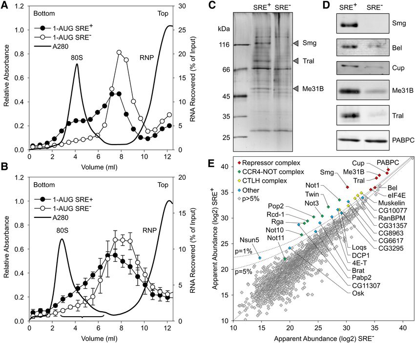
Downloaded from rnajournal.cshlp.org on May 31, 2021 - Published by Cold Spring Harbor Laboratory Press
Translational repression of Drosophila nanos mRNA
causing repression and degradation of hundreds of maternal RESULTS
mRNAs (Tadros et al. 2007; Chen et al. 2014a). Smg regulates
Composition of the SRE-dependent repressor complex
nos by binding two Smaug recognition elements (SREs) in the
nos 3′ UTR and recruits the CCR4–NOT complex, which cat- For an analysis of the constituents of the SRE-dependent re-
alyzes mRNA deadenylation (Semotok et al. 2005; Jeske et al. pressor complex, gradient centrifugation was used as a first
2006; Zaessinger et al. 2006). For translational repression, purification step. In Figure 1, radiolabeled, m7G-capped lu-
Smg binds the protein Cup (Nelson et al. 2004), and a ciferase RNAs were used carrying a nos 3′ UTR fragment with
miRNA-independent repressive role of Ago1 has also been two SREs, either wild type (SRE+) or with an inactivating
reported (Pinder and Smibert 2013), but the mechanism of point mutation in each (SRE−). The RNAs had “internal”
repression is not fully understood (Jeske et al. 2011). poly(A) tails, which stimulate translation like a 3′ -terminal
Deadenylation and translational repression of nos can be tail, but are protected from SRE-dependent deadenylation
observed in extracts from early Drosophila embryos (Jeske by flanking 3′ sequences (Fig. 1A; Jeske et al. 2011). As re-
et al. 2006, 2011). Deadenylation and repression both depend ported (Jeske et al. 2011), these RNAs develop the full extent
on the SREs and, by inference, on Smg, but are independent of translational repression only upon a preincubation with
of each other. Smg-associated Cup inhibits translation by embryo extract under conditions that do not permit transla-
binding the cap-binding translation initiation factor eIF4E tion (Fig. 1B,D; Supplemental Table S1). After a first preincu-
and competitively displacing eIF4G (Nelson et al. 2004; bation, aliquots of the RNAs were sedimented through
Jeske et al. 2011). However, the 5′ cap as well as eIF4E and sucrose gradients (Fig. 1B,C), and the peak fractions were as-
eIF4G are dispensable for SRE-dependent repression (Jeske sayed for translation in embryo extract either directly or after
et al. 2011); thus, an additional repression mechanism a second preincubation. The results revealed strong transla-
must exist. In support of this, the SRE-dependent repressor tional repression independently of the second preincubation
complex contains the proteins Me31B and Trailer hitch (Fig. 1B,D; Supplemental Table S1). Thus, the repressor
(Tral) in addition to Smg, Cup, and eIF4E (Jeske et al. complex formed during the first preincubation survived the
2011). Me31B and its orthologs are DEAD-box family RNA long fractionation procedure, in agreement with its known
helicases/RNA-dependent ATPases and involved in transla- kinetic stability (Jeske et al. 2011). The RNA sedimented fast-
tional repression in flies (Nakamura et al. 2001; Tritschler er than in earlier experiments showing SRE-dependent inhi-
et al. 2009), yeast (Dhh1p) (Coller and Parker 2005), and ver- bition of 48S complex formation (Jeske et al. 2011), probably
tebrates (DDX6/p54/RCK) (Minshall et al. 2001; Chen et al. due to differences in experimental conditions. With precau-
2014b; Mathys et al. 2014). Tral (S. cerevisiae Scd6p; C. ele- tions taken to suppress RNase activity in the extract (see
gans CAR1; vertebrate Rap55 or Lsm14) associates with Materials and Methods), RNA stability was not significantly
Me31B and also represses translation (Audhya et al. 2005; different between the repressed SRE+ RNA and the SRE−
Boag et al. 2005; Wilhelm et al. 2005; Tanaka et al. 2006; control (Fig. 1E,F).
Weston and Sommerville 2006; Nissan et al. 2010; For the actual analysis of the repressor complex, similar
Hubstenberger et al. 2013; Ayache et al. 2015). RNAs as in Figure 1 were used (1-AUG nos and 1-AUG nos
Formation of the SRE-dependent repressor complex in SRE−) that only differed by containing a shorter ORF and
embryo extract is ATP-dependent and slow, requiring 20– no 5′ cap; the cap is irrelevant for repression and stability of
30 min. Once formed, the complex is kinetically unusually the repressor complex (Jeske et al. 2011). The RNAs were ran-
stable, with an estimated t1/2 of ∼4 h. These observations sug- domly biotinylated. After incubation in extract, these shorter
gest that the repressor complex is not governed by a simple RNAs showed an SRE-dependent difference in sedimenta-
association–dissociation equilibrium. Presumably due to tion, the SRE+ RNA sedimenting in the 80S region ahead of
the stability of the complex, repression is ∼20- to 50-fold, the control; this was more visible in an analytical gradient
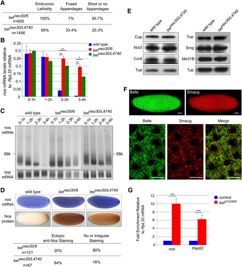
that is, ≥95% of the SRE-containing RNA is turned off. (Fig. 2A) than in the preparative experiment (Fig. 2B).
Importantly, SRE-dependent repression acts on translation Gradient fractions were selected as shown in Figure 2B,
initiation driven by the CRPV IRES. As this IRES can directly pooled from several runs and concentrated. Equal quantities
associate with ribosomes, independently of any initiation fac- of the SRE+ and SRE− RNPs, based on trace-labeling of the
tors, the repressor complex likely affects either ribosome as- RNA, were affinity-purified on streptavidin beads, and pro-
sociation or elongation (Jeske et al. 2011). teins eluted by SDS were analyzed by gel electrophoresis
Here we report a systematic analysis of the composition of (Fig. 2C) followed by liquid chromatography/tandem mass
the SRE-dependent repressor complex. In addition to the spectrometry (LC/MS/MS). Proteins detected were evaluated
previously known proteins, the DEAD-box protein Belle by label-free quantification (MaxQuant) (Cox and Mann
(Bel) was found in the complex. Genetic experiments con- 2008) based on the intensities of the MS signals and spectral
firmed that Bel participates in nos regulation in vivo. counts, corrected for the molecular mass of each protein. In
Me31B and Tral bind in multiple copies along the repressed Figure 2E, the apparent abundance of each protein is plotted
RNA, presumably sequestering it in a form that is inaccessible for the SRE+ RNA against the control. All proteins are listed in
for ribosomes. Supplemental Table S2.
www.rnajournal.org 1553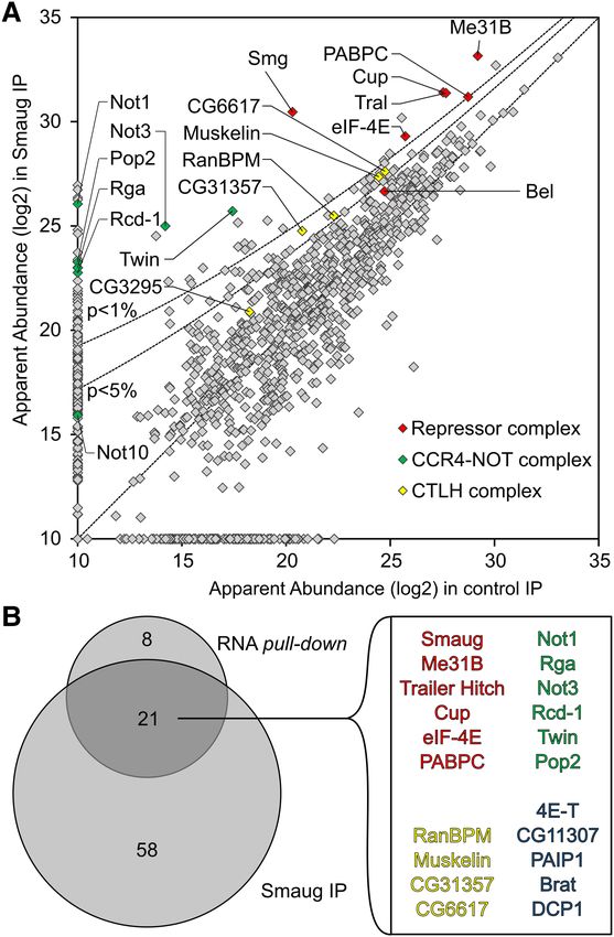
Downloaded from rnajournal.cshlp.org on May 31, 2021 - Published by Cold Spring Harbor Laboratory Press

Götze et al.

                                                                                 As expected, Smg was among the most abundant proteins
                                                                              and most strongly enriched in the SRE+ RNP. Cup, Tral, and
                                                                              Me31B formed a tight cluster with an apparent abundance
                                                                              even higher than Smg and enriched in the SRE+ RNP.
                                                                              Three additional proteins were also abundant and enriched
                                                                              in the SRE+ RNP: First, enrichment of eIF4E-1 (Hernández
                                                                              et al. 2005) agrees with previous results (Jeske et al. 2011).
                                                                              As the RNA was not capped, the protein’s presence was pre-
                                                                              sumably due to protein–protein interactions, e.g., with Cup
                                                                              (Nelson et al. 2004; Chekulaeva et al. 2006). Second, the pres-
                                                                              ence of PABPC was expected due to the internal poly(A) tail.
                                                                              An SRE-dependent enrichment of the protein agrees with the
                                                                              observation that a poly(A) tail facilitates repression (Jeske
                                                                              et al. 2006, 2011) and with the presence of PABPC in
                                                                              DDX6 complexes purified under stringent conditions
                                                                              (Ayache et al. 2015; Bish et al. 2015). In contrast, Western
                                                                              analyses of RNP complexes isolated by a simple pull-down
                                                                              procedure consistently showed the PABPC content to be in-
                                                                              dependent of the SREs (Fig. 2D; Jeske et al. 2011). The pro-
                                                                              cedure leading to the MS analysis took considerably longer
                                                                              than a simple pull-down and might thus reveal a more stable
                                                                              association of PABPC with the repressed RNA compared to
                                                                              the control. Third, a novel component, the RNA-dependent
                                                                              ATPase Belle (Bel) (Johnstone et al. 2005) was identified. Its
                                                                              specific association with the SRE+ RNA was confirmed by
                                                                              Western blot (Figs. 2D, 5).
                                                                                 The CCR4–NOT complex is responsible for Smg-depen-
                                                                              dent deadenylation (Semotok et al. 2005; Zaessinger et al.
                                                                              2006) and associates with SRE-containing RNAs (Jeske
                                                                              et al. 2011). Satisfyingly, all core components of the complex
                                                                              (Not1, Ccr4/Twin, Caf1/Pop2, Not2/Rga, Not3, and Caf40/
                                                                              Rcd-1) formed a cluster of similar SRE-specific enrichment
                                                                              and roughly similar abundance (Fig. 2E). However, reduced
                                                                              abundance of all subunits compared to the Smg/Cup cluster
FIGURE 1. Reporter RNAs maintain their repressed state during gradi-          suggests that the CCR4–NOT complex is not part of the sta-
ent centrifugation. (A) Cartoon of luciferase reporter RNAs. (B) Scheme       ble core of the repressor complex. Several other proteins were
of the assay. Black triangles indicate addition of embryo extract, and        also enriched in the SRE+ RNP, but less so than either the
drop symbols indicate samples withdrawn for translation and luciferase
assays. Numbers refer to the data shown in D. (C) Repressor complexes         Smg/Cup cluster or the CCR4–NOT complex (Fig. 2E;
formed on radiolabeled reporter RNAs were separated by sucrose-gradi-         Supplemental Table S3). These proteins include the con-
ent sedimentation. Distributions of the two RNAs are overlaid. UV ab-         served CTLH (C terminal to LisH [Lissencephaly type-1-
sorption indicates the positions of free RNPs and the 80S ribosome.           like homology motif]) complex (Francis et al. 2013) and
(D) As shown in B, luciferase RNAs were tested for translational repres-
sion either directly or after preincubation in embryo extract (samples 1      the Cup paralog 4E-T (Kamenska et al. 2014, 2016). Dcp1,
and 2 in B). A second set of samples was preincubated in extract and          which has been found to be associated with a Me31B–Tral–
then separated by gradient centrifugation. Aliquots from the peak frac-       Cup complex (Tritschler et al. 2008) was present, but the cat-
tions as in C were assayed for translation in embryo extract either with      alytic subunit of the decapping complex, Dcp2, was not de-
or without a second preincubation in fresh extract (samples 3 and 4 in
B). Luciferase activities in these assays are listed in Supplemental Table    tected at all. A low-level presence of Oskar may be related
S1. (E) RNAs were purified from equal volumes of the peak fractions           to its role in derepression of nos in the pole plasm.
of gradients as in C, and equal aliquots were assayed for translation in         Pat1 (HPat or Patr-1 in Drosophila) and EDC3, which
rabbit reticulocyte lysate, which does not exhibit SRE-dependent repres-
                                                                              compete with Tral for the same surface of Me31B
sion (Jeske et al. 2006). Thus, similar luciferase yields indicated similar
RNA recoveries for both RNAs. Error bars represent the standard devia-        (Tritschler et al. 2008, 2009; Haas et al. 2010; Jonas and
tion of three independent experiments. (F ) Radiolabeled luciferase RNA       Izaurralde 2013; Sharif et al. 2013), were present at much
from the sucrose gradient shown in Figure 1C was purified and analyzed        lower levels than Tral and weakly enriched on the SRE+
by denaturing gel electrophoresis and phosphorimaging. Numbers above
the lanes indicate fraction numbers of the sucrose gradient. Note that
                                                                              RNA (Supplemental Fig. S1C). Ypsilon schachtel (Yps) and
the inclusion of “short RNA” (see Materials and Methods) strongly sta-        Exuperantia (Exu) have been found in Me31B-containing
bilized the RNA compared to earlier experiments (Jeske et al. 2011).          RNPs (Nakamura et al. 2001; Wilhelm et al. 2005), but Yps

1554        RNA, Vol. 23, No. 10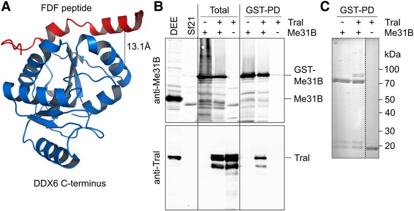
Downloaded from rnajournal.cshlp.org on May 31, 2021 - Published by Cold Spring Harbor Laboratory Press

                                                                                       Translational repression of Drosophila nanos mRNA

FIGURE 2. Analysis of the SRE-dependent repressor complex. (A) Radiolabeled, biotinylated RNAs (1-AUG nos and 1-AUG nos SRE−) were incu-
bated for assembly of a repressor complex and separated on a sucrose gradient as in B, but the volume loaded was smaller (0.2 mL versus 1 mL). (B)
Radiolabeled, biotinylated RNAs (1-AUG nos and 1-AUG nos SRE−) were separated on a preparative sucrose gradient (see Materials and Methods).
Fractions pooled for the analysis of the repressor complex are indicated by the bracket. Error bars represent the standard deviation (n = 4). (C)
Corresponding fractions from a total of 12 gradients from four independent experiments each for the SRE+ RNA and the SRE− control were pooled,
and RNPs were purified on streptavidin beads. Equal amounts based on trace-labeling of the RNA were analyzed by SDS-PAGE and silver staining.
Arrowheads indicate bands enriched in the SRE+ RNP that might correspond to Smaug (109 kDa), Trailer Hitch (69 kDa), and Me31B (52 kDa).
(D) Specific association of proteins with the SRE+ RNA was confirmed by Western analysis in an independent pull-down assay. Smg, Cup,
Me31B, and PABPC served as controls for Bel. (E) Proteins in the purified RNP fractions were analyzed by mass spectrometry and label-free quan-
tification. Apparent protein abundance in SRE+ versus SRE− RNP was plotted on a log2 scale. Proteins enriched in the SRE+ RNP beyond P = 0.05 and
NOT11 are labeled. The complete list of proteins represented in E is found in Supplemental Table S2. Different sets of proteins in the same data are
highlighted in Supplemental Figure S1, and additional enriched proteins are listed in Supplemental Table S3.

was not enriched in the repressor complex (Supplemental                     et al. 2010; Barckmann et al. 2015) was not present in our
Fig. S1C). Western blotting confirmed an equal association                  constructs.
with both RNAs (data not shown). Exu was not detected,                         In an independent experiment, Smg was immunoprecipi-
consistent with the absence of nos from immunoprecipitated                  tated from extract that had not been treated with RNase.
Exu–Yps complexes (Wilhelm et al. 2000).                                    Proteins were identified by LC/MS/MS and compared to a
   As expected (Nelson et al. 2004; Jeske et al. 2011), eIF4G               preimmune serum control. The results supported those of
was moderately depleted from the repressed RNP. All other                   the streptavidin purification: Core components of the repres-
initiation factors were less abundant than eIF4E and eIF4G                  sor complex were enriched with Smg; only the enrichment of
and not enriched in either RNP (Supplemental Fig. S1A).                     Belle was weak. All core subunits of the CCR4–NOT complex
Ribosomal proteins were depleted from the SRE+ RNP                          and four subunits of the CTLH complex were also enriched
(Supplemental Fig. S1B,D). Ago 1 has been reported to par-                  (Fig. 3; Supplemental Table S4).
ticipate in SRE-dependent repression (Pinder and Smibert
2013), but was not enriched in the SRE+ RNP. Other proteins
                                                                            Repressed nos mRNA exists as a monomeric RNP
involved in small RNA pathways were not enriched either
(Supplemental Fig. S1C). This is not unexpected as the region               The repressed RNPs sedimented rapidly, comparable to ribo-
of the nos 3′ UTR most strongly targeted by piRNAs (Rouget                  somes (Fig. 2A). In the case of oskar mRNA, oligomerization

                                                                                                                  www.rnajournal.org         1555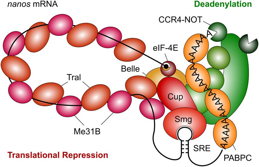
Downloaded from rnajournal.cshlp.org on May 31, 2021 - Published by Cold Spring Harbor Laboratory Press

Götze et al.

                                                                         between SRE+ RNA and SRE− control was observed (Fig.
                                                                         4B). Calculations (see figure legend) indicated that endoge-
                                                                         nous nos RNA present in the extract would have been detect-
                                                                         able if it had been associated with the bait RNA. Thus, the
                                                                         repressed nos RNP does not oligomerize with other re-
                                                                         pressed RNPs. The lack of nos oligomerization is consistent
                                                                         with in vivo data (Little et al. 2015). No SRE-dependent en-
                                                                         richment of other RNAs was observed, making it unlikely
                                                                         that trans-acting RNAs are involved in SRE-dependent re-
                                                                         pression in vitro.

                                                                         Multiple copies of Me31B and Tral associate
                                                                         with the repressed RNA
                                                                         Me31B orthologs can oligomerize on their own or when
                                                                         bound to RNA, and the ability of protein variants to oligo-
                                                                         merize correlates with their ability to repress translation.
                                                                         The proteins appear to bind in multiple copies along RNA
                                                                         in vivo (Minshall and Standart 2004; Ernoult-Lange et al.
                                                                         2012).
                                                                            In order to determine whether oligomerization of repres-
                                                                         sor proteins on the reporter RNAs might play a role in
                                                                         SRE-dependent translational repression, we estimated the
                                                                         stoichiometries of proteins in the repressor complex: Three
                                                                         different biotinylated radiolabeled RNAs were used, each
                                                                         containing two copies of the SREs: SREonly (200 nt), the
                                                                         1-AUG nos RNA used for the MS analysis (630 nt) and the
                                                                         luciferase reporter RNA (1956 nt); corresponding SRE−
FIGURE 3. MS analysis of proteins coprecipitated with Smg. (A)           RNAs served as controls. All RNAs were allowed to assemble
Quantitative MS data were plotted for the Smg immunoprecipitation        repressor complexes before affinity purifications were carried
versus a preimmune control. Proteins that were also significantly en-
riched in the streptavidin pull-down of the SRE-dependent repressor
                                                                         out. The quantities of immobilized RNAs were determined
complex are highlighted as in Figure 2E. P-value cutoffs are indicated   from their specific radioactivities, and amounts of associated
as lines. (B) Venn diagram comparing proteins enriched beyond P =        proteins were estimated by Western blotting and comparison
0.05 in the Smg immunoprecipitation and in the streptavidin pull-        to standard curves of purified recombinant material. Repre-
down (Fig. 2E). The 21 proteins in the overlap are listed. Belle,
NOT10, and the CTLH complex subunit CG3295 had P-values higher
                                                                         sentative data are shown in Figure 5A and Supplemental Fig-
than 0.05. All proteins enriched in the Smg IP are listed in             ure S2, and a summary of the average stoichiometries is
Supplemental Table S4.                                                   presented in Figure 5B. RNA association of Smg was SRE-
                                                                         dependent, but independent of RNA length: The stoichiom-
                                                                         etry was between 1 and 2 for all three RNAs. Within the
of the repressed RNPs contributes to their rapid sedimenta-              accuracy of the experiment, this was equimolar with the
tion (Chekulaeva et al. 2006; Besse et al. 2009). However,               SREs (see legend to Supplemental Fig. S2). Binding of Cup
when a biotinylated SRE+ RNA was incubated in embryo                     was also approximately stoichiometric with the SREs. Bel
extract together with a second SRE+ RNA, lacking biotin                  bound independently of RNA length, but tended to be less
and distinguishable by size, streptavidin pull-down resulted             abundant; with the longest RNA, specific binding was no lon-
in the purification of only the biotinylated RNA; no associa-            ger distinguishable from background. In contrast to Smg,
tion with the second RNA was seen (Fig. 4A). We conclude                 Cup, and Bel, both Tral and Me31B clearly bound in a
that Smg-dependent repression does not involve RNA                       length-dependent manner, in excess of Smg and the SREs
oligomerization.                                                         and approximately equimolar to each other. As these data in-
   As an unbiased test for a potential association of the SRE+           dicate binding of multiple copies of Me31B and Tral along the
RNA with other RNAs, total RNA was isolated from purified                RNA, it is unclear whether the amounts associated with the
repressor complexes and from SRE− controls and analyzed by               SRE− RNAs should be subtracted as background or not. With-
deep sequencing. Although sequencing was targeted to small               out background subtraction, the stoichiometry of Me31B/Tral
RNAs, nos sequences were also recovered; these were limited              binding to RNA was near one copy of Me31B and Tral per 100
to the part contained in the bait RNA, and no difference                 nucleotides.

1556       RNA, Vol. 23, No. 10
Downloaded from rnajournal.cshlp.org on May 31, 2021 - Published by Cold Spring Harbor Laboratory Press
Translational repression of Drosophila nanos mRNA
ditions of unchecked endogenous nuclease activity, was mod-
erately more stable than an SRE− control (Jeske et al. 2011).
These data strongly argue in favor of sequestration of the
RNA by a protein complex.
The stoichiometries indicate that Me31B and Tral cooper-
ate as a defined subcomplex within the repressor complex.
Indeed, treatment of a Smg immunoprecipitate with the
cross-linker BuUrBu (Müller et al. 2010) identified a cross-
link between Tral and Me31B consistent with the interaction
dependent on the FDF motif of Tral (Fig. 6A; Supplemental
Fig. S3; Tritschler et al. 2008, 2009). Thus, this interaction is
likely to be relevant within the context of the repressor com-
plex. When Tral and GST-Me31B were coexpressed in insect
cells by means of baculovirus vectors, Tral was copurified
with GST-Me31B on glutathione beads, suggesting the exis-
tence of a stable complex (Fig. 6B,C). Copurification was
not affected by elevated salt concentration or RNase A.
The components of the repressor complex were abundant
among the soluble proteins of the embryo extract, as estimat-
ed by quantitative Western blotting (with an error of approx-
imately two; see Materials and Methods): Smg was present at
0.08 μM; Cup, 2 μM; Bel, 0.8 μM; Me31B, 3.5 μM, Tral, 7.6
μM. We estimate that extracts were approximately twofold
diluted compared to egg content. In comparison, an
mRNA concentration of roughly 0.4 μM in a Drosophila
egg can be estimated on the basis of an egg volume of 0.01
μL (Azevedo et al. 1996), a total RNA content of 0.19 μg
per egg (Hough-Evans et al. 1980), and the assumption
that 2% of this is mRNA with an average length of 3000 nt.
FIGURE 4. SRE-containing RNAs do not oligomerize. (A) A biotiny- The ratio of protein to RNA concentration is consistent
lated RNA of 200 nt (SREonly; SRE+ or SRE−) and a nonbiotinylated with Smg acting on a sizeable fraction of maternal mRNAs
RNA of 630 nt (AUGonly; SRE+ or SRE−) were incubated together in (Tadros et al. 2007; Chen et al. 2014a) and with Cup partic-
embryo extract under conditions permitting assembly of the repressor ipating in translational repression exerted by other RNA
complex. Streptavidin pull-downs were performed to enrich the bio-
tinylated RNA together with potentially associated RNAs. RNA was elut- binding proteins, e.g., Bruno (Nakamura et al. 2004;
ed in formamide loading buffer at 95°C. The lanes labeled “RNA” show Wilhelm et al. 2005; Chekulaeva et al. 2006). The abundance
the purified RNAs used, “input” shows the RNAs after incubation in ex- of both Me31B and Tral is consistent with the two proteins
tract, “FT” is the flow-through of the pull-down, and “elution” shows
the bound fraction. The figure shows one experiment of two. (B)
binding in multiple copies and as a complex to repressed
RNA was purified from affinity-purified SRE+ and SRE− RNPs and mRNAs. The high concentration of Me31B, exceeding that
deep-sequenced. Reads mapping to the nos gene are displayed. For the of mRNA, is consistent with data in other organisms
experiment, bait RNAs were used at 10 nM. The abundance of nos (Ernoult-Lange et al. 2012 and references cited therein).
has been estimated as 2 nM (Trcek et al. 2015). With an approximately
twofold dilution upon extract preparation and an additional twofold
The MS data suggest that the CCR4–NOT complex is not
dilution in the assay, endogenous nos sequences should have been part of the core repressor complex. Association of the CCR4–
detectable if an association with the bait RNA had taken place. NOT complex with the 630 nt RNA was examined by quan-
titative Western blotting. In agreement with the MS analysis,
CCR4, Caf1, and Not2 bound the RNA in an SRE-dependent
“Coating” of RNA by Me31B and Tral may form an inert, manner, but were clearly substoichiometric (Fig. 5E and data
“masked” RNP that is at the core of translational repression, not shown).
sterically preventing ribosome access to the RNA. For want of
a reagent more comparable in size to a ribosome (3 × 10⁶
Bel is required for nos mRNA translational repression
Da), accessibility of the repressed RNA was probed with the
in vivo
endonuclease RNase I (as an MPB fusion protein; 72,000
Da). The repressed SRE+ RNA proved to be considerably Belle is a DDX3-type RNA helicase. These proteins have been
more resistant to the nuclease than the SRE− control (Fig. reported to be involved in translation, but both activating and
5C,D). This agrees with an earlier observation that an SRE+ repressive roles have been described. To examine a direct role
RNA, when simply incubated in embryo extract under con- of Bel in nos mRNA control in the embryo, we used two
www.rnajournal.org 1557Downloaded from rnajournal.cshlp.org on May 31, 2021 - Published by Cold Spring Harbor Laboratory Press Götze et al. FIGURE 5. The SRE-dependent repressor complex sequesters the RNA through multiple copies of Me31B and Tral. (A) Three biotinylated RNAs of different lengths but each containing two SREs were used, together with matching SRE− controls, for repressor complex formation in embryo extract and streptavidin pull-down. Bound proteins were analyzed by Western blotting. Known amounts of recombinant proteins were used as standards. Analyses of Smg and Tral are shown as representative examples. (B) Stoichiometries of bound proteins were estimated from experiments as in A. Signals for SRE+ and mutant controls are shown. The horizontal lines mark a 1:1 molar ratio of protein to RNA. Error bars represent the standard deviation from three to five independent experiments ([∗ ] P ≤ 0.05; [∗∗ ] P ≤ 0.01; [∗∗∗ ] P ≤ 0.001). Additional data are presented in Supplemental Figure S2. (C) An RNase I protection experiment was carried out as described in Materials and Methods. (D) Quantification of experiments as shown in C (average of n = 4 with three independent batches of embryo extract). Error bars represent the standard deviation. Data were fitted to a first-order decay with the last time point of both RNAs omitted. The half-life of the SRE+ RNA was 1.7-fold longer than that of the SRE− control. (E) The as- sociation of Caf1 and Not2 with the SRE+ RNA and SRE− control was examined as in A. Three streptavidin pull-down experiments were carried out with the 630 nt RNA and independent batches of embryo extract. Western blotting and comparison to standard curves was carried out for the proteins indicated. The average amount of Smg recovered was 200 ± 100 fmol. Tral was recovered at 1000 ± 150 fmol in the SRE+ sample and at 500 ± 120 fmol in the SRE− sample (data not shown). All three subunits of the CCR4–NOT complex were present below the smallest amount in the standard curves (50 fmol). In a separate Western blot, signals for Caf1, Not2 and Ccr4 were below 12.5 fmol (data not shown). strong or null alleles of bel, bel 6 and bel L4740, which cause with stronger alleles (Johnstone et al. 2005; Ihry et al. larval lethality. In addition, the hypomorphic allele bel neo30 2012). Consistent with this, transheterozygous belneo30/6 and was used, which leads to female sterility when combined belneo30/L4740 females were sterile: When crossed with wild- 1558 RNA, Vol. 23, No. 10
Downloaded from rnajournal.cshlp.org on May 31, 2021 - Published by Cold Spring Harbor Laboratory Press

                                                                                    Translational repression of Drosophila nanos mRNA

                                                                                                    Western blots were in agreement with
                                                                                                    the staining pattern of the majority of
                                                                                                    embryos, showing increased levels in 0–
                                                                                                    2 h belneo30/L4740 embryos and reduced
                                                                                                    levels in 0–2 h belneo30/6 mutant embryos
                                                                                                    (Supplemental Fig. S4B). Defects in nos
                                                                                                    regulation in bel mutant embryos did
                                                                                                    not result from reduced levels of other
                                                                                                    components of the repressor complex
                                                                                                    or of the CCR4–NOT complex (Fig. 7E).
                                                                                                       These results show that Bel partici-
                                                                                                    pates in the repression of nos mRNA in
                                                                                                    the somatic part of the embryo and
                                                                                                    thus imply that Bel is present there. A
FIGURE 6. Me31B and Tral form a complex. (A) Structure of a complex between the C-terminal GFP-tagged Bel protein has been report-
domain of DDX6 and an EDC3 peptide containing the FDF motif (PDB 2WAX) (Tritschler et al.
2009). Tral uses the same motif to bind Me31B. The black line represents the cross-link identified ed to be distributed throughout the syn-
(Supplemental Fig. S3), with the Cα − Cα distance indicated. (B) Sf21 cells were infected with cytial embryo (Johnstone et al. 2005).
baculoviruses expressing GST-Me31B, Tral, or both as indicated. “Total” refers to an SDS lysate. Immunostaining of embryos with anti-
Purifications on glutathione beads were carried out from native lysates. Proteins were analyzed by Bel and anti-Smg antibodies validated
Western blotting for Me31B (top) or Tral (bottom). Drosophila embryo extract (DEE) and non-
infected SF21 cells served as controls. (C) Glutathione bead eluates were analyzed by SDS poly- the cytoplasmic distribution of Bel
acrylamide gel electrophoresis and Coomassie staining.                                              throughout the embryo and its partial
                                                                                                    colocalization with Smg (Fig. 7F).
                                                                                                       In an independent experiment to ask
type males, they produced embryos (referred to as belneo30/6                   whether nos mRNA is bound to Bel in embryos, we used
and belneo30/L4740 embryos) that failed to eclose (Fig. 7A).                   the GFP protein-trap bel allele bel CC00869, in which GFP is in-
    neo30/6
bel          embryos showed a stronger phenotype than                          serted in frame in the N-terminal part of Bel (Buszczak et al.
belneo30/L4740, most of them being fragile and having short                    2007). RNA immunoprecipitation with anti-GFP antibody
or no dorsal appendages.                                                       showed an enrichment of nos mRNA over Rpl32 mRNA in
   To address a role of Bel in nos mRNA deadenylation and                      0–2 h embryos expressing GFP-Bel (bel CC00869) compared
decay, we quantified nos mRNA by RT-qPCR in wild-type                          to control embryos. Another Smg target, Hsp83 (Semotok
and bel mutant embryos spanning 1 h intervals during the                       et al. 2005), was also enriched (Fig. 7G).
first 4 h of embryogenesis. nos mRNA decay was prominent                          Taken together, these results support a functional role of
after 2 h in wild-type embryos, but was strongly impaired in                   Bel in the nos repressor complex in vivo, acting on both trans-
bel mutant embryos (Fig. 7B). Accordingly, poly(A) test as-                    lational repression and deadenylation.
says, used to measure nos mRNA poly(A) tail lengths in
embryos up to 4 h of development, showed that deadenyl-
ation was inhibited in bel mutant embryos (Fig. 7C; Supple-
                                                                               DISCUSSION
mental Fig. S4A). In situ hybridization of 0–2 h embryos
suggested that nos mRNA stabilization might even start                         We have identified seven stoichiometric components of the
before 2 h of embryogenesis, since the staining was darker                     SRE-dependent repressor complex that are likely to explain
in bel mutants than in wild-type embryos (Fig. 7D).                            its ATP-dependent formation, high stability and repressive
Translational repression of nos was also impaired in bel mu-                   potency: Smg, which directly recognizes the SREs; Cup,
tant embryos: Immunostaining with anti-Nos antibody re-                        which associates with Smg; the DEAD-box ATPase Me31B
vealed ectopic, increased Nos levels in belneo30/L4740 embryos                 and its partner Tral; a second DEAD-box ATPase, Bel; and
(Fig. 7D). belneo30/6 embryos showed heterogeneous staining:                   finally the cap-binding initiation factor eIF4E and, with less
A large proportion (80%) were irregularly or not stained, but                  certainty, the cytoplasmic poly(A) binding protein, PABPC.
the remaining 20% showed again high levels of ectopic Nos                      The repressor complex analyzed is functional since transla-
protein throughout the embryo (Fig. 7D). The heterogeneity                     tion of the RNA on which it has assembled is fully repressed
in belneo30/6 embryos could be due to earlier defects during                   (Fig. 1D). Thus, assembly of the seven proteins identified
oogenesis (Johnstone et al. 2005) or to a potential gain-of-                   constitutes the slow step of translation repression. The
function nature of the bel 6 allele: bel 6 has a stop codon after              same complex likely facilitates deadenylation, since Smg
the first third of the coding sequence, which encodes a 4E-BP                  and the SREs are also important for deadenylation of nos
domain (Yarunin et al. 2011; Ihry et al. 2012). Thus, a trun-                  by CCR4–NOT. Accordingly, all core components of the
cated protein in bel 6 might dominantly affect translation                     CCR4–NOT complex were associated with the repressor
through binding to eIF4E. Analyses of Nos protein levels by                    complex.

                                                                                                              www.rnajournal.org        1559
Downloaded from rnajournal.cshlp.org on May 31, 2021 - Published by Cold Spring Harbor Laboratory Press Götze et al. FIGURE 7. Bel is required for nos mRNA translational repression in vivo. (A) Phenotypic quantification of embryos coming from belneo30/6 or belneo30/L4740 mutant females crossed with wild-type males. Numbers refer to the embryos examined. (B) nos mRNA quantification using RT- qPCR in wild-type and bel mutant embryos spanning 1 h intervals up to 4 h of development. RpL32 was used as a control mRNA for normalization. Means are from three to four biological replicates. The error bars represent SEM. (∗ ) P < 0.05; (∗∗ ) P < 0.01 using the bilateral Student’s t-test. (C) PAT assays measuring nos mRNA poly(A) tail lengths in wild-type and bel mutant embryos spanning 1 h intervals up to 4 h of development. PAT assay profiles using ImageJ are shown in Supplemental Figure S4A. sop encodes a ribosomal protein and was used as a control mRNA. (D) In situ hybrid- ization of nos mRNA (top panels) and immunostaining with anti-Nos (bottom panels) of wild-type and bel mutant 0–2 h embryos. Quantification of immunostaining is indicated below the images. (E) Western blots of wild-type and bel mutant 0–2 h embryos probed with antibodies against six com- ponents of the nos repressor complex, including the CCR4–NOT complex. Anti-α-tubulin (Tub) was used as a loading control. (F ) Confocal images of syncytial embryos co-stained with rabbit anti-Bel and guinea pig anti-Smg. Bottom panels show a higher magnification. Quantification using the Pearson correlation coefficient (PCC) indicated significant partial colocalization (PCC = 0.52). Anterior is to the left. The scale bars represent 30 and 10 μm in top and bottom panels, respectively. (G) Quantification of nos and Hsp83 mRNAs using RT-qPCR in anti-GFP immunoprecipitations from bel CC00869 embryos that express GFP-Bel and control (wild type) embryos that do not express GFP. RpL32 mRNA was used for normalization. mRNA levels in control embryos were set to one. Means are from two biological replicates quantified in triplicates. The error bars represent SEM. (∗∗∗ ) P < 0.001 using the bilateral Student’s t-test. The CCR4–NOT complex can also contribute to transla- for translational repression. The assay of the repressor com- tional repression, independently of its deadenylase activity plex only tests for constituents incorporated in the slow step, (Cooke et al. 2010; Braun et al. 2011; Chekulaeva et al. though; as the translation assay requires incubation of the 2011; Kuzuoğlu-Öztürk et al. 2016). However, CCR4–NOT gradient-purified repressed RNP with embryo extract, we was clearly substoichiometric and thus may not be essential cannot exclude that other components of the extract may 1560 RNA, Vol. 23, No. 10
Downloaded from rnajournal.cshlp.org on May 31, 2021 - Published by Cold Spring Harbor Laboratory Press

                                                                               Translational repression of Drosophila nanos mRNA

associate with the stable complex and participate in its repres-    Xenopus oocyte mRNPs (Weston and Sommerville 2006).
sive activity, i.e., a protein that is not among the stable core    The presence of Me31B and its partner Tral in the repressor
components of the repressed RNP may still play a role in            complex is also consistent with previous reports of these two
repression.                                                         proteins interacting and causing translational repression (see
   Five subunits of the conserved CTLH complex were en-             Introduction). DDX6-type proteins bind RNA even in the
riched in the purified repressor complex, but substoichio-          absence of ATP (Dutta et al. 2011; Ernoult-Lange et al.
metric with respect to the core components. The yeast               2012; Sharif et al. 2013). Tral presumably contributes directly
edition of the complex is a ubiquitin ligase (Santt et al.          to RNA coating, as it contains two types of potential RNA
2008; Chen et al. 2017). Smg is degraded during cell cycle          binding domains, an N-terminal Lsm domain and two
14 (Dahanukar et al. 1999; Benoit et al. 2009), and most other      RGG domains. Evidence for RNA binding by Tral orthologs
core constituents of the repressor complex also strongly            has been published (Audhya et al. 2005; Tanaka et al. 2006).
decrease during the maternal-to-zygotic transition (Gouw            As the complex affords protection even against a relatively
et al. 2009). The CTLH complex might be involved in the             small endonuclease, we propose that it prevents translation
degradation of these proteins.                                      by sterically excluding ribosomes, in agreement with the orig-
   The DEAD-box RNA helicase Bel was the only newly dis-            inal idea of masking (Spirin 1966, 1994). Specificity of repres-
covered constituent of the repressor complex. Enrichment of         sion for the nos RNA depends on sequence-specific binding
the protein was less pronounced compared to the other core          of Smg, but on the basis of biochemical similarities we sus-
components, but genetic data confirmed that Bel is required         pect that other repressors may use similar mechanisms
for both translational repression and deadenylation of nos          (Chekulaeva et al. 2006; Minshall et al. 2007).
mRNA in vivo. Bel orthologs Ded1p and DDX3 are known                   A conceptual assembly of the repressor complex (Fig. 8)
to be involved in translation, but their precise role is unclear,   starts with Smg binding to the SREs. Smg binds Cup, which,
since both depletion and overexpression inhibit translation         in turn, associates with the Lsm domain of Tral (Tritschler
(for reviews, see Soto-Rifo and Ohlmann 2013; Sharma                et al. 2008; Igreja and Izaurralde 2011). Tral uses its FDF mo-
and Jankowsky 2014). Bel and its orthologs can be localized         tif to bind Me31B (Tritschler et al. 2008, 2009; Igreja and
in RNP granules containing repressed mRNAs and promote              Izaurralde 2011; this paper), but Me31B can also directly in-
granule formation (Soto-Rifo and Ohlmann 2013; Sharma               teract with Cup (Nishimura et al. 2015; Ozgur et al. 2015;
and Jankowsky 2014). Bel and C. elegans LAF-1 have been             Kamenska et al. 2016). The mechanism of Me31B•Tral poly-
suggested to play a role in the translational repression of spe-    merization remains to be analyzed. Cup also brings in eIF4E
cific mRNAs, bruno and tra-2, respectively, but evidence for a      (Wilhelm et al. 2003; Nakamura et al. 2004; Nelson et al.
direct role has been lacking so far (Goodwin et al. 1997;           2004; Zappavigna et al. 2004; Igreja and Izaurralde 2011;
Yarunin et al. 2011). A cooperation of Ded1p with Dhh1p             Kinkelin et al. 2012). Bel may join the complex via interac-
(Me31B) in translational repression is suggested by genetic         tions with eIF4E (Sharma and Jankowsky 2014) or Me31B
and physical interactions (Tseng-Rogenski et al. 2003;              (Drummond et al. 2011). Candidates for recruiting the
Beckham et al. 2008; Drummond et al. 2011).                         CCR4–NOT complex include Me31B (Chen et al. 2014b;
   The idea that maternal mRNA in unfertilized eggs is not          Mathys et al. 2014; Rouya et al. 2014; Ozgur et al. 2015;
translated because it is masked by a “protective protein            Waghray et al. 2015) and Smg (Semotok et al. 2005;
coat” was proposed more than 50 years ago (Spirin 1966).
Whereas mechanisms have been analyzed that repress mater-
nal mRNAs by targeting, directly or via the poly(A) tail, the 5′
cap function (Wilhelm and Smibert 2005; Lasko 2011;
Barckmann and Simonelig 2013), proteins coating and
sequestering the RNA have not been identified with certainty.
Circumstantial biochemical evidence has supported the con-
cept, though: Repressed RNPs formed in vitro sediment rap-
idly, suggesting association of the RNA with many proteins
and tight packaging (Chekulaeva et al. 2006; this paper).
Repressed RNAs are also moderately resistant to nucleases
(Chekulaeva et al. 2006; Jeske et al. 2011; this paper).
Repression of CRPV IRES-dependent translation, which is
independent of all initiation factors, is consistent with exclu-
sion of ribosomes (Jeske et al. 2011). Here we present evi-
dence suggesting that the protective protein coat is formed         FIGURE 8. Model of the SRE-dependent repressor complex. The car-
                                                                    toon is based on the results of this paper and the references cited in the
by a complex of Me31B and Tral. The SREs nucleate the as-
                                                                    Discussion. Note that the accuracy of our Western blots is limited, thus
sembly of multiple copies of a Me31B•Tral complex on the            the stoichiometry of protein binding depicted in the figure should not be
RNA. The ortholog of Me31B is a component of stored                 interpreted narrowly.

                                                                                                           www.rnajournal.org          1561
Downloaded from rnajournal.cshlp.org on May 31, 2021 - Published by Cold Spring Harbor Laboratory Press
Götze et al.
Zaessinger et al. 2006). Me31B and Tral are both present in wrong RNA being repressed, but presumably also in a nos
the embryo at very high concentrations, and micromolar mRNA active in the wrong place. Polymerization of the
concentrations of Me31B or Tral (Scd6p) can inhibit transla- Me31B•Tral complex along the RNA would constitute a
tion nonspecifically in vitro (Coller and Parker 2005; Nissan fool-proof mechanism guaranteeing that the repressive ac-
et al. 2010). It will be interesting to find out how assembly of tion of the SREs is strictly intramolecular.
the stable Me31B•Tral oligomer is restricted to SRE-contain-
ing mRNAs.
The presence of two ATP-dependent RNA helicases, Bel MATERIALS AND METHODS
and Me31B, in the repressor complex probably accounts
for its ATP-dependence. (Note that the apparent ATP-de- RNA
pendence of Smg- and miRNA-dependent deadenylation All RNA constructs (SRE only; 1-AUG nos; luciferase reporter; all
has recently been shown to be a misinterpretation of the with two wild-type SREs or with a point mutation in each SRE)
data [Niinuma and Tomari 2017]. ATP-dependence of re- have been previously described (Jeske et al. 2006, 2011). RNAs
pressor complex formation thus needs to be reexamined, were synthesized with T3 RNA polymerase. Biotin-16-UTP (Jena
but is unlikely to suffer from the same misinterpretation.) Bioscience) and [α-32P]-UTP were incorporated during transcrip-
Me31B and/or Bel might also be responsible for the kinetic tion at a reduced concentration of UTP. For incorporation of a sim-
ilar number of biotin molecules per RNA, UTP was adjusted
stability of the repressor complex. An attractive model is pro-
according to the number of uridines in the RNA (Luc: 1 mM, 1-
vided by the exon junction complex (EJC), which is frozen on AUG: 0.25 mM, SREonly: 0.1 mM) at a constant concentration of
the RNA because its central component, the DEAD-box biotin-16-UTP (20 μM). When desired, an m7G cap was also incor-
ATPase eIF4AIII, is locked in a post-hydrolysis state by other porated cotranscriptionally. RNAs were gel-purified.
EJC constituents (Ballut et al. 2005; Nielsen et al. 2009). Due “Short RNA” was produced by partial hydrolysis of yeast RNA: 75
to the cooperativity of ATP and RNA binding, this fixes the mg of yeast total RNA was dissolved in 5 mL 20 mM Tris–HCl, pH
EJC on the RNA. Ded1 and two other DEAD-box helicases 8.0. 200 μL of 2.5 M NaOH was added, and the mixture incubated
tested were able to form very long-lived complexes with for 50 min at 40°C. Two hundred microliters of 5 M HCl was added
RNA in the presence of ATP analogs (Liu et al. 2014). and incubation continued for 10 min at 40°C. After addition of 600
Thus, this “clamping” function may be a general feature of μL 3 M sodium acetate, the RNA was purified by phenol/chloroform
DEAD-box helicases. We speculate that a component of the extraction and isopropanol precipitation.
repressor complex may inhibit the dissociation of ATP or
its hydrolysis products from Me31B and/or Bel to prevent Embryo extract and in vitro translation
the disintegration of the repressor complex.
Polymerization of Me31B and Tral along the RNA, nucle- Extracts were prepared as previously described (Jeske and Wahle
ated by Smaug binding in the 3′ UTR, conceptually solves a 2008) except that embryos (Canton S) were 15 to 135 min old,
and the lysate was centrifuged twice (20,000g, 30 min, 4°C).
problem that, to our knowledge, has barely been discussed
Aliquots were frozen in liquid N2 and stored at −80°C.
in the literature, although it is faced by all 3′ UTR-bound pro-
Luciferase reporter RNAs were incubated at 25°C in 40% embryo
tein complexes repressing translation initiation: Cartoons de- extract, 16 mM Hepes pH 7.4, 50 mM potassium acetate, 1 mM
picting the mechanism of action of such complexes magnesium acetate, 0.8 mM ATP, 0.25 mg/mL yeast tRNA, 0.2
invariably show an interaction of the 3′ end with the 5′ mg/mL “short RNA,” 0.08 g/L creatine kinase, 1 mM DTT, 80 U/
end, accompanied by the formation of an RNA loop. Any mL RNase inhibitor. Reactions were started with or without prein-
such interaction has to be intramolecular, i.e., the 3′ UTR- cubation by the addition of 20 mM phosphocreatine and amino ac-
bound repressor complex has to find the 5′ end of its ids (20 μM each), incubated for 30 min at 25°C and stopped on ice.
“own” mRNA in the face of competition from “foreign” 5′ Luciferase activity was assayed with the Promega kit.
ends. (For an interesting alternative, see Macdonald et al.
2016.) One possibility for such an intramolecular interaction
Purification of the repressor complex
to occur would be “through space”: The two opposite ends of
the flexible mRNA molecule diffuse randomly through the Radiolabeled, biotinylated RNAs (10 nM) were incubated under
cytoplasm. As they are tethered to each other via the RNA conditions in which no translation takes place (“preincubation con-
body, an intramolecular interaction would be favored by a ditions”; 60% embryo extract, 26 mM Hepes-KOH pH 7.4, 81 mM
high local concentration of the cis 5′ end with respect to potassium acetate, 1.6 mM magnesium acetate, 1.3 mM ATP, 1 mM
DTT, 80 U/mL RNase inhibitor, 0.2 mg/mL “short RNA”) for 25
the regulatory 3′ UTR site. However, the efficiency with
min at 25°C. The inclusion of “short RNA” improved RNA stability
which this leads to an intramolecular interaction depends
and complex recovery. Aliquots (1 mL) were loaded on 5%–45%
on variables like the length of the RNA and the concentration sucrose gradients (12 mL per tube in TL buffer: 16 mM Hepes-
of competing 5′ ends. One would suspect that a more reliable KOH pH 7.4, 50 mM potassium acetate, 1 mM magnesium acetate,
mechanism should have evolved, in particular with a repres- 0.8 mM ATP) and centrifuged for 3 h at 40,000 rpm, 4°C (Beckman
sor complex as stable as the one described here: Any trans in- SW40Ti). Gradients were harvested from the bottom in 20 fractions.
teraction established by mistake would not only result in a Fractions 5–10 were pooled, frozen in liquid N2 and stored at −80°C.
1562 RNA, Vol. 23, No. 10Downloaded from rnajournal.cshlp.org on May 31, 2021 - Published by Cold Spring Harbor Laboratory Press
Translational repression of Drosophila nanos mRNA
Pools from three gradients were combined and concentrated in separated in 33 equally sized bins along the diagonal axis. The dis-
Amicon centrifugal filters. Streptavidin beads (GE Healthcare; 30 tance of each protein from the diagonal in each bin follows a normal
μL packed volume) were blocked with TL buffer containing 0.1 distribution with a mean of 0 for all nonspecifically bound proteins.
mg/mL yeast RNA and 0.1 mg/mL methylated BSA and washed The distances were fitted against the normal distribution to obtain
with TL buffer. Beads were incubated with equal amounts, based σ². To estimate P-values for each protein enrichment, the σ²-values
on trace-labeling of the RNA, of the concentrated pools and 0.1 were fitted with the equation y = a × b −c·(bin-45). The squared differ-
mg/mL yeast RNA for 15 min at room temperature, pelleted and re- ences to the model were multiplied with the number of proteins per
suspended in 200 μL of TL buffer with yeast RNA as described bin to weight the data in the nonlinear least squares fit. Data are
above. Beads were pelleted, resuspended in the same buffer and cen- available via ProteomeXchange with identifier PXD006596.
trifuged through a 30% sucrose cushion in TL buffer (200 μL). They
were washed once more with the same buffer in a fresh tube, once
with wash buffer (50 mM Hepes–KOH pH 7.4, 150 mM KCl, 54 Analysis of RNA associated with the repressor complex
mM potassium acetate, 1 mM magnesium acetate, 30 μg/mL hepa-
rin, 0.1 mg/mL yeast RNA), and once with wash buffer without Repressor complex was isolated as described above from 400 μL of
RNA. Proteins were eluted in 10 mM Tris–HCl pH 8.0, 0.5% SDS reaction mixture without gradient centrifugation, and no yeast
at 80°C for 10 min. RNA was used during the pull-down and washing procedures.
For “simple” pull-down assays, the same procedure was used, but RNA was eluted with TRIzol for 10 min at 80°C. Five hundred nano-
gradient centrifugation was omitted. grams of total RNA was used in the small RNA protocol with the
TruSeq Small RNA Sample Prepkit v2 (Illumina) according to the
manufacturer’s instructions. The barcoded libraries were size re-
stricted between 140 and 165 bp, purified and quantified using
Mass spectrometry the Library Quantification Kit (Illumina/Universal, KAPA Biosys-
Streptavidin-purified repressor proteins from four preparations (12 tems). Library pooling, cluster generation, high-throughput se-
gradients) for each RNA were pooled and separated in an SDS–poly- quencing of 2 × 100 bp and demultiplexing of raw reads was done
acrylamide gel. Each gel lane was cut into 12 pieces, and the proteins according to Stokowy et al. (2014).
were in-gel digested with trypsin (Shevchenko et al. 2006). Disul- Reads were stripped of the 3′ linker (TGGAATTCTCGGGTGCC
fides were reduced with DTT and cysteines alkylated with iodoace- AAGGAACTCCAGTCAC) using Cutadapt, and the resulting RNA
tamide. Peptides were analyzed by LC/MS/MS on an U3000 RSLC sequences were mapped to the Drosophila melanogaster genome us-
Nano-HPLC system coupled to an Orbitrap Fusion Tribrid mass ing Bowtie (100% match; release 5). Reads were first annotated to
spectrometer equipped with a nano-electrospray ionization source tRNA, rRNA, snoRNA, snRNA, and miRNAs. piRNAs were the re-
(Thermo Fisher Scientific). The samples were loaded onto a trapping maining reads that were 23–29 nt in length. piRNAs were mapped to
column (Acclaim PepMap C8, 300 μm × 5 mm, 5 μm, 100 Å) TE using Bowtie with up to three mismatches. Uniquely mapped
and washed for 15 min with 0.1% trifluoroacetic acid (TFA) at a piRNAs were mapped to piRNA clusters using cluster coordinates
flow rate of 30 μL/min. Trapped peptides were eluted on the sepa- from Brennecke et al. (2007). mRNA-derived small RNAs were
ration column (Acclaim PepMap C18, 75 μm × 250 mm, 2 μm, uniquely mapped reads that mapped in sense orientation to genes.
100 Å), which had been equilibrated with 99% A (0.1% formic Small RNA counts were normalized to 1 million mapped reads.
acid). Peptides were separated with a linear gradient: 0%–35% B
(100% acetonitrile, 0.08% formic acid) for 90 min at 40°C and a
flow rate of 300 nL/min. Full MS data were acquired in the orbitrap RNase protection assay
(R = 60,000), MS/MS spectra (HCD, 30% normalized collision ener- Two nanomolars of radiolabeled 1-AUG-RNA (SRE+ or SRE−) were
gy) were recorded in the linear trap for 5 sec (most intense signals). incubated under preincubation conditions (without DTT and
MS data were analyzed with MaxQuant 1.5.2.8 (Cox and Mann RNase inhibitor) for 25 min at 25°C. Seventy microliters of the re-
2008) (RRID: SCR:014485). For protein identification, data were action were mixed with 35 μL of RNase If (NEB) at a final concen-
searched against the Uniprot proteome (www.uniprot.org) of D. tration of 0.66 U/μL. Fifteen-microliter aliquots of the reaction were
melanogaster (20,042 protein entries; accessed January 19, 2015). stopped at different time points in SDS-containing 2× proteinase K
The inverted sequences of all proteins were used for decoy analysis. buffer with 20 μg Proteinase K, 20 μg glycogen and an unrelated ra-
Mass accuracy was set to 20 ppm and 0.5 Da for precursor and frag- diolabeled RNA as extraction control. After incubation at 37°C for
ment ions, respectively. Carbamidomethylation was set as fixed 30 min, the sample was ethanol precipitated and analyzed on a dena-
modification, and methionine oxidation and N-terminal acetylation turing 5% polyacrylamide gel.
were set as variable modifications. The search included common
contaminating proteins, but these were omitted from the plots
shown. Raw files from the analysis of 12 gel pieces of one lane Western blots and immunostaining
each for WT and MUT were combined into one experiment for
MaxQuant analysis. For calculation of the apparent protein abun- The Western blots in Figure 7 and Supplemental Figure S4 and
dance, the resulting peptide intensities for each protein were multi- immunostaining of embryos were performed as previously de-
plied by the number of peptide spectral matches (PSMs) and scribed (Benoit et al. 2005). In other experiments, SDS–polyacryl-
normalized to the molecular weight of the protein. The calculated amide gels were blotted overnight in 25 mM Tris, 192 mM glycine
apparent abundance values were plotted, on a log2 scale, for proteins onto PVDF membranes and blocked in 5% milk in TBST. After pri-
bound to SRE-containing RNA versus mutant RNA. For estimation mary antibody incubation, blots were washed with TBST and incu-
of the P-value for SRE-dependent enrichment, the proteins were bated with fluorescently labeled secondary antibodies (IR-Dye; LI-
www.rnajournal.org 1563You can also read