Physicochemical and bio-metabolite characterizations of chitosan isolated from American cockroach Periplaneta americana and cricket Acheta ...
←
→
Page content transcription
If your browser does not render page correctly, please read the page content below
Page |1
BIOMED Natural and Applied Science
2022;2(1):25-36
ISSN: 2789-178X Arocjournal.com
 RESEARCH ARTICLE

 Physicochemical and bio-metabolite characterizations of
 chitosan isolated from American cockroach (Periplaneta
 americana) and cricket (Acheta domesticus)
 Zainab Garba Umar1, Moses E. Abalaka1, Safiya Yahaya Daniyan1, Hausatu Babayi1 and
 Kamoru Abdulazeez Adeniyi2

 1Department of Microbiology, Federal University of Technology, P.M.B. 65, Minna, Niger State, Nigeria
 2Department of Biological Sciences, Federal University Dutse, P.M.B. 7056, Dutse, Jigawa State, Nigeria

 Correspondence should be addressed to Z.G.U; Email: zainabgarba52@yahoo.com

 https://doi.org/10.53858/bnas02012536

 ABSTRACT
 Background: Chitosan is gaining increased research interest due to its wide range of potential
 applications in the field of biotechnology, medicine and pharmacology, agriculture, textiles, cosmetics
 and wastewater treatments. This study was designed specifically to explore locally available chitosan
 from American cockroaches (Periplaneta americana) and cricket (Acheta domesticus) as an alternative
 source of chitosan for industrial application. Methods: Chitosan was extracted by alkaline deacetylation
 of chitin and were physicochemically characterized using Fourier Transformed Infrared Spectroscopy
 (FTIR), scanning electron microscope (SEM), degree of deacetylation (DD), solubility and Gas
 Chromatography-Mass Spectrometry. Results: The yielding proportion of Chitosan obtained from P.
 americana and A. domesticus were 99.7 and 90.6%. The cockroach and cricket chitosan have the same
 degree of deacetylation of 90.3 % and 88.5%respectively. The FTIR Spectroscopy of the chitosan from
 P. americana and A. domesticus revealed that the obtained chitosan was in alpha polymorphic form.
 This fact was confirmed by the scanning electron microscope (SEM) which shows the morphology of the
 chitosans from both insects. Chitosan from Cockroach and crickets compares favourably and were found
 to exhibit some similarities. It was established that P. americana chitosan was more soluble in dilute
 acetic acid with about 102.1% solubility, while A. domesticus chitosan had 93%. The GCMS analysis
 revealed the presence of a class of compounds like benzene derivative, alkene derivative, bicycle
 derivative, bicycle alcohol derivative, multiple cyclic compounds, saturated tricycle and derivatives of
 chloroalkane, fatty acid, saturated, and saturated alcohol. Conclusion: Based on all these qualities
 possessed by the chitosan of both insects, they can replace commercial chitin and chitosan in terms of
 utilization and applications. Therefore, both insects are promising alternative sources of chitin and
 chitosan.
 Keywords:Chitosan; Periplaneta americana; Acheta domesticus; Deacetylation; FTIR; GCMS; SEM.
 Received: 08 January 2022, Revised: 02 February 2022, Published: 14 March 2022
 Citation: Umar, Z.G., Abalaka, M.E., Daniyan, S.Y., Babayi, H., and Adeniyi, K.A. (2022). Physicochemical and bio-metabolite
 characterizations of chitosan isolated from American cockroach (Periplaneta americana) and cricket (Acheta domesticus). BIOMED
 Natural and Applied Science, 2(1):25-36, https://doi.org/10.53858/bnas02012536

 1.0 Introduction

 In all biomedical applications today, polymers are playing an important role. Many notable advances in
 technology have followed exploitation of the properties offered by new polymeric materials like blends
 and composites [1,2]. Biopolymers include the polysaccharides such as cellulose, starch, the
 carbohydrate polymers produced by bacteria and fungi and animal protein-based biopolymers such as
 chitin, chitosan, wool, silk, gelatin and collagen, Biopolymers, especially the carbohydrate origin, have
 been found to have a very promising industrial application in various forms [3,4]. Most commercial
 polysaccharides (e.g., cellulose, dextran, pectin, alginic acid, agar–agar, agarose, starch, carrageenan,
 and heparin) are either neutral or acidic, but chitosan is a basic polysaccharide [5].

 BIOMED Natural and Applied Science 2022;2(1):25-36 https://doi.org/10.53858/bnas02012536
 ISSN: 2789-178X
Page |2

Chitosan is a copolymer that consists of β-(1 → 4)-linked 2-acetamido-2-deoxy-D glucopyranose and 2-
amino-2-deoxy-Dglucopyranose units [5]. This is generally obtained by alkaline deacetylation of chitin,
which is the main component of the exoskeleton of crustaceans, such as shrimps, prawns, crabs, krills
squids, crayfish, lobsters e.t.c [6]. Chitin is a naturally abundant mucopolysaccharide and is second to
cellulose in terms of the amount produced annually by biosynthesis. The structure of chitosan
(deacetylated chitin) is very similar to cellulose [7]; however, less attention has been paid to chitin than
cellulose, primarily due to its inertness. Hence, it remains an essentially unutilized resource [8].
Deacetylation of chitin yields chitosan, which is a relatively reactive compound and is produced in
numerous forms, such as powder, paste, film and fibre [9].

 Chitosan is soluble in dilute aqueous acetic, lactic, malic, formic and succinic acids[10]. Chitosan is
polycationic at pH < 6 and it readily interacts with negatively charged molecules, such as proteins,
anionic polysaccharides (e.g., alginate and carrageenan), fatty acids, bile acids and phospholipids [11].
Nonetheless, chitosan may also selectively chelate metal ions such as iron, copper, cadmium and
magnesium [10]. Much attention has been paid to chitosan-based biomedical materials, because of its
unique properties such as biodegradability, non-toxicity, anti-bacterial effect and biocompatibility [12].

Chitosan can be found in nature in the cell wall of fungi (of the class Zygomycetes), mushrooms and in
insect cuticles. Generally, chitin is found in the exoskeleton of arthropods (crustaceans, insects,
myriapods and arachnids) [13]. The natural biopolymer occurs naturally in three polymorphic forms with
different orientation of the microfibrils known as alpha (α), beta (β)and gamma (γ) chitin [14]. Chitin
and chitosan have excellent properties, such as biodegradability, biocompatibility, no toxicity and
absorption [15]. Chitin and chitosan are now attracting great interest because of their wide range of
potential applications in the field of biotechnology, medicine and pharmacology, agriculture, textiles,
cosmetics and wastewater treatments [10,16]

Chitosan has a number of commercial and possible biomedical uses. It can be used in agriculture as a
seed treatment and biopesticide, helping plants to fight off fungal infections [17]. In winemaking, it can
be used as a fining agent, also helping to prevent spoilage. In industry, it can be used in a self-healing
polyurethane coating [14]. In medicine, it is useful in bandages to reduce bleeding and as an
antibacterial agent; it can also be used to help deliver drugs through the skin [18]. More controversially,
chitosan has been asserted to have used in limiting fat absorption, which would make it useful for
dieting, but there is evidence against this [13]. Other uses of chitosan that have been researched include
use as a soluble dietary fibre [19]. This study was designed specifically to explore locally available
chitosan from cockroach and cricket as an alternative source of chitosan, chemically characterizing the
extracted chitosan using FTIR spectroscopy, GCMS, SEM analysis, Degree of deacetylation and
Percentage Solubility.

2.0 Materials and methods

2.1 Collection and identification of insects

The Acheta domisticus and Periplaneta americana were collected from Bosso area, behind Federal
University of Technology, Minna. Nigeria They were taken to the Department of animal biology for
authentication by two independent Entomologist. The body of the insects were rubbed with 80-85%
alcohol to preserve the body from drying, decaying and breaking into pieces. Then stored in a big jar
that has an air tight lid until required.

2.2 Preparation of the extract

The insects were sorted into cockroach and cricket species and their exoskeleton were prepared for the
study by first removing the internal organs and scraping any loosed tissue. The materials were then
washed, dried and crushed into powder using mortar and pestle.

2.3 Isolation of chitin and synthesis of Chitosan

The isolation of chitin and synthesis of chitosan from Acheta domisticus and Periplaneta americana was
carried out as described in previous studies [16] [20-22]. The exoskeleton does not exist as standalone
biopolymer, but rather in a conglomeration with other biomaterials, mainly proteins, lipids and inorganic
salt. In the process, treatment of the exoskeleton with 5% HCl dissolves the calcium salts and lipids and

BIOMED Natural and Applied Science 2022;2(1):25-36 https://doi.org/10.53858/bnas02012536
ISSN: 2789-178X
Page |3

other minor inorganic constituents to yield chitin. Treating the resultant chitin with 40% NaOH solution
at 120°Cfor 2 hours with stirring for homogeneity, yielded the desired chitosan [16] [20]. During the
alkaline treatment, a concomitant hydrolysis of the acetamido groups of chitin takes place, this results
in the formation of Chitosan. The resultant chitosan were washed thoroughly with distilled water and
oven dried at 50 °C for 24 hours [21,22].

2.4 Purification of the chitosan

The extracts were purified by dissolving in 2% acetic acid and re precipitated out using 20% NaOH
solution. The pure chitosan was then washed to neutrality and oven dried for 24 hours and then
grounded into powder with an electrical blender. The process of deacetylation of chitin to chitosan were
confirmed using the method described previously [23]. Briefly, 5ml of I2 / KI solution was added to each
testube which gives a yellow colouration to the solution. To this solution concentrated sulphuric acid
was added, the colour changes from yellow/brown to dark purple indicated the presence of chitosan.

2.5 Characterization of chitosan

2.5.1 Fourier Transformed Infra-Red Spectroscopy (FTIR)

The FTIR spectra of chitosan was measured with Shimadzu FTIR Spectrophotometer. The spectra was
average scan of 45 scans recorded at resolution of 16 (1/cm) in the range of 4000– 500cm-1 spectral
region. The absorbance of the spectral peak observed was compared with the absorbance of the
reference peak to determine the degree of deacetylation of the sample [22].

2.5.2 Scanning Electron Microscope (SEM)

Scanning electron microscope Quanta 200, FEGESEM was used to examine the surface morphology of
the chitosan powder from the Periplaneta americana and Acheta domesticus.

2.5.3 Degree of deacetylation

The degree of deacetylation (DD) of chitosan obtained from cockroach and cricket were determined by
comparing the absorbance of the Amide peak to that of the hydroxyl group. This was used to calculate
the degree of deacetylation of the samples using formular below

DD = 100 – [A1659 / A3428 * 100/1.33

Where A1659 and A3428, are the absolute heights of the absorption bands of Amide and Hydroxyl
groups respectively and 1.33 is a constant [24]

2.5.4 Solubility of chitosan

The chitosan samples from cockroach and cricket were tested for solubility in aqueous solution of acetic
acid. One gram of chitosan powder from the cockroach and cricket was weighted and labeled as A and
B for each chitosan in a centrifuge tube (of known weight) respectively and dissolved with 10ml of 10%
acetic acid solution for 30minutes using an incubator shaker at 240rpm and 25°C. The solution was then
immersed in boiling water bath for10minutes, cooled to room temperature and then centrifuged at
10,000rmp for 10 minutes. The supernatants were decanted and the un dissolved particles were washed
in distilled water and then centrifuged at 10,000rmp and the supernatant was removed and the
undissolved particles were dried at 60°C for24hrs: finally, weight and the percentage solubility were
calculated as shown below [24]:
 initial weight of tube+chitosan−final weight tube+chitosan
Percentage solubility =
 initial weight of tube+chitosan−initial weight of tube

2.5.5 Gas chromatography- mass spectrometer (GC-MS)

The GC-MS analysis was carried out on a GC-MS (Model: QP2010 PLUS Shimadzu, Japan) comprising a
AOC-20i auto-sampler and gas chromatograph interfaced to a mass spectrometer (GC-MS) The
instrument is equipped with a VF 5 ms fused silica capillary column of 30 m length, 0.25 mm diameter
and 0.25 μm film thickness. The temperatures employed were; column oven temperature 80ºC,
Injection Temp 250ºC at a pressure of 108.0 kPa, with total flow and column flow of 6.20 ml/min and

BIOMED Natural and Applied Science 2022;2(1):25-36 https://doi.org/10.53858/bnas02012536
ISSN: 2789-178X
Page |4

1.58 ml/min respectively. The linear velocity was 46.3 cm/sec and a purge flow of 3.0 ml/min. The GC
program ion source and interface temperature were 200.00ºC and 250.00ºC respectively with solvent
cut time of 2.50 min. The MS program starting time was 3.00min which ended at 30.00 min. with event
time of 0.50 sec, scan speed of 1666 μl/sec, scan range 40-800u and an injection volume of 1 μl of the
plant extract (split ratio 10:1). The total running time of GC-MS was 30 min. The relative percentage of
the extract was expressed as percentage with peak area normalization [25] .

3.0 Results and Discussion

3.1 Chitosan yield from Periplaneta americana and Acheta domesticus

The yielding proportion of Chitosan obtained from Periplaneta americana and Acheta domesticus at
various stages of extraction are depicted in Table 1. The extraction was initiated by demineralization
process, where 10 and 15.77% of the samples were removed as minerals (comprising CaCO3 and other
minor inorganic salts). This was followed by the deacetylation process in the presence of NaoH alkaline
solution, at a very high temperature, yielding 55.39 % and 51.25 % of chitin to chitosan and 99.7 and
90.6% of the exoskeleton to chitosan from P. americana and A. domesticus respectively. The present
study indicated that chitosan can be successfully extracted from Periplaneta americana (cockroach) and
Acheta domesticus (cricket).

 Kaya et al. [13] obtained a much lower yield when they worked on the chitosan from male and female
grasshoppers, where they obtained the percentage yield as 36.4 % and 18.44 % respectively. This may
be because they used 50 grams of exoskeleton which if increased could increase the yield. The work of
Ladchumanadas et al. [26] got a similar result when they extracted chitosan from shrimp (396 g) and
crabs (204 g). Even though crustaceans are bigger in size than cockroach and cricket (hence less amount
of chitosan in the exoskeleton of the insects) the result of this study has shown that they are a good
source of chitosan

 Table 1: Percentage yield of Chitosan from Periplaneta americana and Acheta domesticus

 Category Periplaneta Acheta
 americana domesticus
 Exoskeleton 1000g 1000g
 Stage 1: % weight of the sample after 900g (90%) 884.23g (88.42% )
 demineralization
 Stage 2: weight of the product remaining after 498.52g (55.39%) 453.2g (51.25% )
 deacetylation
 weight of final product (chitosan) 997.04g (99.7%) 906g (90.6%)

3.2 Scanning electron microscope profile of chitosan from P. americana and A.domesticus

The morphology of Periplaneta americana and Acheta domesticus chitosan was studied by SEM
micrograph at different magnifications (Figures 1 and 2). The chitin of P. americana had smooth surface
with pores and fibrous veins on it. The chitosan had wrinkles on the surface, which might be as result
of the same harsh conditions of deacetylation (Figures 1A and B). The general surface morphology of
cricket chitosan presents a smooth surface with big pores that can be seen in some areas and fibres
(figure 2). The surface morphology could be as a result of high protein content in the cricket which
maintains the exoskeleton and makes it non porous.

The morphology of chitosan isolated from the insects, is similar to what was reported by Ibitoye et al.
[22] and Chen et al. [27]; their work reported that chitin and chitosan had a combination of rough and
smooth layers of flakes, also big pores can be seen in chitin and chitosan of both insects (cricket and
cockroach).

BIOMED Natural and Applied Science 2022;2(1):25-36 https://doi.org/10.53858/bnas02012536
ISSN: 2789-178X
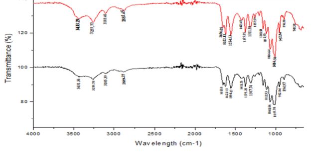
Page |5

Figure 1: Surface morphology of Periplaneta americana chitosan. (A) magnification x 2000 (B) x 400 magnification).

Figure 2: Surface morphology of Acheta domesticus chitosan. (A) magnification x 400 (B) x 2000 magnification).

3.3 FTIR profiles of chitosan from P. americana and A. domesticus

FTIR spectrum of Periplaneta americana and Acheta domesticus displayed the major characteristic band
of chitosan as shown on Figure 3 and Table 2. The spectra showing a peak at 3400 cm-1 indicates
symmetric stretching vibration of OH and amine absorption peak at 2923.88cm-1 indicates the presence
of CH stretch which is characteristic of chitosan. Absorption band at 1650.95 was due to C=O stretching
(amide I) 1095.49 and 1033.77 peaks for C-O stretching. NH bend, C=N stretching (amide II) at 1557
and NH stretching at 3284. Peak at 894.91 is a CH ring stretching a characteristic bond for − 1 −
4 glycosidic linkage which is also a characteristic of chitosan.

The work of Brugnerotto et al., [28]; Anicuta et al., [29] and Ibitoye et al., [22] reported similar result
where they obtained amine absorption peak at 2923.88 cm-1, OH stretching at 3400,C=O stretching at
1650.95, 1033.77 and Peak at 894.91 (CH ring stretching, a characteristic bond for − 1 − 4 glycosidic
linkage )when they carried out FTIR on cockroach chitosan. For the chitosan of A. domesticus, the
spectra showing a peak at 3350 cm-1 indicates symmetric stretching vibration of OH and amine
absorption peak at 1423cm-1 indicates the presence of CH stretch which is characteristic of chitosan.
Absorption band at 1653 was due to C=O stretching (amide I) 1423 and 1375 peaks for C-O stretching.
NH bend, C=N stretching (amide II) at 1554 and NH stretching at 3257. Peak at 896 is a CH ring
stretching, a characteristic bond for − 1 − 4 glycosidic linkage which is also a characteristic of chitosan.

 This results are in line with the study of Kovaleva et al., [30] who obtained a similar result when they
carried out FTIR on the chitosan of cricket (C=O stretching (amide I) 1423 and 1375 peaks for C-O
stretching. NH bend, C=N stretching (amide II) at 1554 and NH stretching at 3257. Peak at 896 is a CH
ring stretching, a characteristic bond for − 1 − 4 glycosidic linkage).

BIOMED Natural and Applied Science 2022;2(1):25-36 https://doi.org/10.53858/bnas02012536
ISSN: 2789-178X
Page |6

 Table 2: The FTIR absorption bands and chemical groups (cm-1) of chitosan in Cricket and Cockroach

 Functional OH NH C=O NH2 CH CH2 CH ring
 group stretching
 Absorption band 3400 3264 1650.95 1557 1095.49, 2923.88, 894.91
 for Cockroach 1033.77 2873
 Absorption band 3350 3257 1653 1554 1423, 1375 1423, 1311 896
 for Cricket

 Figure 3: FTIR Spectra of chitosan from cockroach (red wave) and cricket (black wave).

3.4 Degree of deacetylation of chitosan from Periplaneta americana and Acheta domesticus

The degree of deacetylation of the chitosan obtained from Periplaneta americana and Acheta domesticus
have shown that P. americana had the highest yield and hence have high degree of deacetylation of
90.3 % while A. domesticus has 88.5%. This implies that the chitosan extracted from P. americana and
A. domesticus has high degree of deacetylation and can be used as a source of chitosan.

A deacetylation degree of 55-70% is defined as a low deacetylated degree of chitosan, which is almost
insoluble in water. Deacetylaytion degree of 70-85% is the middle deacetylation degree, which can
partly dissolve in water. 85-95% is a high deacetylation degree of chitosan, which has good solubility in
water, and 95-100% is called ultra-high deacetylation degree of chitosan, which is usually difficult to
attain. Meanwhile, reducing the molecular weight of chitosan by degradation is beneficial to increasing
its solubility in water [31].

Kaya et al. [13], reported a similar result when they worked on chitosan, obtained from male and female
grasshopper and reported that male specie had the highest yield and had high degree of deacetylation
of 98.63 %. While the female grasshopper has 89.43%. It was established that the degree of
deacetylation (DD) of the chitosan extracted from male and female grasshopper are comparable (98-
89%) and are higher than that of commercial chitosan from Shrimp, prawn, or crab [10,16]

3.5 Solubility of chitosan from Periplaneta americana and Acheta domesticus

It was established that P. americana chitosan was more soluble in dilute acetic acid with about 102.1%
solubility, while A. domesticus chitosan had 93%. However, it was postulated that solubility below 87.0%
is considered as low solubility [13]. From the results, it can be said that both insects had high solubility
and therefore can be used conveniently as an alternative source of chitosan. The result is in conformity
with Murat et al. [13] who worked on chitosan from grasshoppers and reported that male chitosan is
more soluble in dilute acetic acid with about 75% solubility, while female chitosan has 46.34%.

BIOMED Natural and Applied Science 2022;2(1):25-36 https://doi.org/10.53858/bnas02012536
ISSN: 2789-178X
Page |7

3.6 GCMS characterization of chitosan from P. americana and A. domesticus

 The GC-MS analysis carried out on the cricket chitosan (Table 3) revealed the presence of a total of 18
compounds (Table 3). The chromatogram of cricket chitosan is shown in Figure 4. The compounds
identified include: cyclohexane,dimethylacetal, cyclohexane,1,5-diisopropyl-2,3-dimethyl, 3,7,11,15-
Tetramethyl-2-hexadecen-1-ol, Pentadecanoic acid,14-methyl-methylester, Palmitic acid,
linolelacidic,methylester 11-octadecanoic acid,methylester, stearic acid,methylester, oleic acid,
octadecanoic acid, 2-piperidinone,N-[4-bromo-n-butyl]-palmitin,1,2-di,-2-aminoethylhydrogen
phospha, 1-dodecanol,3,7,11-trimethyl, eicosanoic acid methyl ester, 9-octadecenal, oxalic acid,
hexadecylhexyl ester, 2-methyl-Z, Z-3,13-Octadecadienol and linoleoyl chloride.

The various bioactive components present in the chitosan obtained from cockroach are shown in Table
4 and observed in Figure 5. A total of 17 compounds including cyclohexane, dimethyl acetal,
pentadecanoic acid, 14-methyl-,methyl ester, hexadecanoic acid, 7-methyl-Z-tetradecen-1-ol acetate,
linolelaidic acid, methyl ester, 11-octadecenoic acid, methyl ester, stearic acid methyl ester, oleic acid,
octadecanoic acid, hexadecanohydrazide, decyl fluoride, arachic acid, octadecenylaldehyde, oxalic acid,
13-docosenoic acid, hexadecanoic acid,2,,3-dihydroxypropyl ester, linoleoyl chloride were identified

 Figure 4: Gas chromatogram of cricket chitosan

 Figure 5: Gas chromatogram of cockroach chitosan

BIOMED Natural and Applied Science 2022;2(1):25-36 https://doi.org/10.53858/bnas02012536
ISSN: 2789-178X
Page |8

 Table 3: Bioactive components of identified from cricket (Acheta domesticus) chitosan

 S/n Retention Area Base Molecular Molecular Name of compound Class of
 Time (%) peak weight formular compound
 (g/mol)

 1. 4.4 0.95 101.10 144 C8H16 O Cyclohexanone,dimethyl Benzene
 acetal derivative
 2. 13.5 0.59 97.23 196 C14H28 Cyclohexane,1,5- Alkene
 diisopropyl-2,3-dimethyl derivative
 3. 15.2 0.63 43.11 296 C20H40O 3,7,11,15-Tetra methyl- Bicyclo
 2-hexadecen-1-ol derivative
 4. 16.8 4.17 79.29 270 C17H34O2 Pentadecanoic acid,14- Bicyclo
 methyl-methyl ester alcohol
 derivative
 5. 17.9 16.68 43.18 256 C16H32O2 Palmitic acid Multiple cyclic
 compound
 6. 19.8 4.29 67.14 294 C19H34O2 Linolelaidic acid, methyl Saturated
 ester tricyclo
 derivative
 7. 19.9 5.22 55.22 296 C19H36O2 11-Octadecenoic acid, Benzene
 methyl ester derivative
 8 20.3 2.29 74.15 298 C19H38O2 Stearic acid, methyl ester Unsaturated
 bicyclo
 derivative
 9 20.8 36.68 41.50 282 C18H34O2 Oleic acid Chloro alkane
 derivative
 10 21.0 9.78 43.23 284 C18H36O2 Octadecanoic acid or Saturated
 stearic acid fatty acid
 11 21.9 2.08 43.27 233 C9H16Br NO 2-Pieperidinone,N-[4- Alkane
 bromo-n-butyl] derivative
 12 22.4 0.91 43.10 691 C37H74NO8P Palmitin,1,2-di,-2- Un saturated
 aminoethyl hydrogen alcohol
 derivative
 13 22.5 1.01 55.39 228 C15H32O 1-dodecanol,3,7,11- Unsaturated
 trimethyl alcohol
 derivative
 `
 14 22.7 0.64 74.10 326 C21H42O2 Eicosanoic acid, methyl Fatty acid
 ester
 15 23.8 7.44 43.23 266 C18H34O 9-octadecenal or octa Alkane
 aldehyde derivative
 16 24.0 1.77 43.10 398 C24H46O4 Oxalic acid, Saturated
 hexadecylhexylester alcohol
 derivative
 17 24.2 3.00 55.79 280 C19H36O 2-Methyl-Z,Z-3,13- Unsaturated
 octadecadienol alcohol
 derivative

 18 26.1 1.85 55.59 298 C18H31ClO Linoleoyl chloride Unsaturated
 alcohol
 derivative

BIOMED Natural and Applied Science 2022;2(1):25-36 https://doi.org/10.53858/bnas02012536
ISSN: 2789-178X
Page |9

Table 4: Bioactive components of identified from American cockroach (Periplaneta americana) chitosan

 S/n Retention Area Base M.W M.F Compound Names Class of
 Time (%) peak (g/mol) compound

 1. 4.4 0.68 105.05 120 C9H12 0-Ethylmethylbenzene Benzene
 derivative
 2. 16.8 2.48 105.05 120 C9H12 1-Hepten-5-yne, 2-methyl-3- Alkene
 methylene derivative
 3. 18.0 19.32 66.00 132 C10H12 Cyclobuta[1,2,3,4]dicyclopentene Bicyclo
 derivative
 4. 19.5 0.70 105.05 134 C9H10O 2-indanol Bicyclo
 alcohol
 derivative
 5. 19.9 2.43 66.00 134 C10H14 3a,4,5,6,7,7a-Hexahydro-4,7- Multiple
 methanoindene cyclic
 compound
 6. 19.9 3.23 67.05 136 C10H16 Tricyclo[4.2.1.1(2,5)]decane Saturated
 tricyclo
 derivative
 7. 20.3 1.47 128.00 128 C10H8 4-Phenylbut-3-ene-1-yne Benzene
 derivative
 8 20.9 39.48 115.05 142 C11H10 1,6-Methano[10]annulene Unsaturated
 bicyclo
 derivative
 9 21.1 10.47 43.00 232 C14H29 Cl 1-Chloroteradecane Chloro
 alkane
 derivative
 10 21.9 3.71 57.05 226 C16H34 5-Propyltridecane Alkane
 derivative
 11 22.4 2.17 57.05 268 C19H40 2,6-Dimethylheptadecane Alkane
 derivative
 12 23.3 1.25 43.00 250 C18H34 1-Octadecyne Un
 saturated
 alcohol
 derivative
 13 23.8 6.33 43.00 296 C20H40O 3,7,11,15-Tetramethyl-2- Unsaturated
 hexadecen-1-ol alcohol
 derivative
 `
 14 24.0 1.09 43.00 256 C16H32O2 n-Hexadecanoic acid Fatty acid
 15 24.2 2.05 43.00 170 C12H26 3,4,5,6-tetramethyloctane Alkane
 derivative
 16 24.4 1.24 43.00 186 C12H26O, 2-Butyloctanol Saturated
 alcohol
 derivative
 17 26.1 1.90 71.05 296 C20H40O 3,7,11,15-Tetramethyl-2- Unsaturated
 hexadecen-1-ol alcohol
 derivative

The GCMS result of cockroach and cricket from this study, coincides with the study of Mahmoud et al.
(2020) explored simplified methods for insect’s chitin extraction and obtained benzene derivative, fatty
acid, alkene derivative, bicycle derivative, bicyclo alcohol derivative, unsaturated alcohol derivatives,
chloro alkane and multiple cyclic compound. Takeshi et al. [32] carried out GCMS on chitin and chitosan
from molluscan shells, they found out that chitosan contained fatty acid, alkane derivative, saturated
alcohol, unsaturated alcohol, chloroalkane derivative which are also present in this present study.

 Multiple cyclic compounds act by binding to chemicals on the membrane that surrounds the insect nerve
cells which causes distortion in the membrane structure. Substances like the fatty acids also disrupts
enzymes that the body of the insect uses to take up food and oxygen which cause the body of the insect
to stop functioning. Unsaturated alcohol derivatives are known to cause repeated stimulations at the

BIOMED Natural and Applied Science 2022;2(1):25-36 https://doi.org/10.53858/bnas02012536
ISSN: 2789-178X
P a g e | 10

receptor site which result in multiple nerve impulses and seizures. Chloro alkane and unsaturated bicyclo
derivatives work against insects nervous system by either blocking nerve impulses or by over stimulating
them [33][34].

Benzene derivatives inhibits the function of the genetic makeup of bacteria there by killing the organism.
[35]. The toxicity decreases with increase in the methyl substitution and in some cases it increases with
increase in methyl substitution. GC-MS spectrum confirmed the presence of various components with
different retention time as illustrated in figure 1 and 2. The mass spectrometer analyzes the compounds
elucidated at different times to identify the nature and structure of the compounds. The large compound
fragments in to small compounds giving rise to the appearance of peaks at different M/Z ratios the mass
spectra are finger print of that compound which can be identified from the data library.

The present study helps to predict the formular and characterize 18 compounds in the chitosan from
cricket and 17 compounds from the chitosan from cockroach. Further investigation may lead to isolation
of bioactive compounds and their structural elucidation and screening of pharmacological activity will be
helpful for further drug development.

4.0 Conclusion

This study reveals that chitin and chitosan can be extracted from cockroach and cricket. The presence
of various bioactive compounds detected after GC-MS analysis using the chitosan from cricket and
cockroach justifies the use of the chitosan for various uses like bio medical, cosmetic and waste water
management. However, isolation of individual phytochemical constituents and subjecting it to biological
activity will be definitely giving fruitful results and will open a new area of investigation of individual
components and their pharmacological potency. From these results it could be concluded that chitosan
from cricket and cockroach contains various bioactive compounds. Evaluation of pharmacological activity
is under progress. Therefore, it is recommended as insects of pharmaceutical importance

Acknowledgement: Authors are appreciative for the support from the Head, Department of
microbiology and laboratory technicians for providing enabling environment and expertise for the
accomplishment of this investigation.

Authors Contributions: The work was conducted in collaboration of all authors. All authors
read and approved the final version of the manuscript.

Competing Interests: The authors have no conflict of interest.

Funding: This research received no external funding”

Ethics Approval Statement: Not applicable

References

1. Chee, K. Determination of polymer-polymer miscibility by viscometry. European Polymer Journal
 1990, 26, 423-426.

2. Kittur, F.; Prashanth, K.H.; Sankar, K.U.; Tharanathan, R. Characterization of chitin, chitosan
 and their carboxymethyl derivatives by differential scanning calorimetry. Carbohydrate polymers
 2002, 49, 185-193.

3. Crini, G. Historical review on chitin and chitosan biopolymers. Environmental Chemistry Letters
 2019, 17, 1623-1643.

4. Azofeifa, D.E.; Arguedas, H.J.; Vargas, W.E. Optical properties of chitin and chitosan
 biopolymers with application to structural color analysis. Optical materials 2012, 35, 175-183.

5. Kurita, K. Chitin and chitosan: functional biopolymers from marine crustaceans. Marine
 biotechnology 2006, 8, 203-226.

BIOMED Natural and Applied Science 2022;2(1):25-36 https://doi.org/10.53858/bnas02012536
ISSN: 2789-178X
P a g e | 11

6. Vinodh, R.; Sasikumar, Y.; Kim, H.-J.; Atchudan, R.; Yi, M. Chitin and chitosan based biopolymer
 derived electrode materials for supercapacitor applications: A critical review. Journal of
 Industrial and Engineering Chemistry 2021, 104, 155-171.

7. Kardas, I.; Struszczyk, M.H.; Kucharska, M.; van den Broek, L.A.; van Dam, J.E.; Ciechańska,
 D. Chitin and chitosan as functional biopolymers for industrial applications. The European
 polysaccharide network of excellence (EPNOE) 2012, 329-373.

8. Monteiro Jr, O.A.; Airoldi, C. Some thermodynamic data on copper–chitin and copper–chitosan
 biopolymer interactions. Journal of Colloid and Interface Science 1999, 212, 212-219.

9. Minagawa, T.; Okamura, Y.; Shigemasa, Y.; Minami, S.; Okamoto, Y. Effects of molecular weight
 and deacetylation degree of chitin/chitosan on wound healing. Carbohydrate polymers 2007,
 67, 640-644.

10. Mohammadkhani, G.; Kumar Ramamoorthy, S.; Adolfsson, K.H.; Mahboubi, A.; Hakkarainen,
 M.; Zamani, A. New Solvent and Coagulating Agent for Development of Chitosan Fibers by Wet
 Spinning. Polymers 2021, 13, 2121.

11. Zhao, Y.; Park, R.-D.; Muzzarelli, R.A. Chitin deacetylases: properties and applications. Marine
 drugs 2010, 8, 24-46.

12. Nam, Y.S.; Park, W.H.; Ihm, D.; Hudson, S.M. Effect of the degree of deacetylation on the
 thermal decomposition of chitin and chitosan nanofibers. Carbohydrate Polymers 2010, 80,
 291-295.

13. Kaya, M.; Lelešius, E.; Nagrockaitė, R.; Sargin, I.; Arslan, G.; Mol, A.; Baran, T.; Can, E.; Bitim,
 B. Differentiations of chitin content and surface morphologies of chitins extracted from male and
 female grasshopper species. PloS one 2015, 10, e0115531.

14. Stevens, C.V. Chitin and Chitosan: properties and applications. 2019.

15. Rinaudo, M. Chitin and chitosan: Properties and applications. Progress in polymer science 2006,
 31, 603-632.

16. Marei, N.H.; Abd El-Samie, E.; Salah, T.; Saad, G.R.; Elwahy, A.H. Isolation and characterization
 of chitosan from different local insects in Egypt. International journal of biological
 macromolecules 2016, 82, 871-877.

17. Kmiec, M.; Pighinelli, L.; Tedesco, M.; Silva, M.; Reis, V. Chitosan-properties and applications
 in dentistry. Adv Tissue Eng Regen Med Open Access 2017, 2, 00035.

18. Koide, S. Chitin-chitosan: properties, benefits and risks. Nutrition research 1998, 18, 1091-
 1101.

19. Islam, S.; Bhuiyan, M.; Islam, M. Chitin and chitosan: structure, properties and applications in
 biomedical engineering. Journal of Polymers and the Environment 2017, 25, 854-866.

20. Pakizeh, M.; Moradi, A.; Ghassemi, T. Chemical extraction and modification of chitin and
 chitosan from shrimp shells. European Polymer Journal 2021, 159, 110709.

21. Badawy, R.M.; Mohamed, H.I. Chitin extration, composition of different six insect species and
 their comparable characteristics with that of the shrimp. J. Am. Sci 2015, 11, 127-134.

22. Ibitoye, E.; Lokman, I.; Hezmee, M.; Goh, Y.; Zuki, A.; Jimoh, A. Extraction and physicochemical
 characterization of chitin and chitosan isolated from house cricket. Biomedical Materials 2018,
 13, 025009.

23. Wanule, D.; Balkhande, J.; Ratnakar, P.; Kulkarni, A.; Bhowate, C. Extraction and FTIR analysis
 of chitosan from American cockroach, Periplaneta americana. Extraction 2014, 3, 299-304.

BIOMED Natural and Applied Science 2022;2(1):25-36 https://doi.org/10.53858/bnas02012536
ISSN: 2789-178X
P a g e | 12

24. Laka, M.; Chernyavskaya, S. Preparation of chitosan powder and investigation of its properties.
 In Proceedings of Proceedings-Estonian Academy of Sciences Chemistry; p. 78.

25. Lawal, B.; Shittu, O.K.; AbdulRasheed-Adeleke, T.; Ossai, P.C.; Ibrahim, A.M. GC-MS
 determination of bioactive constituents of giant African Snail (Archachatina maginata)
 Haemolymph. IOSR Journal of Pharmacy and Biological Sciences (IOSR-JPBS) 2015, 10, 59-64

26. Ladchumananandasivam, R.; da Rocha, B.G.; Belarmino, D.D.; Galv, A.O. The use of
 exoskeletons of shrimp (Litopenaeus vanammei) and crab (Ucides cordatus) for the extraction
 of chitosan and production of nanomembrane. 2012.

27. Chen, S.; Wei, X.; Sui, Z.; Guo, M.; Geng, J.; Xiao, J.; Huang, D. Preparation of Antioxidant and
 antibacterial chitosan film from Periplaneta americana. Insects 2021, 12, 53.

28. Brugnerotto, J.; Lizardi, J.; Goycoolea, F.; Argüelles-Monal, W.; Desbrieres, J.; Rinaudo, M. An
 infrared investigation in relation with chitin and chitosan characterization. Polymer 2001, 42,
 3569-3580.

29. Anicuta, S.-G.; Dobre, L.; Stroescu, M.; Jipa, I. Fourier transform infrared (FTIR) spectroscopy
 for characterization of antimicrobial films containing chitosan. Analele Universită Ńii din Oradea
 Fascicula: Ecotoxicologie, Zootehnie şi Tehnologii de Industrie Alimentară 2010, 2010, 1234-
 1240.

30. Kovaleva, E.; Pestov, A.; Stepanova, D.; Molochnikov, L. Characterization of chitin and its
 complexes extracted from natural raw sources. In Proceedings of AIP conference proceedings;
 p. 050007.

31. Pacheco-Torgal, F.; Ivanov, V.; Karak, N.; Jonkers, H. Biopolymers and biotech admixtures for
 eco-efficient construction materials; Woodhead Publishing: 2016.

32. Furuhashi, T.; Beran, A.; Blazso, M.; Czegeny, Z.; Schwarzinger, C.; Steiner, G. Pyrolysis GC/MS
 and IR spectroscopy in chitin analysis of molluscan shells. Bioscience, biotechnology, and
 biochemistry 2009, 73, 93-103.

33. Cui, G.; Guo, J.; Zhang, Y.; Zhao, Q.; Fu, S.; Han, T.; Zhang, S.; Wu, Y. Chitosan
 oligosaccharide derivatives as green corrosion inhibitors for P110 steel in a carbon-dioxide-
 saturated chloride solution. Carbohydrate polymers 2019, 203, 386-395.

34. Ozkan, G.; Baydar, H.; Erbas, S. The influence of harvest time on essential oil composition,
 phenolic constituents and antioxidant properties of Turkish oregano (Origanum onites L.).
 Journal of the Science of Food and Agriculture 2010, 90, 205-209.

35. Odokuma, L.; Oliwe, S. Toxicity of substituted benzene derivatives to four chemolithotrophic
 bacteria isolated from the New Calabar River. Global Journal of Environmental Sciences 2003,
 2, 72-77.

 Submit your article to AROC JOURNALS
 -AROC in Pharmaceutical and Biotechnology
 -AROC in Agriculture
 -AROC in Bioinformatics
 -AROC in Food and Nutrition
 -AROC in Natural Product Research
 -BIOMED Natural and Applied Science
 Via https://arocjournal.com/

Copyright © 2022 Umar et al. This is an open access article distributed under the terms and conditions of the
Creative Commons Attribution License (CC BY) which allowed unrestricted download, distribution and reused as
long as the original authors are properly cited.
BIOMED Natural and Applied Science 2022;2(1):25-36 https://doi.org/10.53858/bnas02012536
ISSN: 2789-178X
You can also read