Novel homozygous mutations in the transcription factor NRL cause non-syndromic retinitis pigmentosa
←
→
Page content transcription
If your browser does not render page correctly, please read the page content below
Molecular Vision 2022; 28:48-56                                        © 2022 Molecular Vision
        Received 2 March 2021 | Accepted 15 May 2022 | Published 17 May 2022

Novel homozygous mutations in the transcription factor NRL
cause non-syndromic retinitis pigmentosa
    Mohammed E. El-Asrag,1,2 Marta Corton,3 Martin McKibbin,1,4 Almudena Avila-Fernandez,3 Moin D.
    Mohamed,5 Fiona Blanco-Kelly,3 Carmel Toomes,1 Chris F. Inglehearn,1 Carmen Ayuso,3 Manir Ali1

    (The first two authors contributed equally to this work.)
    1
     Division of Molecular Medicine, Leeds Institute of Medical Research, St. James’s University Hospital, University of Leeds,
    Leeds, UK; 2Department of Zoology, Faculty of Science, Benha University, Benha, Egypt; 3Department of Genetics & Genomics,
    Instituto de Investigación Sanitaria-Fundación Jiménez Díaz University Hospital- Universidad Autónoma de Madrid (IIS-FJD,
    UAM), Centre for Biomedical Network Research on Rare Diseases (CIBERER), Madrid, Spain; 4The Eye Department, St. James’s
    University Hospital, Leeds, UK; 5Ophthalmology Department, St Thomas' Hospital, London, UK

         Purpose: To describe the clinical phenotype and genetic basis of non-syndromic retinitis pigmentosa (RP) in one family
         and two sporadic cases with biallelic mutations in the transcription factor neural retina leucine zipper (NRL).
         Methods: Exome sequencing was performed in one affected family member. Microsatellite genotyping was used for
         haplotype analysis. PCR and Sanger sequencing were used to confirm mutations in and screen other family members
         where they were available. The SMART tool for domain prediction helped us build the protein schematic diagram.
         Results: For family MM1 of Pakistani origin, whole-exome sequencing and microsatellite genotyping revealed ho-
         mozygosity on chromosome 14 and identified a homozygous stop-loss mutation in NRL, NM_006177.5: c.713G>T,
         p.*238Lext57, which is predicted to add an extra 57 amino acids to the normal protein chain. The variant segregated with
         disease symptoms in the family. For case RP-3051 of Spanish ancestry, clinical exome sequencing focusing on the morbid
         genome highlighted a homozygous nonsense mutation in NRL, c.238C>T, p.Gln80*, as the most likely disease candidate.
         For case RP-1553 of Romanian ethnicity, targeted-exome sequencing of 73 RP/LCA genes identified a homozygous
         nonsense mutation in NRL, c.544C>T, p.Gln182*. The variants were either rare or absent in the gnomAD database.
         Conclusions: NRL mutations predominantly cause dominant retinal disease, but there have been five published reports of
         mutations causing recessive disease. Here, we present three further examples of recessive RP due to NRL mutations. The
         phenotypes observed are consistent with those in the previous reports, and the observed mutation types and distribution
         further confirm distinct patterns for variants in NRL causing recessive and dominant diseases.

     Retinitis pigmentosa (RP) is a group of inherited disor-              malformation, hearing loss and intellectual disability [4]. RP
ders of the retina that are characterized initially by the loss            is genetically heterogeneous, with autosomal dominant, auto-
of rod photoreceptor function followed by cone photorecep-                 somal recessive and X-linked patterns of inheritance. To date,
tors. The age of onset and severity vary among affected                    mutations in 93 genes have been shown to cause the condition
individuals. Night blindness is often the earliest symptom,                (RetNet). These genes encode proteins that are required for
and clinical hallmarks include attenuated retinal vessels                  the development and maintenance of photoreceptor structure
in the peripheral retina, bone spicule pigments throughout                 and its matrix membranes, visual transduction, ciliary traf-
the fundus and absent α- and β-waves following electroreti-                ficking and photoreceptor outer segment shedding. Recent
nography [1,2]. RP is the most common cause of hereditary                  advances in technology have helped to identify the genetic
blindness, with an incidence of approximately 1 in 4,000 in                causes of up to 70% of RP patients, with the remaining group
the population [3]. The majority of cases are non-syndromic,               still of unknown etiology [5,6].
though about a quarter are syndromic, associated with                           The neural retina leucine zipper gene (NRL, OMIM
non-ocular conditions such as obesity, polydactyly, renal                  162080) maps to human chromosome 14q11.2 [7] and encodes
                                                                           a 237 amino acid protein that belongs to the Maf subfamily
Correspondence to: Manir Ali, Division of Molecular Medicine,              of transcription factors, which are required for cell differ-
Leeds Institute of Medical Research, St. James’s University                entiation [8]. The protein has a Maf transactivation and a
Hospital, University of Leeds, UK; Phone: 44 113 343 8420; FAX:            bZIP (basic leucine zipper) DNA-binding and dimerization
44 113 343 8603; email: m.ali@leeds.ac.uk                                  domain. During mammalian retinal development, NRL is

                                                                      48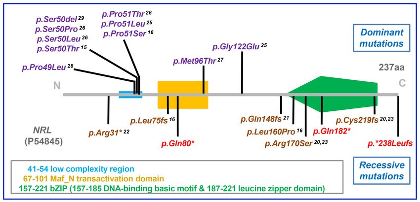
Molecular Vision 2022; 28:48-56                                                 © 2022 Molecular Vision

required for rod photoreceptor differentiation and synergis-                 were removed using Picard, and the mean depth of reads per
tically interacts with CRX, NR2E3 and other transcription                    base was noted. Insertions, deletions and single nucleotide
factors to regulate the activity of photoreceptor-specific                   variants were noted in VCF format using the Unified Geno-
genes such as rhodopsin [9-11]. The transgenic knockout                      typer function of GATK.
mouse (Nrl−/−) has an unusual retinal photoreceptor layer
with shorter outer and inner segments, a functionally rod-less                    The variant list was annotated with ANNOVAR software
retina and super-normal cone function mediated by S-cones                    and filtered to include only non-synonymous coding vari-
[12]. The transgenic overexpression of Nrl in mouse retina                   ants, insertions and deletions affecting the coding sequence
induces postmitotic photoreceptor precursors to drive differ-                and any variants within 5 bp of splice donor and acceptor
entiation toward rods instead of cones [13]. This function of                sites, with a minimum read depth of 10 and a minor allele
Nrl to induce rod development while repressing the S-cone                    frequency ≤1% in the Exome Variant Server, as well as the
pathway acts through direct activation of the transcription                  1000 Genomes and Genome Aggregation databases. The
factor Nr2e3 [14]. Mutations in NRL have been associated                     pathogenicity of missense variants was assessed using the
with both dominant and recessively inherited retinal disease                 following prediction software: PolyPhen2, SIFT and CADD.
in humans [15,16]. The study reported herein describes novel                 For the analysis of whole exome and clinical exome data, the
recessive mutations in NRL causing non-syndromic RP in one                   variant list was compared against the known retinal dystrophy
family and two sporadic cases.                                               genes in RetNet (accessed December 2021).

                                                                                 To detect homozygous regions from exome sequencing
                           METHODS                                           data, the AgileGenotyper software was used [19]. The
                                                                             exported variant list was analyzed using AutoSNPa, and the
Patient recruitment and sampling: Ethical approval for                       homozygous regions were visualized against a circular ideo-
the study was obtained from the committees of the Leeds                      gram of Chromosomes 1 to 22 using AgileMultiIdeogram.
Teaching Hospitals National Health Service Trust ([17]/
YH/0032) and the Fundación Jiménez Díaz University                           Microsatellite genotyping: Oligonucleotide primer pairs
Hospital in Madrid (reference number PIC 134–2016_FJD).                      spanning polymorphic short tandem repeats were selected
Consent was obtained from the individuals and their guard-                   from the UCSC database. The PCR was performed with the
ians where appropriate. The patients were diagnosed with                     primers on genomic DNA, and the products were genotyped
non-syndromic RP following detailed clinical examination                     on the ABI3130xl Genetic Analyzer (Applied Biosystems,
by an experienced ophthalmologist and after taking a clinical                Warrington, U.K.). Allele sizing was achieved using the
history. Blood samples were collected and stored in BD                       GeneMapper v.4 software (Applied Biosystems).
vacutainer® EDTA blood collection tubes (BD Biosciences,
                                                                             PCR and Sanger sequencing: The PCR was typically
Oxford, U.K.). Genomic DNA was extracted from peripheral
                                                                             performed on 30 ng of genomic DNA in 10 μl volumes
blood cells according to standard protocols using a QIAamp
                                                                             according to standard protocols, which included 40 cycles
DNA blood midi kit (Qiagen Limited, Manchester, U.K.).
                                                                             of 94 °C for 30 s, 55–60 °C for 45 s and 72 °C for 45 s. An
Exome sequencing: Targeted enriched libraries were prepared                  aliquot of the reaction product was visualized by agarose gel
using either SureSelect Human Whole Exome reagent v.5                        electrophoresis, and the remaining mixture was treated with
(Agilent Technologies, Santa Clara, CA), Clinical Exome                      ExoSAP-IT (GE Healthcare, Chalfont St. Giles, U.K.). The
solution (Sophia Genetics, Boston, MA) covering the exons of                 treated PCR product was cycle sequenced using the BigDye
4,490 known disease-related genes or a customized Haloplex                   Terminator v.3.1 reaction mix (Applied Biosystems) and run
reagent (Agilent) of the exons of 73 genes associated with RP/               on an ABI3130xl Genetic Analyzer (Applied Biosystems).
LCA as previously described [17]. Libraries were sequenced                   The sequencing output was analyzed using the Sequence
using a paired-end protocol on the HiSeq2500 or NextSeq500                   Analysis v.5.2 software package (Applied Biosystems)
platforms (Illumina, Little Chesterford, U.K.).                              according to the manufacturer’s instructions.

     The quality of the output sequencing data was evaluated                 Bioinformatics: Genomic DNA sequence information was
using FASTQ tools on the Galaxy platform [18]. After quality                 downloaded from the NCBI website. Oligonucleotide primer
control filtering, the sequencing data was aligned against the               pairs were designed to amplify across the variant of interest
reference human genome (hg19/GRCh37) using Bowtie2 and                       using Primer3 software. The primers used in this study are
processed in SAM/BAM format using SAMtools and the                           listed in Appendix 1. The SMART tool was used to predict
Genome Analysis Toolkit (GATK). Potential PCR duplicates                     protein domains in the amino acid sequence.

                                                                        49
Molecular Vision 2022; 28:48-56 © 2022 Molecular Vision
RESULTS Clinical details and molecular analysis of family MM1: The
proband (IV:2) in family MM1 is one of three affected indi-
One family of Pakistani origin (MM1) and two sporadic cases, viduals in a Pakistani family with consanguineous parents.
one of Spanish ancestry (RP-3051) and the other of Romanian She has had problems with her vision since early childhood
descent (RP-1553), were investigated in this study (Figure and had surgery for strabismus when she was seven years old.
1). All the affected subjects were diagnosed with early-onset At 19 years old, she was registered as visually impaired with
retinitis pigmentosa (Table 1). None of the examined patients grossly constricted visual fields and best corrected acuity
showed extraocular features. Where a family history of of 6/24 and 6/36. Ophthalmic examination revealed typical
consanguinity was known, it suggested an autosomal reces- clumped pigment deposits, and posterior subcapsular cata-
sive pattern of inheritance for disease transmission. racts developed gradually in both eyes. At 43 years old, the
Figure 1. Families analyzed in
this study. The structures of the
pedigrees are shown for A: family
MM1, B: case RP-3051 and C:
case RP-1553. The proband in each
pedigree is highlighted with an
arrow. Microsatellite genotyping
in family MM1 around the NRL
locus is shown with allele sizes and
haplotypes for three microsatellite
markers, D14S1280, D14S608 and
D14S599, located at 26.7, 28.8 and
34.7Mb from the top of chromo-
some 14 using the hg19/GRCh37
human reference genome. Haplo-
type analysis (red block) confirmed
homozygosity in family MM1
around the NRL locus only in the
affected cases. The NRL mutation
(located at 24.6 Mb) is stated as
M1, M2 and M3. Genomic DNA
was only available from members
to whom a genotype has been
assigned. Note that only the affected
individuals are homozygous for the
mutation.
50Molecular Vision 2022; 28:48-56 © 2022 Molecular Vision
Table 1. Clinical details of retinal dystrophy patients examined in this study.
Best
Age at corrected
Family examination visual acuity Fundus Additional
ID Ethnicity Subject Gender (years) (OD & OS) examination Diagnosis Findings
Pigmentary Retinitis Posterior subcap-
MM1 Pakistani IV:2 F 19 6/24 & 6/36 retinopathy pigmentosa sular opacification
43 6/36 & 6/60
Pigmentary Retinitis
IV:3 M 20 6/9 & 6/36 Amblyopia (OS)
retinopathy pigmentosa
Posterior subcap-
33 NA
sular opacification
Pigmentary Retinitis Photophobia,
RP-3051 Spanish II:6 M 57 NA
retinopathy pigmentosa dyschromatopsia
Pigmentary Retinitis Nystagmus,
RP-1553 Romanian II:1 F 21 6/60 & 6/60
retinopathy pigmentosa complete scotoma
The gender, age at examination, corrected visual acuity, ophthalmoscopy results, diagnosis and any additional findings are summarized.
M=male, F=female, OD=right eye, OS=left eye, NA=not available.
best corrected acuity was 6/36 and 6/60. Her younger brother genetic testing at 57 years of age with a clinical diagnosis of
(IV:3), when examined at 20 years old, had amblyopia in his RP. He had night blindness since childhood, loss of visual
left eye and best corrected visual acuity of 6/9 and 6/36, with acuity since the third decade, and was now experiencing
a fundus consistent with typical RP changes. At 33 years old, photophobia and slight dyschromatopsia. A second sibling
when he was last examined, he had no response to electroreti- manifested a similar presentation of RP, but clinical data were
nography and had developed posterior subcapsular cataracts. unavailable for that person.
The other affected case (IV:5) in the family was not available
Clinical exome sequencing of the index case II:6
for further examination.
followed by filtration analysis identified a novel homozygous
Whole exome sequencing of the genomic DNA from nonsense mutation, c.238C>T, p.Gln80* (ClinVar accession
subject IV:2 identified 60,603 total variants that differed from SCV001478094) in the NRL gene, as the most likely cause
the reference genome sequence. After filtration, 119 variants of RP in the patient. This variant, which was absent in the
remained, of which 25 were homozygous changes. After gnomAD database, was verified by Sanger sequencing in
comparing the variant list against the known RetNet genes, patient genomic DNA (Figure 2B).
a single candidate—a novel homozygous stop-loss mutation, Clinical details and molecular analysis of case RP-1553: The
NM_006177.5: c.713G>T, p.*238Lext57 (ClinVar accession patient (II:1) in the family RP-1553 is the only affected child
SCV001478096) in the transcription factor gene NRL—was of a Romanian couple who are not known to be related. She
identified as the best candidate. AgileMultiIdeogram analysis had visual impairment since two years old and was diagnosed
of the WES data highlighted a 3.4 Mb homozygous region with early-onset RP at five years of age. Her most recent
that contained the NRL gene (Appendix 2), and microsatel- examination at 21 years old showed nystagmus, complete
lite genotyping with polymorphic markers confirmed this loss of the visual field, best corrected visual acuity of 6/60 in
homozygosity (Figure 1A). The NRL variant was present both eyes, non-recordable electroretinography and typical RP
once in 115,494 alleles in the gnomAD database, and Sanger presentation upon funduscopy.
sequencing (Figure 2A) confirmed that the mutation segre-
gated with the disease phenotype in the family (Figure 1A) Genomic DNA from patient II:1 underwent targeted
as expected for a recessive condition. exome sequencing for the 73 RP/LCA genes. Following
variant prioritization, a novel homozygous mutation in NRL,
Clinical details and molecular analysis of case RP-3051: The c.544C>T p.Gln182* (ClinVar accession SCV001478095),
index case (II:6) in the family RP-3051 is the sixth child of was found as the most likely disease candidate. This variant
a consanguineous Spanish couple from a small village with was confirmed by Sanger sequencing the DNA of the patient
a relatively high level of endogamy. He was referred for (Figure 2C) and was absent from the gnomAD database.
51Molecular Vision 2022; 28:48-56 © 2022 Molecular Vision
DISCUSSION recessive or dominantly transmitted retinal disease in humans
(see Figure 3). The type of mutation, null allele or missense,
Mutations in the NRL gene have been reported to cause either and the location of missense mutations in the protein
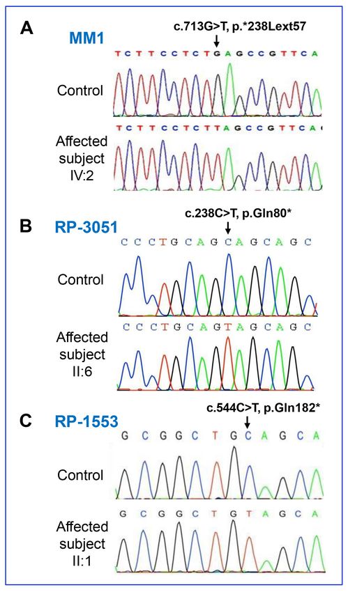
Figure 2. Conf ir mator y NR L
sequence analysis. Chromatograms
of the NRL sequence variants from
a normal control individual and
affected subjects, IV:2 from family
MM1 (A), II:6 from family RP-3051
(B) and II:1 from family RP-1553
(C). Note that homozygous muta-
tions c.713G>T in family MM1,
c.238C>T in family RP-3051 and
c.544C>T in family RP-1553 are
shown.
52Molecular Vision 2022; 28:48-56 © 2022 Molecular Vision
Figure 3. Mapping the NRL mutation spectrum onto the protein sequence. The domains in the 237 amino acid NRL protein are indicated.
NRL mutations identified in dominant disease [15,16,25-29] are presented in purple text above the protein domain representation, and
recessive disease [16,20-23] are shown in brown text below the illustration. The novel mutations described in this paper are highlighted in
red in the diagram.
sequence determine the inheritance pattern of disease trans- all the functional domains of the protein, it is most likely
mission. There are only five published reports of NRL muta- that the extra amino acids at the C-terminus are problematic.
tions causing recessive disease [16,20-23]. These mutations Hypothetically, should the mutant transcript escape early
are all either null alleles caused by nonsense or frameshift decay, the extra amino acids would presumably interfere with
mutations or are missense mutations that map to the bZIP NRL dimerization and protein interactions to impede normal
domain, which is required for DNA-binding, dimerization of function.
NRL and interaction with the homeodomain of CRX [10]. To date, the NRL mutations causing dominant retinal
Nonsense and frameshift mutations are likely to cause disease disease are all missense mutations apart from a single amino
either by nonsense-mediated decay of the mutant transcript acid in-frame deletion [15,16,25-29]. In vitro assays have
or, if a truncated protein is synthesized, due to complete or shown that these mutations exhibit reduced phosphoryla-
partial loss of the bZIP domain that would be detrimental tion of NRL but enhanced transcriptional activation of the
to NRL function. However, missense mutations in the bZIP rhodopsin promoter, resulting in the gaining of function in
domain are also damaging by preventing DNA binding and excess of normal promoter activation [15,24,27,28]. Although
reducing transcription activation [24]. These mutations there- other rod-specific promoters have not yet been directly inves-
fore cause recessive disease by loss of normal protein func- tigated, this excessive activation of mutant NRL presumably
tion. Here, we report three further novel mutations causing affects several NRL target gene promoters that have been
recessive disease, consistent with this pattern. The p.Gln80* found to be important for maintaining rod photoreceptor
mutation removes the C-terminal end of the minimal trans- function [30].
activation domain, the DNA-binding basic domain and the The clinical presentation and phenotype of the patients
leucine zipper dimerization domain, whereas the p.Gln182* is different for recessive and dominant NRL retinopathy. To
removes only the leucine zipper domain. Despite the absence date, 25 patients have been studied with the dominant form
of segregation analysis of these mutations, the loss of key of disease [15,16,25-29] and, including the patients described
functional domains would certainly be consistent with loss herein, 12 subjects have been described with recessive disease
of NRL function. Though the p.*238Lext57 stop-loss muta- [16,20-23]. Night blindness from early childhood is a common
tion, which segregated with disease in the family, contains symptom, followed by variable amounts of progressive visual
53Molecular Vision 2022; 28:48-56 © 2022 Molecular Vision
field constriction and reduced visual acuity. Other features APPENDIX 2. HOMOZYGOSITY MAPPING IN
co-existing in patients include nystagmus in 4/12 recessive FAMILY MM1 USING WES DATA.
[22,23], this paper] and 1/25 dominant cases [16], strabismus To access the data, click or select the words “Appendix 2.”
or amblyopia in 5/12 recessive [16,23], this paper] and 0/25 MultiIdeogram of the WES data of patient IV:2 showing
dominant subjects and posterior subcapsular cataracts in the homozygous regions in blue. The NRL gene (arrowed) is
more elderly, which was seen in 4/12 recessive [22,23], this located on chromosome 14 at ~24.6Mb (hg19) and maps
paper] and 3/25 dominant patients [26]. For recessive NRL within the homozygous region 21,860,360–25,288,227.
retinopathy, also called clumped pigment retinal degen-
eration, which is a subtype of autosomal recessive RP, these ACKNOWLEDGMENTS
signs coincide with the early fundal appearance of clusters of
clumped pigmented deposits in the periphery that is accom- We thank the patients who participated in this study. We
panied by chorioretinal atrophy and attenuated arterioles acknowledge Dr. Raluca O. Ionescu from Hospital Univer-
[16,22,23]. However, for dominant disease, fundal abnormali- sitario Clinico San Carlos in Madrid for patient recruit-
ties tend to appear in the third decade with attenuated vessels ment. This work was supported by grants from the Instituto
de Salud Carlos III (ISCIII) from the Spanish Ministry of
and bone spicule–shaped pigment deposits that are more typi-
Health, including CIBERER (06/07/0036), IIS-FJD Biobank
cally seen in patients diagnosed with RP [25-29]. Although
PT13/0010/0012, and FIS (PI16/00425 and PI19/00321); and
patients with NRL retinopathy show markedly reduced rod
from the regional government of Madrid, RAREGenomics-
and cone functions on electroretinography, patients with
CM (CAM, B2017/BMD-3721), all partially supported by
dominant disease also show loss of short wavelength (blue- or
FEDER (European Regional Development Fund). MC is
S-) cone function [16,28] whereas recessive disease patients
supported by the Miguel Servet Program (CPII17 00006)
show no detectable rod function and a relatively enhanced
from ISCIII. MC, AA-F, FB-K and CA are supported by
S-cone function [16,22,23]. This enhanced S-cone feature
funding from the Spanish National Organization of the Blind
in recessive NRL retinopathy is similar to what is observed
(ONCE) and the Ramon Areces Foundation. MEE-A was
in Nrl-knockout mice [12] and patients with retinopathy due
funded by an Egyptian government scholarship during this
to NR2E3 mutations [31]. It would be interesting to deter-
work. CT, CFI and MA are supported by funding from Fight
mine whether the patients described in this report share this
for Sight and Retina UK.
enhanced S-cone phenotype, though limited patient access
has precluded further study.
REFERENCES
To summarize, we describe one family and two sporadic
cases with different novel homozygous NRL mutations 1. Hartong DT, Berson EL, Dryja TP. Retinitis pigmentosa.
Lancet 2006; 368:1795-809. [PMID: 17113430].
accounting for the disease phenotype. Previously, only five
families and six mutations had been described with this form 2. Verbakel SK, van Huet RAC, Boon CJF, den Hollander AI,
Collin RWJ, Klaver CCW, Hoyng CB, Roepman R, Klevering
of RP, so the results contribute to the mutation spectrum
BJ. Non-syndromic retinitis pigmentosa. Prog Retin Eye Res
for this condition. The phenotypes observed are consistent 2018; 66:157-86. [PMID: 29597005].
with those in previous reports, and the observed mutation
3. Haim M. Epidemiology of retinitis pigmentosa in Denmark.
types and distribution further confirm distinct patterns for Acta Ophthalmol Scand Suppl 2002; 233:1-34. [PMID:
variants causing recessive and dominant disease. Identifying 11921605].
the genetic cause of disease in these patients provides more 4. Pierrottet CO, Zuntini M, Digiuni M, Bazzanella I, Ferri P,
accurate genetic counselling for the families and stratifies Paderni R, Rossetti LM, Cecchin S, Orzalesi N, Bertelli M.
the patients into distinct groups as future treatments become Syndromic and non-syndromic forms of retinitis pigmentosa:
available. a comprehensive Italian clinical and molecular study reveals
new mutations. Genet Mol Res 2014; 13:8815-33. [PMID:
25366773].
APPENDIX 1. OLIGONUCLEOTIDE PRIMER 5. Riera M, Navarro R, Ruiz-Nogales S, Mendez P, Bures-Jelstrup
PAIRS FOR THE ANALYSIS OF THE NRL A, Corcostegui B, Pomares E. Whole exome sequencing
MUTATIONS. using Ion Proton system enables reliable genetic diagnosis of
inherited retinal dystrophies. Sci Rep 2017; 7:42078-[PMID:
To access the data, click or select the words “Appendix 28181551].
1.” The DNA sequences, PCR product size and annealing 6. Haer-Wigman L, van Zelst-Stams WA, Pfundt R, van den
temperature of each primer pair are shown. Born LI, Klaver CC, Verheij JB, Hoyng CB, Breuning MH,
54Molecular Vision 2022; 28:48-56 © 2022 Molecular Vision
Boon CJ, Kievit AJ, Verhoeven VJ, Pott JW, Sallevelt SC, pigmentosa in Spanish patients. Invest Ophthalmol Vis Sci
van Hagen JM, Plomp AS, Kroes HY, Lelieveld SH, Hehir- 2015; 56:2173-82. [PMID: 25698705].
Kwa JY, Castelein S, Nelen M, Scheffer H, Lugtenberg D,
18. Blankenberg D, Gordon A, Von Kuster G, Coraor N, Taylor J,
Cremers FP, Hoefsloot L, Yntema HG. Diagnostic exome
Nekrutenko A. Manipulation of FASTQ data with Galaxy.
sequencing in 266 Dutch patients with visual impairment.
Bioinformatics 2010; 26:1783-5. [PMID: 20562416].
Eur J Hum Genet 2017; 25:591-9. [PMID: 28224992].
19. Carr IM, Bhaskar S, O’Sullivan J, Aldahmesh MA, Sham-
7. Yang-Feng TL, Swaroop A. Neural retina-specific leucine
seldin HE, Markham AF, Bonthron DT, Black G, Alkuraya
zipper gene NRL (D14S46E) maps to human chromosome
FS. Autozygosity mapping with exome sequence data. Hum
14q11.1-q11.2. Genomics 1992; 14:491-2. [PMID: 1427865].
Mutat 2013; 34:50-6. [PMID: 23090942].
8. Swaroop A, Xu JZ, Pawar H, Jackson A, Skolnick C, Agarwal
N. A conserved retina-specific gene encodes a basic motif/ 20. Neveling K, Collin RWJ, Gilissen C, van Huet RAC, Visser L,
leucine zipper domain. Proc Natl Acad Sci USA 1992; Kwint MP, Gijsen SJ, Zonneveld MN, Wieskamp N, de Ligt
89:266-70. [PMID: 1729696]. J, Siemiatkowska AM, Hoefsloot LH, Buckley MF, Kellner
U, Branham KE, den Hollander AI, Hoischen A, Hoyng
9. Rehemtulla A, Warwar R, Kumar R, Ji X, Zack DJ, Swaroop C, Klevering BJ, van den Born LI, Veltman JA, Cremers
A. The basic motif-leucine zipper transcription factor Nrl FPM, Scheffer H. Next-generation genetic testing for reti-
can positively regulate rhodopsin gene expression. Proc Natl nitis pigmentosa. Hum Mutat 2012; 33:963-72. [PMID:
Acad Sci USA 1996; 93:191-5. [PMID: 8552602]. 22334370].
10. Mitton KP, Swain PK, Chen S, Xu S, Zack DJ, Swaroop A. 21. Beryozkin A, Shevah E, Kimchi A, Mizrahi-Meissonnier L,
The leucine zipper of NRL interacts with the CRX home- Khateb S, Ratnapriya R, Lazar CH, Blumenfeld A, Ben-Yosef
odomain. A possible mechanism of transcriptional synergy T, Hemo Y, Pe’er J, Averbuch E, Sagi M, Boleda A, Gieser L,
in rhodopsin regulation. J Biol Chem 2000; 275:29794-9. Zlotogorski A, Falik-Zaccai T, Alimi-Kasem O, Jacobson SG,
[PMID: 10887186]. Chowers I, Swaroop A, Banin E, Sharon D. Whole exome
11. Cheng H, Khanna H, Oh ECT, Hicks D, Mitton KP, Swaroop sequencing reveals mutations in known retinal disease genes
A. Photoreceptor-specific nuclear receptor NR2E3 functions in 33 out of 68 Israeli families with inherited retinopathies.
as a transcriptional activator in rod photoreceptors. Hum Sci Rep 2015; 5:13187-[PMID: 26306921].
Mol Genet 2004; 13:1563-75. [PMID: 15190009]. 22. Newman H, Blumen SC, Braverman I, Hanna R, Tiosano B,
12. Mears AJ, Kondo M, Swain PK, Takada Y, Bush RA, Saunders Perlman I, Ben-Yosef T. Homozygosity for a recessive loss-
TL, Sieving PA, Swaroop A. Nrl is required for rod photo- of-function mutation of the NRL gene is associated with a
receptor development. Nat Genet 2001; 29:447-52. [PMID: variant of enhanced S-cone syndrome .Invest Ophthalmol
11694879]. Vis Sci 2016; 57:5361-71. [PMID: 27732723].
13. Oh EC, Khan N, Novelli E, Khanna H, Strettoi E, Swaroop A. 23. Littink KW, Stappers PTY, Riemslag FCC, Talsma HE, van
Transformation of cone precursors to functional rod photo- Genderen MM, Cremers FPM, Collin RWJ, van den Born
receptors by bZIP transcription factor NRL. Proc Natl Acad LI. Autosomal recessive NRL mutations in patients with
Sci USA 2007; 104:1679-84. [PMID: 17242361]. enhanced S-cone syndrome. Genes (Basel) 2018; 9:68-.
14. Oh ECT, Cheng H, Hao H, Jia L, Khan NW, Swaroop A. Rod 24. Kanda A, Friedman JS, Nishiguchi KM, Swaroop A. Reti-
differentiation factor NRL activates the expression of nuclear nopathy mutations in the bZIP protein NRL alter phos-
receptor NR2E3 to suppress the development of cone photo- phorylation and transcriptional activity. Hum Mutat 2007;
receptors. Brain Res 2008; 1236:16-29. [PMID: 18294621]. 28:589-98. [PMID: 17335001].
15. Bessant DAR, Payne AM, Mitton KP, Wang Q-L, Swain PK, 25. Hernan I, Gamundi MJ, Borràs E, Maseras M, García-
Plant C, Bird AC, Zack DJ, Swaroop A, Bhattacharya SS. Sandoval B, Blanco-Kelly F, Ayuso C, Carballo M. Novel
A mutation in NRL is associated with autosomal dominant p.M96T variant of NRL and shRNA-based suppression and
retinitis pigmentosa. Nat Genet 1999; 21:355-6. [PMID: replacement of NRL mutants associated with autosomal
10192380]. dominant retinitis pigmentosa. Clin Genet 2012; 82:446-52.
[PMID: 21981118].
16. Nishiguchi KM, Friedman JS, Sandberg MA, Swaroop A,
Berson EL, Dryja TP. Recessive NRL mutations in patients 26. Gao M, Zhang S, Liu C, Qin Y, Archacki A, Jin L, Wang Y,
with clumped pigmentary retinal degeneration and relative Liu F, Chen J, Liu Y, Wang J, Huang M, Liao S, Tang Z,
preservation of blue cone function. Proc Natl Acad Sci USA Guo AY, Jiang F, Liu M. Whole exome sequencing identifies
2004; 101:17819-24. [PMID: 15591106]. a novel NRL mutation in a Chinese family with autosomal
dominant retinitis pigmentosa. Mol Vis 2016; 22:232-42.
17. Fernandez-San Jose P, Corton M, Blanco-Kelly F, Avila-
[PMID: 27081294].
Fernandez A, Lopez-Martinez MA, Sanchez-Navarro I,
Sanchez-Alcudia R, Perez-Carro R, Zurita O, Sanchez- 27. DeAngelis MM, Grimsby JL, Sandberg MA, Berson EL, Dryja
Bolivar N, Lopez-Molina MI, Garcia-Sandoval B, Riveiro- TP. Novel mutations in the NRL gene and associated clinical
Alvarez R, Ayuso C. Targeted next-generation sequencing findings in patients with dominant retinitis pigmentosa.
improves the diagnosis of autosomal dominant retinitis Arch Ophthalmol 2002; 120:369-75. [PMID: 11879142].
55Molecular Vision 2022; 28:48-56 © 2022 Molecular Vision
28. Martinez-Gimeno M, Maseras M, Baiget M, Beneito M, Anti- Swaroop A. Transcriptional regulation of rod photoreceptor
ñolo G, Ayuso C, Carballo M. Mutations P51U and G122E in homeostasis revealed by in vivo NRL targetome analysis.
retinal transcription factor NRL associated with autosomal PLoS Genet 2012; 8:e1002649-[PMID: 22511886].
dominant and sporadic retinitis pigmentosa. Hum Mutat 31. Haider NB, Jacobson SG, Cideciyan AV, Swiderski R, Streb
2001; 17:520-[PMID: 11385710]. LM, Searby C, Beck G, Hockey R, Hanna DB, Gorman S,
29. Qin Y, Liu F, Yu S, Yang L, Gao M, Tang Z, Guo AY, Zhang Duhl D, Carmi R, Bennett J, Weleber RG, Fishman GA,
M, Li P, Liu M. Identification of a novel NRL mutation in a Wright AF, Stone EM, Sheffield VC. Mutation of a nuclear
Chinese family with retinitis pigmentosa by whole-exome receptor gene, NR2E3, causes enhanced S cone syndrome,
sequencing. Eye (Lond) 2017; 31:815-7. [PMID: 28106895]. a disorder of retinal cell fate. Nat Genet 2000; 24:127-31.
30. Hao H, Kim DS, Klocke B, Johnson KR, Cui K, Gotoh N, Zang [PMID: 10655056].
C, Gregorski J, Gieser L, Peng W, Fann Y, Seifert M, Zhao K,
Articles are provided courtesy of Emory University and the Zhongshan Ophthalmic Center, Sun Yat-sen University, P.R. China.
The print version of this article was created on 17 May 2022. This reflects all typographical corrections and errata to the article
through that date. Details of any changes may be found in the online version of the article.
56You can also read