Neonatal hyperoxia inhibits proliferation and survival of atrial cardiomyocytes by suppressing fatty acid synthesis
←
→
Page content transcription
If your browser does not render page correctly, please read the page content below
RESEARCH ARTICLE

                                          Neonatal hyperoxia inhibits proliferation
                                          and survival of atrial cardiomyocytes by
                                          suppressing fatty acid synthesis
                                          Ethan David Cohen,1 Min Yee,1 George A. Porter Jr.,1 Erin Ritzer,1 Andrew N. McDavid,2
                                          Paul S. Brookes,3 Gloria S. Pryhuber,1 and Michael A. O’Reilly1
                                          Department of Pediatrics, 2Department of Biostatistics & Computational Biology, and 3Department of Anesthesiology,
                                          1

                                          School of Medicine and Dentistry, The University of Rochester, Rochester, New York, USA.

                                                  Preterm birth increases the risk for pulmonary hypertension and heart failure in adulthood. Oxygen
                                                  therapy can damage the immature cardiopulmonary system and may be partially responsible for
                                                  the cardiovascular disease in adults born preterm. We previously showed that exposing newborn
                                                  mice to hyperoxia causes pulmonary hypertension by 1 year of age that is preceded by a poorly
                                                  understood loss of pulmonary vein cardiomyocyte proliferation. We now show that hyperoxia also
                                                  reduces cardiomyocyte proliferation and survival in the left atrium and causes diastolic heart failure
                                                  by disrupting its filling of the left ventricle. Transcriptomic profiling showed that neonatal hyperoxia
                                                  permanently suppressed fatty acid synthase (Fasn), stearoyl-CoA desaturase 1 (Scd1), and other
                                                  fatty acid synthesis genes in the atria of mice, the HL-1 line of mouse atrial cardiomyocytes,
                                                  and left atrial tissue explanted from human infants. Suppressing Fasn or Scd1 reduced HL-1 cell
                                                  proliferation and increased cell death, while overexpressing these genes maintained their expansion
                                                  in hyperoxia, suggesting that oxygen directly inhibits atrial cardiomyocyte proliferation and survival
                                                  by repressing Fasn and Scd1. Pharmacologic interventions that restore Fasn, Scd1, and other fatty
                                                  acid synthesis genes in atrial cardiomyocytes may, thus, provide a way of ameliorating the adverse
                                                  effects of supplemental oxygen on preterm infants.

                                          Introduction
                                          Approximately 10% of births occur before 37 weeks of gestation and are, thus, considered preterm. Chil-
                                          dren who are born preterm face an increased risk of developing airway hyperreactivity, chronic wheezing,
                                          emphysema, and other respiratory diseases as children and young adults compared with those born at term
                                          (1–5). Now that more and more severely preterm infants are reaching adulthood, it is becoming clear that
                                          these individuals are also predisposed to pulmonary vascular disease and heart failure (6, 7). Most strikingly,
                                          people who were born extremely preterm are 4.6 times more likely to develop pulmonary hypertension and
                                          17 times as likely to suffer heart failure as young adults than people who were born at term (8). While prior
                                          studies have reported a loss of pulmonary capillaries, altered ventricular shape, and decreased aortic size in
Conflict of interest: The authors have    young adults born preterm (9–13), the root causes of pulmonary hypertension, heart failure, and other car-
declared that no conflict of interest     diovascular diseases in the survivors of preterm birth remain poorly understood. Recent echocardiographic
exists.                                   imaging, MRI, and CT imaging studies indicate that preterm infants have larger left atria and lower diastolic
Copyright: © 2021, Cohen et al. This is   function than term infants (14–17). These changes may be an early and direct response to oxygen exposure
an open access article published under    because diastolic heart failure has been seen in preterm infants on supplemental oxygen therapies (18).
the terms of the Creative Commons              We and other investigators have been using rodents to understand how early-life exposure to high
Attribution 4.0 International License.    levels of oxygen (hyperoxia) causes cardiovascular disease. In our model, newborn mice are exposed
Submitted: June 1, 2020                   to room air or hyperoxia (100% oxygen) from birth to P4 and recovered in room air. Mice exposed to
Accepted: January 27, 2021                neonatal hyperoxia develop pulmonary diseases like those of former preterm infants, including alveolar
Published: March 8, 2021                  simplification nonatopic airway hyperreactivity (19) and reduced lung function (20). They also have
                                          persistent inflammation and develop fibrotic lung disease after influenza A infection (21, 22). Addition-
Reference information: JCI Insight.
2021;6(5):e140785.                        ally, hyperoxia-exposed mice develop pathological symptoms of pulmonary hypertension that include
https://doi.org/10.1172/jci.              pulmonary capillary rarefaction, right ventricular hypertrophy, and a 50% mortality by 1 year of age
insight.140785.                           (23). Other investigators have similarly observed cardiovascular disease, including right ventricular

                                                                                                                                                               1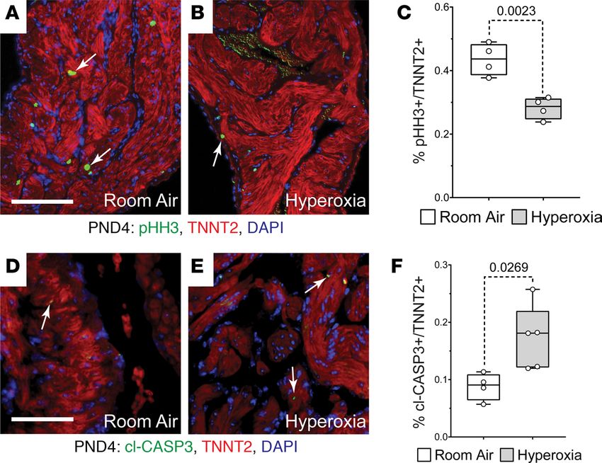
RESEARCH ARTICLE
hypertrophy, hypertension, capillary rarefication, and other types of vascular dysfunction, in animals
exposed to neonatal hyperoxia (24–28). Together, these findings demonstrate that neonatal hyperoxia
can cause pulmonary hypertension and other cardiovascular diseases in adult rodents.
We recently reported that hyperoxia inhibits the proliferation of the cardiomyocytes wrapping the pul-
monary vein and prevents them from expanding to cover its distal branches within the growing lungs of
neonatal mice (29). Since these cells form a contractile sleeve around the pulmonary vein and extending
into the left atria, the early loss of these cells may increase the force needed to drive pulmonary circulation
and, thus, contribute to the development of pulmonary venous congestion and heart failure in aged hyper-
oxia-exposed mice (30). Herein, we report that neonatal hyperoxia inhibits the proliferation of left atrial
cardiomyocytes by suppressing genes required for the de novo synthesis of fatty acids. Failure to expand
these cells results in hypoplastic left atria that lose the ability to effectively fill the left ventricle as mice age,
a finding that may help explain the pathogenesis of diastolic heart failure in the survivors of preterm birth.
Results
Neonatal hyperoxia inhibits left atrial cardiomyocyte proliferation. We previously showed that hyperoxia causes
a loss of the cardiomyocytes wrapping the pulmonary vein by inhibiting their proliferation (29). Since the
pulmonary vein cardiomyocytes are contiguous with the left atria, we investigated whether neonatal hyper-
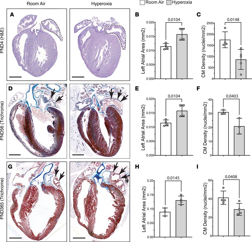
oxia altered left atrial morphology. Counterintuitively, H&E staining revealed that the left atria of mice
exposed to neonatal hyperoxia were larger than those of controls on P4 (dotted lines; Figure 1, A and B).
However, costaining for the cardiomyocyte marker cardiac tropoinin T (TNNT2) and DAPI showed the
number of nuclei per mm2 of TNNT2+ myocardium is reduced in the left atria of P4 hyperoxia-exposed
mice relative to controls (Figure 1C). The left atria of mice exposed to neonatal hyperoxia remained larg-
er and continued to have fewer nuclei in TNNT2+ regions of the left atria than controls on P56 (arrows;
Figure 1, D–F) and P365 (arrows; Figure 1, G–I). Sectioned hearts of P4 hyperoxia-exposed and room air
control mice were next stained for TNNT2 and the mitosis marker phosphorylated histone H3 (pHH3)
or cleaved caspase 3 (cl-CASP3), which labels apoptotic cells. Fewer pHH3+ (arrows; Figure 2, A–C) and
more cl-CASP3+ (arrows; Figure 2, D–F) cardiomyocytes were observed in the left atria of mice exposed to
neonatal hyperoxia than controls. Together, these data suggest that neonatal hyperoxia reduced the prolif-
eration and survival of left atrial cardiomyocytes.
Neonatal hyperoxia causes diastolic dysfunction in adult mice. To determine how neonatal hyperoxia affects
adult heart function, echocardiography was first performed on five P56 mice that were exposed to room
air or hyperoxia between P0 and P4 and an equal number of room air controls (Supplemental Table 1;
supplemental material available online with this article; https://doi.org/10.1172/jci.insight.140785DS1).
Hyperoxia did not affect left ventricular systolic function on P56, but the calculated mass of the left ven-
tricular wall was lower in hyperoxia-exposed mice than controls. Since a prior study showed that hyper-
oxia reduced ventricular cardiomyocyte proliferation (31), the reduced density of the left ventricular wall
observed by echocardiography may reflect lower densities of cardiomyocytes in the left ventricular myo-
cardium. Doppler imaging was used to measure the maximum velocity (Vmax) of blood flowing across
the mitral valve during early diastole when the left ventricle relaxes (E-peak) and late diastole when the left
atrium contracts (A-peak). While hyperoxia did not affect either the E- or A-peak Vmax at this age, 3 of the
5 hyperoxia-exposed mice had E/A ratios > 2, which is above normal range in humans and mice (32–34).
Cardiac function was next examined in 10 hyperoxia-exposed and 8 room air control mice on P365
to determine how the effects of neonatal hyperoxia progress with age (Supplemental Table 2). Four of
the 10 hyperoxia-exposed mice examined at this age had E/A ratios > 2 like those observed on P56.
Moreover, the mean E/A ratio was higher in hyperoxia-exposed mice than controls on P365 (Figure 3A),
although the P value for this change was slightly above the threshold for significance (unpaired 2-tailed t
test, P = 0.051). Interestingly, mean A-peak Vmax was lower in hyperoxia-exposed mice than controls at
this age, while E-peak Vmax was unaffected (Figure 3, B and C). The ratio of E-peak velocity to mitral
annular velocity (E/e’), an indicator of left atrial pressure in early diastole, was also unaffected (room
air, –32.34 ± 8.36, n = 8; hyperoxia, –30.33 ± 5.92, n = 10; P = 0.56). Neonatal hyperoxia may, thus,
specifically disrupt the later active phase of diastole, which is driven by atrial contraction, without affect-
ing early diastole, which is driven by ventricular relaxation. Ejection fraction and fractional shortening
were also significantly reduced in P365 hyperoxia-exposed mice relative to controls (Figure 3, D and E).
Neonatal hyperoxia did not significantly affect mean systolic volume of the left ventricle, but the 2 mice
JCI Insight 2021;6(5):e140785 https://doi.org/10.1172/jci.insight.140785 2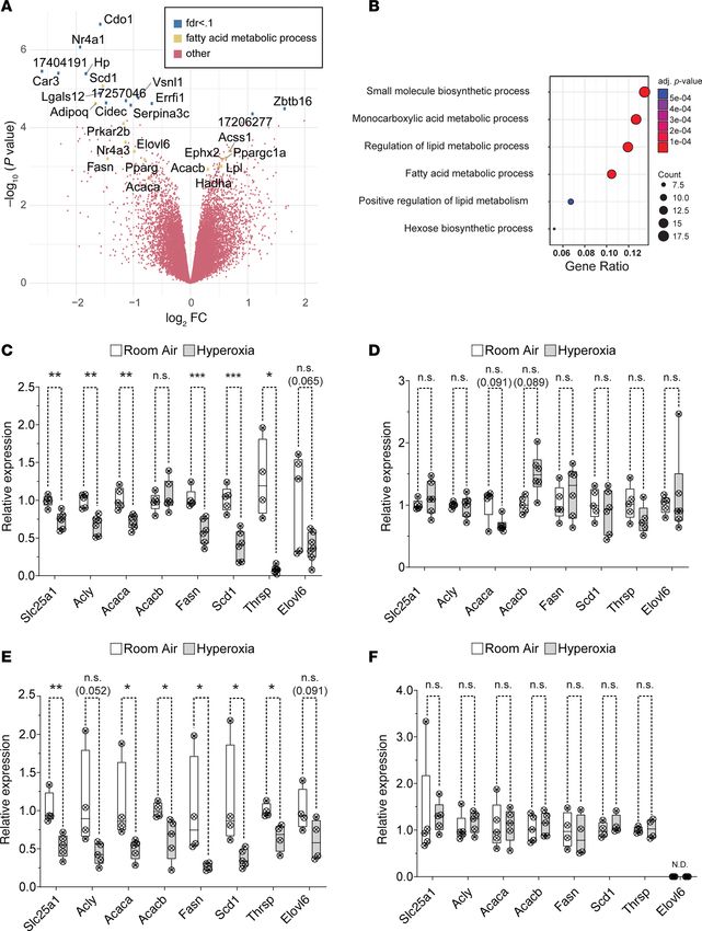
RESEARCH ARTICLE

Figure 1. Neonatal hyperoxia enlarges the left atria of mice but reduces the density of cardiomyocyte (CM) nuclei in the left atrial myocardia. (A) H&E-
stained sections of hearts from P4 mice that were exposed to room air (left) or hyperoxia (right). Dotted lines outline the left atria. (B) Mean area of the
left atria in sections of room air– and hyperoxia-exposed mice on P4. n = 4 mice per condition. (C) Numbers of DAPI-stained nuclei per mm2 of TNNT2+
myocardium. Room air, n= 4; hyperoxia, n = 5 mice per condition. (D and G) Sectioned hearts of P56 (D) and P365 (G) mice exposed to room air (right) or
hyperoxia (right) from P0–P4 stained with Masson’s trichrome. (E and H) Graphs show mean area of the left atria in sections of room air– and hyperox-
ia-exposed mice on P56 (E) and P365 (F). (E) n = 4 per condition. (H) Room air, n = 3; hyperoxia, n = 4. (F and I) Graphs show numbers of DAPI-stained
nuclei per mm2 of TNNT2+ myocardium in room air– and hyperoxia-exposed mice on P56 (F) and P365 (I). (F) n = 3 mice per condition. (I) n = 4 mice per con-
dition. Bars in graphs show mean values, circles show individual data points, and error bars show SDs. The P values between data sets are from unpaired
2-tailed t tests. Scale bars: 400 µm (A) or 100 µM (D and G).

                                   with the highest E/A ratios had hearts with much higher left ventricular systolic volumes than controls
                                   that were identified as statistical outliers (arrows; Figure 3F). The ventricular dilation in the most severe-
                                   ly affected mice may help maintain cardiac output in the setting of diastolic dysfunction. In support of
                                   this idea, the 2 hyperoxia-exposed mice with the highest E/A ratios and systolic volumes also had higher
                                   diastolic volumes, stroke volumes, and cardiac outputs than the remaining hyperoxia-exposed mice (gray
                                   circles; Figure 3, G–I). The mean diastolic volume, stroke volume, and cardiac output of the remaining
                                   mice were also reduced in the remaining mice exposed to neonatal hyperoxia relative to those exposed to
                                   room air when these 2 individuals were excluded from the analysis (white circles, box plots, and P values;
                                   Figure 3, G–I). Together, these data suggest that neonatal hyperoxia causes mice to develop diastolic
                                   dysfunction as adults (P56) that progresses to heart failure by middle age (P365).

JCI Insight 2021;6(5):e140785 https://doi.org/10.1172/jci.insight.140785                                                                                  3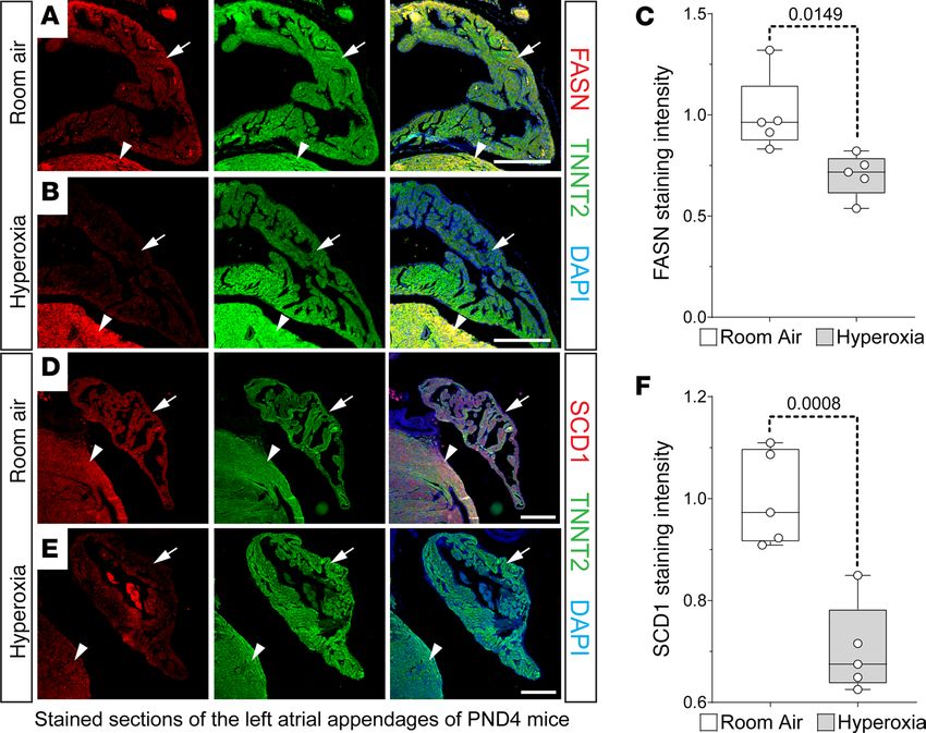
RESEARCH ARTICLE

                                 Figure 2. Hyperoxia reduces postnatal proliferation and survival of left atrial cardiomyocytes in neonatal mice. (A
                                 and B) Sections of left atrial appendages from P4 control (A) and hyperoxia-exposed (B) mice stained with phosphor-
                                 ylated Histone H3 (pHH3, green), cardiac troponin T (TNNT2, red), and DAPI (blue). Arrows point to pHH3+ cardiomyo-
                                 cytes. (C) Percentage of pHH3+ cardiomyocytes in P4 mice exposed to room air or hyperoxia. n = 4 mice per condition. (D
                                 and E) Sectioned left atrial appendages of P4 control (D) and hyperoxia-exposed (E) mice stained for the active, cleaved
                                 form of Caspase 3 (cl-Casp3), TNNT2, and DAPI. Arrows point to cl-Casp3+ cardiomyocytes. (F) Percentage of cl-Casp3–
                                 labeled CMs in the left atria of P4 hyperoxia-exposed and control mice. Room air, n = 4; hyperoxia, n = 5 mice per
                                 condition. Box plots show median, second quartiles, and third quartiles; whiskers indicate range, and circles represent
                                 individual data points. P values are results of unpaired 2-tailed t tests. Scale bars: 200 μm.

                                      Neonatal hyperoxia inhibits the de novo fatty acid synthesis pathway in the atria of mice. An Affymetrix
                                 array was used to identify hyperoxia-induced changes in gene expression responsible for the inhibition
                                 of left atrial cardiomyocyte proliferation. RNA was isolated from the atria of 4 hyperoxia-exposed and 3
                                 room air control mice on P4 and hybridized to the array. Out of 39,000 transcripts examined, only 158
                                 differed by ≥ 1.5-fold with a P < 0.05 and FDR < 0.3 (Figure 4A). Neonatal hyperoxia increased expres-
                                 sion of 53 genes (Supplemental Table 3) and inhibited expression of 105 genes (Supplemental Table 4).
                                 Gene ontology (GO) analysis identified several overlapping sets of genes involved in biosynthetic pro-
                                 cesses, monocarboxylic acid metabolism, lipid metabolic processes, and the regulation of lipid metabo-
                                 lism (Figure 4B). The genes involved in fatty acid metabolism that were upregulated in the atria of hyper-
                                 oxia-exposed mice relative to controls included peroxisome proliferator–activated receptor γ coactivator
                                 1-α (Ppargc1α), a transcriptional coregulator that binds to peroxisome proliferator–activating receptor α
                                 and other transcription factors to promote mitochondrial biogenesis (35). Hyperoxia also upregulated
                                 acetyl-CoA carboxylase 2 (Acacb), a mitochondrial localized isoform of acetyl-CoA carboxylase that pro-
                                 duces malonyl-CoA for mitochondrial biogenesis and regulation of β-oxidation (36), hydroxyacyl-CoA
                                 dehydrogenase trifunctional multienzyme complex subunit α (Hadha), part of the mitochondrial com-
                                 plex that catalyzes the β-oxidation of long-chain fatty acids (37), and lipoprotein lipase (Lpl), which
                                 cleaves lipids from dietary lipoproteins (38).
                                      Neonatal hyperoxia reduced expression of several other genes involved in fatty acid metabolism.
                                 These included the core enzymes in the de novo fatty acid synthesis pathway, such as solute carrier family
                                 25 member 1 (Slc25a1), the channel that transports citrate produced by the TCA cycle out of the mito-
                                 chondria and into the cytoplasm (39, 40); ATP-citrate lyase (Acly), the enzyme that converts cytoplasmic
                                 citrate to acetyl-CoA (41); and acetyl-CoA carboxylase 1 (Acaca), the cytoplasmic isoform of acetyl-CoA
                                 carboxylase that produces malonyl-CoA for fatty acid synthesis (42). In addition, hyperoxia reduced
                                 mRNA for fatty acid synthase (Fasn),the enzyme that synthesizes the 16-carbon saturated fatty acid palmi-
                                 tate from acetyl-CoA and malonyl-CoA (43); elongation of very long chain fatty acids 6 (Elovl6), the enzyme

JCI Insight 2021;6(5):e140785 https://doi.org/10.1172/jci.insight.140785                                                                               4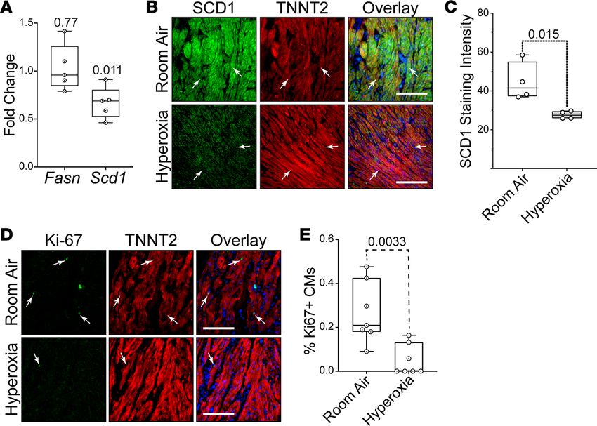
RESEARCH ARTICLE

                                 Figure 3. Neonatal hyperoxia causes mice to develop dilated heart failure later in life. (A–C) Graphs show mean E/A
                                 ratios (A), A-peak velocities (B), and E-peak velocities (C) of blood flow across the mitral valves of control and hyperox-
                                 ia-exposed mice on P365. E/A ratios are plotted on a log10 scale. (D and E) Graphs show mean ejection fraction (D) and
                                 fractional shortening (E) of control and hyperoxia-exposed mice on P365. (F) Graph shows mean volume of the left ven-
                                 tricle during systole for PD365 control and hyperoxia-exposed mice. Arrows point to 2 hyperoxia-exposed mice with left
                                 ventricles that were drastically enlarged relative to controls and other hyperoxia-exposed mice. These 2 mice had the
                                 highest E/A ratios of all mice. (G–I) Box plots show the mean diastolic volume (G), stroke volume (H), and cardiac out-
                                 put (I) of the left ventricles in P365 hyperoxia-exposed and control mice after the 2 mice with enlarged left ventricular
                                 systolic volumes were excluded. Gray circles are values for excluded mice. Box plots show median, second quartiles, and
                                 third quartiles; whiskers show range, and circles show individual data points. F tests were used to determine if values
                                 for control and hyperoxia-exposed mice had equal variances. The P values are from unpaired 2-tailed t tests.

                                 that extends palmitate to produce the 18 carbon saturated fatty acid stearate (44); stearoyl-CoA desaturase 1
                                 (Scd1), which converts palmitate and stearate to the mono-unsaturated fatty acids palmitoleate and oleate
                                 (45); and thyroid hormone-inducible hepatic protein (Thrsp), which binds FASN and increases its activity (46).
                                 Neonatal hyperoxia also downregulated cell death inducing DFFA like effector c (Cidec), a protein required
                                 for the storage of triglycerides as lipid droplets (47), and adiponectin (Adipoq), an adipokine that protects
                                 cardiomyocytes from oxidative stress and apoptosis following ischemic injury (48).
                                      The reduced expression of fatty acid synthesis genes in the atria of mice that were exposed to neonatal
                                 hyperoxia was intriguing, since this pathway is required for the proliferation and survival of both cancer-
                                 ous and noncancerous cells (49–53). The expression of fatty acid synthesis genes was, thus, examined in
                                 an independent set of mice to confirm whether neonatal hyperoxia suppresses their expression in the atria.
                                 Quantitative PCR (qPCR) showed that Slc25a1, Acly, Acaca, Fasn, Scd1, and Thrsp were all expressed at

JCI Insight 2021;6(5):e140785 https://doi.org/10.1172/jci.insight.140785                                                                                  5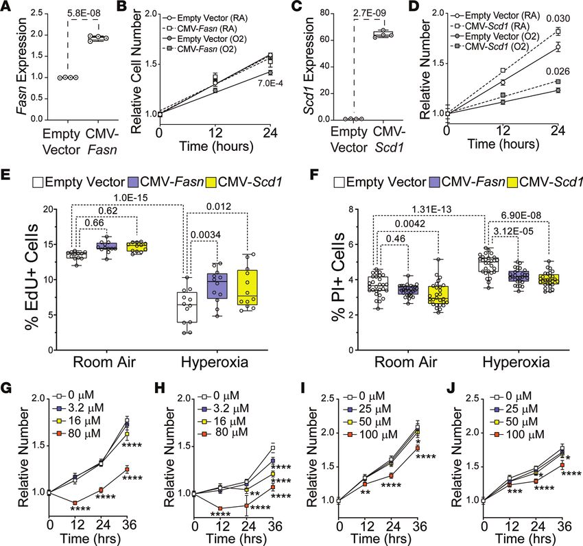
RESEARCH ARTICLE

Figure 4. Neonatal hyperoxia suppresses genes needed for fatty acid synthesis in atria of mice. (A) Volcano plot of log2 fold changes versus log10 P
values. Genes with FDR < 0.1 or annotation in the fatty acid metabolism are highlighted. Genes with FDR < 0.1 are marked by blue dots; genes involved in
fatty acid metabolism are marked by green dots. (B) Gene ontology (GO) analysis of differentially expressed genes. (C–F) Results of qPCR for Slc25a1, Acly,
Acaca, Acacb, Fasn, Scd1, Thrsp, and Elovl6 in atria (C and E) and ventricles (D and E) of hyperoxia-exposed and control mice on P4 (C and D) and P56 (E and
F). (A and B) Room air, n = 3; hyperoxia, n = 4. (C–E) Room air, n = 5; hyperoxia, n = 6. (F) Slc25a1, Acly, Acaca, AcacB: room air, n = 5; hyperoxia, n = 5; Scd1,
Fasn, Thrsp, Elovl6: room air, n = 4; hyperoxia, n = 4. (C–F) Box plots show median, second quartiles, and third quartiles; whiskers show range. Circles show
values for individual control and hyperoxia-exposed mice, respectively. (C–F) *P < 0.05, **P < 0.01, ***P < 0.005, using unpaired 2-tailed t tests.

JCI Insight 2021;6(5):e140785 https://doi.org/10.1172/jci.insight.140785                                                                                         6
RESEARCH ARTICLE
lower levels in the atria of P4 mice exposed to neonatal hyperoxia mice compared with those of controls
(Figure 4C). Mean expression of Elovl6 was also lower in hyperoxia-exposed mice relative to controls,
but the P value for this change was above the threshold set for significance (P = 0.065). In contrast, none
of these genes were significantly affected in the ventricles on P4 (Figure 4D). Acacb was examined in the
hearts of hyperoxia-exposed mice, but the increased expression in our microarray was not significant in
the atria or ventricles of hyperoxia-exposed mice by qPCR. Since mice exposed to hyperoxia are returned
to room air on P4, we investigated when expression of these genes returned to control levels. Surprisingly,
mRNA for Slc25a1, Acly, Acaca, Fasn, Scd1, Thrsp, and Elovl6 remained suppressed on P56 (Figure 4E).
These changes were again specific for the atrium, and the expression of Slc25a1, Acly, Acaca, Acacb, Thrsp,
Fasn, and Scd1 in the ventricle was unaffected on P56, while Elovl6 was not detected (Figure 4F). Hyperox-
ia, thus, permanently suppresses fatty acid synthesis genes in the atria but not ventricles.
IHC was used to confirm that neonatal hyperoxia inhibited FASN and SCD1 protein in left atrial cardio-
myocytes. Staining for FASN (red; Figure 5, A and B) and TNNT2 (green; Figure 5, A and B) detected FASN
in the atrial (arrows) and ventricular (arrowheads) cardiomyocytes of P4 mice exposed to room air. In contrast,
FASN staining was reduced in the left atrial cardiomyocytes of hyperoxia-exposed mice (Figure 5C) but unaf-
fected in ventricular cardiomyocytes. Staining for SCD1 (red; Figure 5, D and E) and TNNT2 (green; Figure 5,
D and E) showed that SCD1 was similarly reduced in left atrial cardiomyocytes of P4 mice exposed to neonatal
hyperoxia relative to controls (arrows; Figure 5, D–F). As observed for FASN, SCD1 levels were less affected in
the ventricles of hyperoxia-exposed and room air control mice (arrowheads; Figure 5, D and E).
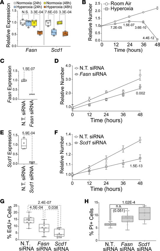
Hyperoxia inhibits fatty acid synthesis genes and proliferation in the HL-1 line of immortalized murine atrial car-
diomyocytes. The HL-1 line of immortalized atrial cardiomyocytes was used to examine the direct effects of
hyperoxia on fatty acid synthesis genes (54). Cells were grown in room air (21% O2, 5% CO2) or hyperoxia
(95% O2, 5% CO2) for 24 or 48 hours before Fasn and Scd1 mRNA were quantified by qPCR (Figure 6A).
Relative to cells grown in room air, Fasn and Scd1 were reduced in HL-1 cells after 48 hours in hyperoxia.
Interestingly, while Scd1 was lower in HL-1 cells after 24 hours of hyperoxia, Fasn was unaffected. However,
this difference in timing is unlikely to reflect a regulatory hierarchy between genes because Scd1 knockdown
does not affect Fasn expression (our unpublished observation). Hyperoxia can, thus, directly affect fatty acid
synthesis genes in atrial cardiomyocytes without the need for other cell types.
To determine if the suppression of fatty synthesis genes in hyperoxia-treated HL-1 cells correlates with
reduced proliferation, asynchronously dividing HL-1 cells were plated at equal density and cultured in
room air or hyperoxia for 0, 12, 24, 36, and 48 hours before being stained with DAPI and counted. HL-1
cells expanded continuously for 48 hours in room air (Figure 6B). In contrast, HL-1 cells grown in hyper-
oxia expanded slower than controls for the first 24 hours, plateaued from 24 to 36 hours, and then declined
in numbers from 36 to 48 hours. These data indicate that hyperoxia directly represses proliferation and fatty
acid synthesis genes in HL-1 cells, as it does in the atria of mice.
Fasn and Scd1 are required for the proliferation and survival of HL-1 atrial cardiomyocytes. To determine if
fatty acid synthesis is required for atrial cardiomyocyte proliferation, HL-1 cells were transfected with non-
targeting (NT) siRNA and siRNA for Fasn or Scd1 and grown in room air for 72 hours. qPCR revealed that
Fasn and Scd1 mRNA were reduced around 75% and 90% in cells transfected with Fasn and Scd1 siRNA,
respectively, when compared with NT siRNA transfected controls (Figure 6, C and D). Asynchronously
dividing Fasn, Scd1, and NT siRNA–treated HL-1 cells were then plated at equal numbers and counted at
12-hour intervals (Figure 6, E and F). Cells transfected with Fasn and Scd1 siRNA grew slower than NT
siRNA transfected controls over 48 hours.
To confirm whether Fasn and Scd1 are required for HL-1 cell proliferation, cells were treated with
DMSO as a vehicle control, the FASN inhibitor G28UCM at 10 μM, or the SCD1 inhibitor A939572
at 10 nM (Tocris Biosciences) and grown in room air (Supplemental Figure 1). While cells in control
media grew continuously for 72 hours, cells in A939572 plateaued after 24 hours and cells in G28UCM
grew slower than controls during the first 24 hours before declining in number from 24 and 72 hours. To
specifically examine the effects of Fasn and Scd1 knockdown on proliferation, Fasn, Scd1, and NT siR-
NA transfected cells were grown for 23 hours, treated with EdU for 1 hour, fixed, and stained with an
anti-EdU antibody and DAPI. The fraction of EdU+ cells was reduced in Fasn and Scd1 siRNA–treated
cells relative to controls (Figure 6G). Propidium iodide (PI) exclusion assays were performed to deter-
mine if Fasn and Scd1 were required for HL-1 cell survival. The percentage of PI+ cells was significantly
higher in Scd1 siRNA transfected cells than controls. The percentage of PI+ cells was also higher in cells
JCI Insight 2021;6(5):e140785 https://doi.org/10.1172/jci.insight.140785 7RESEARCH ARTICLE
Figure 5. Neonatal hyperoxia represses fatty acid synthesis enzymes in murine atrial cardiomyocytes. (A, B, D,
and E). Sections of left atrial appendages from P4 neonates exposed to room air (A and B) or hyperoxia (D and E)
costained for FASN (red, A and B) or SCD1 (red, D and E) and TNNT2 (green, A, B, D, and E). Sections were also stained
with DAPI to label nuclei (blue, A, B, D, and E). Arrows and arrowheads show TNNT2+ cardiomyocytes in left atrial
appendage and LV, respectively. Scale bars: 200 μm. (C and F) Graphs show relative staining intensities for Fasn (C)
and Scd1 (F) measured using NIH ImageJ 2.0/Fiji. (C and F) Room air, n = 5; hyperoxia, n = 5. Box plots show median
values and inner quartiles. Whiskers show the range of values. Circles show values for individual room air– and hyper-
oxia-exposed mice, respectively. F tests were used to determine if samples had equal or unequal variances. P values
are the results of unpaired 2-tailed t tests.
transfected with Fasn siRNA than controls, but the P value of this change was slightly below the thresh-
old for statistical significance (P = 0.051; Figure 6H). Together, these data show that the Fasn and Scd1
are required for atrial cardiomyocyte proliferation and survival.
Fasn and Scd1 overexpression restores proliferation of HL-1 atrial cardiomyocytes in hyperoxia. We next
sought to determine how Fasn and Scd1 overexpression would affect atrial cardiomyocyte proliferation
in hyperoxia. Cells were transfected with Fasn and Scd1 expression vectors (CMV-Fasn and CMV-Scd1,
respectively), as well as empty expression vector. After 24 hours, cells were replated at equal density
and grown in room air or hyperoxia for 0, 12, and 24 hours before being stained and counted. Cells
transfected with CMV-Fasn expressed approximately 2-fold more Fasn than empty vector transfect-
ed cells (Figure 7A). As expected, control transfected cells grew slower in hyperoxia than in room
air (Figure 7B). However, CMV-Fasn transfected cells grew at the same rate as control cells grew in
room air, regardless of whether they were in room air or hyperoxia. Cells transfected with CMV-Scd1
expressed approximately 60-fold more Scd1 than controls (Figure 7C) and grew faster than control cells
in room air and hyperoxia (Figure 7D). Fasn and Scd1 overexpression, thus, partially restores HL-1 cell
growth in hyperoxia.
To specifically examine the effects of Fasn and Scd1 overexpression on proliferation, HL-1 cells were
transfected with control, Fasn, and Scd1 plasmids; grown in room air or hyperoxia for 23 hours; and
treated with EdU to label cells in S-phase (Figure 7E). While numbers of EdU+ cells were unaffected by
Fasn and Scd1 overexpression in room air, more Fasn- and Scd1-overexpressing cells were EdU+ in hyper-
oxia than controls. Cells transfected with control, Fasn, and Scd1 plasmids were also grown in room
air or hyperoxia for 48 hours and subject to PI exclusion assays to determine if Fasn and Scd1 promote
survival in hyperoxia (Figure 7F). Consistent with the increased numbers of cl-CASP3+ cells in the atria
JCI Insight 2021;6(5):e140785 https://doi.org/10.1172/jci.insight.140785 8RESEARCH ARTICLE
Figure 6. Suppression of fatty acid synthesis contributes to reduced HL-1 cell proliferation in hyperoxia. (A) qPCR
for Fasn (left) and Scd1 (right) in HL-1 cells grown in room air or hyperoxia for 24 hours (white and blue, respectively)
or 48 hours (yellow and red, respectively). (B) Numbers of HL-1 cells grown in room air (white circles) and hyperoxia
(gray squares) for 48 hours relative to their density at 0 hours. (C and E) qPCR for Fasn (C) and Scd1 (E) in HL-1 cells
transfected with Fasn and Scd1 siRNAs. Controls were transfected with nontargeting (NT) siRNA. (D and F) Num-
bers of HL-1 cells transfected with control siRNA (white circles in D and F) and Fasn (Gray squares in D) or Scd1 (Gray
squares in F) siRNAs and grown for 48 hours in room air relative to 0 hrs. (G) HL-1 cells transfected with NT, Fasn,
and Scd1 siRNAs, grown in room air for 22 hours and treated with EdU for 2 hours before staining. Graph shows
percentages of EdU+ cells. (H) HL-1 cells transfected with NT, Fasn, and Scd1 siRNAs were grown in room air for 48
hours, stained with PI, and imaged. Graph shows numbers of PI+ NT, Fasn, and Scd1 siRNA transfected cells. (A, C,
and E) n = 4 transfections per condition. Each time/condition: (G and H), n = 12. (B, D, and F) Error bars show 95%
CI; lines and P values are results of linear regressions. (A, C, E, G, and H) Box plots are median, second quartiles,
and third quartile; whiskers indicate range, and markers show individual replicates. P values are from unpaired
2-tailed t tests (C and E) or 1-way ANOVA with Holm-Sidak corrections (A, G, and H).
of P4 hyperoxia-exposed mice, 48 hours of hyperoxia increased the percentage of control transfected
cells stained by PI. While Scd1 overexpression caused a modest reduction in the numbers of PI+ cells in
room air, Fasn did not. However, Fasn- and Scd1-overexpressing cells were both less likely to be PI+ in
hyperoxia than controls. Restoring Fasn and Scd1 expression can, thus, partially restore the survival and
proliferation of atrial cardiomyocytes in hyperoxia.
JCI Insight 2021;6(5):e140785 https://doi.org/10.1172/jci.insight.140785 9RESEARCH ARTICLE
Figure 7. Fasn and Scd1 overexpression increases HL-1 cell proliferation in hyperoxia. (A and C) Fasn (A) and Scd1 (C)
mRNA in HL-1 cells 48 hours after transfection with empty vector or Fasn (A) and Scd1 (C) expression vectors. n = 4 trans-
fections per condition. (B and D) Expansion of Fasn (B), Scd1 (D), and control transfected HL-1 cells over 24 hours. Markers
are mean fold change in cell number for control (circles), Fasn (squares with dashed line, B), and Scd1 (squares with dashed
line, D) grown in room air (white) or hyperoxia (gray). Lines and P values are linear regressions. n = 10 wells per time/condi-
tion. (E) Percentages of EdU+ control (white), Fasn (blue), and Scd1 (yellow) transfected HL-1 cells after 2 hours incubation
in room air (left) and hyperoxia (right). n = 12 wells per condition. (F) Percentages of control (white), Fasn (blue), and Scd1
(yellow) transfected cells labeled after PI staining in room (left) and hyperoxia (right). n = 28 wells per condition. (G and H)
HL-1 cell expansion in media with 0, 3.2, 16, and 80 μM palmitate-BSA in room air (G) or hyperoxia (H) for 36 hours. (I and
J) Expansion of HL-1 cells in media containing 0, 25, 50, and 100 mM oleate-BSA and grown in room air (I) or hyperoxia (J)
for 36 hours. (A, C, E, and F) Box plots show median, second quartiles, and third quartiles; markers represent individual
values; whiskers show range. P values are results of unpaired 2-tailed t tests (A and C) or 1-way ANOVA with Holmes-Sidak
corrections. (B and D) Trendlines and P values linear regressions. (G, H, I, and J) n = 18 wells per time/condition. *P < 0.05,
**P < 0.01, ***P < 0.005, ****P < 0.001 using 2-way ANOVA with Tukey’s multiple comparison tests.
If Fasn and Scd1 promote the growth of HL-1 cells in hyperoxia by increasing the supply of their
products, adding exogenous fatty acids to the culture media may mimic their effects. Cells were, thus,
plated in media containing increasing amounts of BSA-conjugated palmitate (Figure 7, G and H) or
oleate (Figure 7, I and J) and grown in room air (Figure 7, G and I) or hyperoxia (Figure 7, H and J)
for 36 hours. In room air, cells grew at equal rates in media containing low and medium levels of pal-
mitate and oleate conjugated BSA as they did in media containing unconjugated BSA. In contrast, high
concentrations of palmitate and oleate slowed the growth of cells in room air relative to controls. High
levels of palmitate and oleate similarly slowed HL-1 cell growth in hyperoxia relative to controls. As
seen in room air, low and medium concentrations of oleate did not affect HL-1 cell growth in hyperox-
ia. However, HL-1 cells treated with low and intermediate levels of palmitate grew slower in hyperoxia
than cells in control media, suggesting that hyperoxia enhances the effects of palmitate-BSA on their
JCI Insight 2021;6(5):e140785 https://doi.org/10.1172/jci.insight.140785 10RESEARCH ARTICLE
expansion. Palmitate and oleate are, thus, not sufficient for the effects of Fasn and Scd1 overexpression
on atrial cardiomyocyte growth in hyperoxia.
Hyperoxia does not reduce SREBF1 protein in atrial cardiomyocytes. Genes in the de novo fatty acid synthesis
pathway are regulated by sterol-response element binding factor 1 (SREBF1), a transcriptional master regula-
tor of lipogenesis (55). HL-1 cells were, thus, transfected with plasmid that expresses human SREBF1 under
the control of the CMV promoter (CMV-Srebf1) or empty vector and grown in hyperoxia or room for 36 hours.
Performing qPCR with a primer pair recognizing both human and mouse Srebf1 mRNA showed that cells
transfected with CMV-Srebf1 expressed nearly twice as much Srebf1 compared with control cells (Supplemental
Figure 2A). CMV-Srebf1 transfected cells grew slower in room air than those transfected with empty vector
(Supplemental Figure 2B). However, while control transfected cell growth arrested after 12 hours in hyperox-
ia, CMV-Srebf1 transfected cells continued proliferating in hyperoxia up to 36 hours (Supplemental Figure 2C).
To determine if hyperoxia suppresses fatty acid synthesis genes by reducing SREBF1 protein, Western
blotting for SREBF1 was performed on HL-1 cells grown in room air or hyperoxia for 48 hours (Supplemental
Figure 2D; see supplemental material for unedited blots). Two bands of SREBF1 protein were detected in
hyperoxia- and control-treated HL-1 cells, a 120 kd band representing full-length inactive SREBF1 within the
golgi membrane, and a 55 kd band representing the amino-terminus of SREBF1, which localizes to the nucleus
after cleavage. Despite the reduced expression of Fasn, Scd1, and other fatty acid genes in hyperoxia-treated
cells, hyperoxia does not alter the levels of either band of SREBF1 protein (Supplemental Figure 2, F and
G). Lysates of left atria from individual P4 hyperoxia-exposed and control mice were also Western blotted for
SREBF1 protein (Supplemental Figure 2E; see supplemental material for unedited blots). Surprisingly, despite
having lower levels of fatty acid synthesis genes than controls, the left atria of P4 hyperoxia-exposed mice had
higher levels of both forms of SREBF1 than controls (Supplemental Figure 2, H and I). Therefore, hyperoxia
is unlikely to reduce fatty acid synthesis in atrial cardiomyocytes by reducing SREBF1 protein in these cells.
Hyperoxia inhibits fatty acid synthesis genes and proliferation in human left atrial tissue explanted from infant
donors. To determine if changes observed in mice occur in humans, left atrial tissue from term infants who
died shortly after birth due to anencephaly was obtained from the Biorepository for Investigation of Neo-
natal Diseases of the Lung (BRINDL) at the University of Rochester. The muscle was separated from sur-
rounding tissue, cut into ~1 mm3 cubes, grouped, and cultured in room air or hyperoxia for 24 hours before
being used for qPCR or immunostaining. While hyperoxia did not alter the levels of Fasn mRNA, Scd1
expression was lower in hyperoxia-exposed explants than controls (Figure 8A). Immunological staining
showed that SCD1 protein localized to TNNT2+ cardiomyocytes in explants exposed to room air but not
in those exposed to hyperoxia (arrows; Figure 8, B and C). Sectioned explants were stained for the prolif-
eration marker Ki-67 and TNNT2 to determine if hyperoxia reduced the proliferation of human left atrial
cardiomyocytes. Ki67+ nuclei were more frequently observed in the TNNT2+ cardiomyocytes of control
explants than in those exposed to neonatal hyperoxia (arrows; Figure 8, D and E). Hyperoxia, thus, sup-
presses fatty acid synthesis genes and proliferation in human left atrial cardiomyocytes, as it does in mice.
Discussion
There is a significant need to understand why high oxygen exposure at birth is a risk factor for adult
cardiovascular disease. We previously showed that neonatal hyperoxia causes pulmonary hypertension
in adult mice and that this phenotype was preceded by a loss of the cardiomyocytes surrounding the pul-
monary vein due to reduced proliferation (29). We now extend these findings by showing that hyperoxia
similarly inhibits the postnatal proliferation of left atrial cardiomyocytes and causes the myocardium
of this chamber to be hypoplastic. Despite having fewer cardiomyocytes per unit area, the left atria of
hyperoxia-exposed mice were larger than controls, suggesting that the remaining cardiomyocytes hyper-
trophy to compensate for reduced numbers. Pathology was first seen on P56 when most of the hyperox-
ia-exposed mice examine had E/A ratios > 2. By P365, the mean E/A ratio was higher in mice exposed
to neonatal hyperoxia than controls. The velocity of flow across the mitral valve during atrial contraction
was also lower in hyperoxia-exposed mice than controls, suggesting that hyperoxia reduced left atrial
contractility by this age. Interestingly, the 2 mice with the highest E/A ratios (>5) had left ventricles
that were larger than controls during both systole and diastole. The left ventricular dilation in these mice
may be adaptive since it would raise cardiac output in the presence of diastolic dysfunction, reduced
fractional shortening, and lower ejection fraction. In support of this idea, excluding the 2 mice with the
highest E/A ratios from our analyses, diastolic volume, stroke volume, and cardiac output were lower in
JCI Insight 2021;6(5):e140785 https://doi.org/10.1172/jci.insight.140785 11RESEARCH ARTICLE
Figure 8. Hyperoxia suppresses fatty acid synthesis genes in left atrial tissue explanted from human infants.
(A) Results of qPCR for Fasn and Scd1 in left atrial tissue explanted from human infants who died at birth due to
anencephaly and exposed to room air or hyperoxia for 24 hours. n = 5 donors. (B) Sectioned explants exposed to room
air (top) or hyperoxia (bottom) were stained for SCD1 (green), TNNT2 (red), and DAPI (blue). (C) Graph shows staining
intensities for SCD1 in sections of control and hyperoxia-exposed mice determined using NIH ImageJ 2.0/Fiji. n = 4
donors. (D) Sections of explants exposed to room air (top) and hyperoxia (bottom) stained for the proliferation marker
Ki67 (green), TNNT2 (red), and DAPI (blue). (E) Graph shows percentages of TNNT2-expressing cells with Ki67+ nuclei in
explants exposed to room air or hyperoxia. n = 7 donors. (A, C, and E) Circles indicate individual values for explants of
each donor. Boxes show medians and inner quartiles; whiskers represent the range. P values are the results of either
single-sample 1-tailed t test and Wilcox test (A) or unpaired 2-tailed t tests (C and E). (B and D) Scale bars = 100 μm.
the remaining hyperoxia-exposed mice than controls. Mice exposed to hyperoxia during early postnatal
life, thus, develop diastolic dysfunction and heart failure as adults.
Affymetrix arrays, qPCR and IHC revealed that neonatal hyperoxia inhibited genes needed for fatty
acid synthesis in the atrial but not ventricular cardiomyocytes of mice. These included Fasn and Scd1,
which are elevated in highly proliferative tumors, sufficient to drive the expansion of noncancerous cells
when overexpressed and required for the proliferation and survival of noncardiac cells (49, 53, 56–60). We
thus explored whether the repression of Fasn and Scd1 is responsible for the reduced proliferation of left
atrial cardiomyocytes in mice exposed to neonatal hyperoxia. Left atrial cardiomyocytes can be isolated
from neonatal mice, but the yield is low, and the resulting cells lose their proliferative capacity quickly in
culture, making it difficult to examine how genetic manipulations affect their expansion. The HL-1 line of
immortalized atrial cardiomyocytes was, thus, chosen to model the effects of hyperoxia on neonatal atrial
cardiomyocytes, since these cells are differentiated and contractile but still metabolically immature and pro-
liferative (54, 61, 62). Consistent with the loss of atrial CM proliferation and survival in hyperoxia-exposed
mice, exposing HL-1 cells to hyperoxia for 48 hours suppressed Fasn and Scd1 and reduced proliferation
and survival. Inhibitors and siRNAs for Fasn and Scd1 also reduced HL-1 cell proliferation and survival,
while overexpressing these genes partially restored growth in hyperoxia. The repression of Fasn and Scd1
may, therefore, mediate the inhibitory effects of neonatal hyperoxia on the postnatal proliferation and sur-
vival of left atrial cardiomyocyte.
Surprisingly, adding palmitate and oleate to the culture media was not sufficient to reproduce the
effects of Fasn and Scd1 overexpression on the expansion of HL-1 cells in hyperoxia. It is possible that fatty
acids produced de novo are preferred over exogenous fatty acids for synthesizing phospholipids and other
components of new membranes for dividing cells. Alternatively, fatty acid synthesis may prevent potential-
ly toxic metabolic precursors from accumulating. Chemical inhibition and siRNA-mediated knockdown
JCI Insight 2021;6(5):e140785 https://doi.org/10.1172/jci.insight.140785 12RESEARCH ARTICLE
of FASN inhibits oxidative phosphorylation in immortalized melanocytes (49). This loss of mitochondrial
function was partially due to the accumulation of malonyl-CoA, which inhibits the transport of fatty acids
into mitochondria at high concentrations. FASN inhibition also reduces mitochondrial membrane potential
(ΔΨm) and increases production of ROS (63, 64). SCD1 inhibition similarly disrupts mitochondrial func-
tion, increases ROS, and induces apoptosis in many cells, including neonatal ventricular cardiomyocytes
(65, 66). Reduced SCD1 function also activates AMP-dependent protein kinase (AMPK) in many contexts,
including the hearts of Scd1-KO mice (45, 67, 68). AMPK phosphorylates and inhibits the pro-proliferative
kinase mTOR and, thus, inhibits tumor cell proliferation and survival (69). AMPK also reduces fatty acid
synthesis through inhibitory phosphorylation of ACACA, as well as SREBF1 (70), and may play a role in
the hyperoxia-induced suppression of fatty acid synthesis in atrial cardiomyocytes.
Recent echocardiographic imaging, MRI, and CT imaging studies indicate that preterm infants have larg-
er left atria and lower diastolic function than term infants (14–17), suggesting that the heart failure among
young adults born preterm has developmental origins. Serum from preterm infants was shown to have higher
malonyl-carnitine and lower palmitoyl-carnitine levels than serum from term infants (71), suggesting that
preterm birth and reduced FASN activity may be linked. Preterm infants often require mechanical ventilation,
steroids, and other interventions that make it difficult to identify direct effects of supplemental oxygen on
cardiovascular health. We thus sought to determine if hyperoxia suppresses fatty acid synthesis genes and pro-
liferation in explanted human left atrial tissue. Hyperoxia reduced Scd1 expression and the numbers of Ki67+
cardiomyocytes in human atrial explants, confirming that hyperoxia represses at least 1 major component of
the fatty acid synthesis pathway, as well as proliferation in human left atrial cardiomyocytes. These data sug-
gest that the response of left atrial tissue from newborn humans to hyperoxia parallels that of mice and HL-1
cells and that reduced fatty acid synthesis underlies the cardiovascular disease in preterm infants.
Exposure to neonatal hyperoxia did not suppress fatty acid synthesis genes in the ventricles of mice as
it did in the atria. Since the left atrium is the first chamber to encounter oxygen-rich blood from the lungs,
left atrial cardiomyocytes may have evolved to have a different response to hyperoxia than ventricular car-
diomyocytes. The levels Fasn, Scd1, and other fatty acid synthesis genes were also lower in the ventricles
of both hyperoxia-exposed and room air control mice at all times examined, suggesting that fatty acid
synthesis may be less important for proliferation and survival of ventricular than atrial cardiomyocytes.
The suppression of ventricular cardiomyocyte proliferation was hypothesized to reflect a role for oxygen in
promoting their terminal differentiation and maturation (31). However, in our model, Fasn, Scd1, and other
fatty acid synthesis genes continue to be suppressed in the atria of mice exposed to hyperoxia even after
they are returned to room air. If hyperoxia accelerated the normal differentiation of atrial cardiomyocytes
as proposed, fatty acid synthesis genes should return to similar levels in the atria of hyperoxia-exposed and
control mice as the atrial cardiomyocytes of control mice matured. Exposure to hyperoxia in early neonatal
life, thus, permanently reprograms atrial cardiomyocytes metabolism in a maladaptive fashion that may
contribute to the long-term effects of neonatal hyperoxia on cardiovascular health. It is unclear if similar
persistent molecular changes take place in ventricular cardiomyocytes.
Despite the strength of the data presented, it is important to acknowledge the limitations of our studies.
HL-1 cells are a pure population of proliferative atrial cardiomyocytes that can be used to test the function-
al relationship between hyperoxia and fatty acid synthesis genes. However, these cells have been immortal-
ized with the SV40 T antigen and may not faithfully recapitulate the response of primary atrial cardiomy-
ocytes to hyperoxia or the complex interactions between cells within the left atrium. The left atrial tissue
used to demonstrate that hyperoxia affects fatty acid synthesis and cardiomyocyte proliferation in humans
was from anencephalic infants that were born at term and died shortly thereafter instead of preterm infants.
Since oxygen sensitivity is likely to change over the course of gestation, the response of human left atri-
al explants may not accurately reflect how left atrial cardiomyocytes respond to supplemental oxygen in
preterm infants. Moreover, hyperoxia did not suppress Fasn in explanted human atrial tissue as it did in the
atria of mice. Since 1 day of hyperoxia was insufficient to repress Fasn in HL-1 cells, prolonged exposure to
hyperoxia may be needed suppress Fasn in atrial explants. Alternatively, ex vivo studies may not recapitu-
late the effects of hyperoxia seen in vivo. Future studies will be required to determine if restoring fatty acid
synthesis will alleviate the effects of neonatal hyperoxia on left atrial cardiomyocytes in the hearts of mice
or if fatty acid synthesis genes are reduced in banked heart tissue from preterm infants.
In conclusion, the data herein suggest that exposure to hyperoxia in early postnatal life initiates the
development of adult diastolic dysfunction by permanently suppressing Fasn, Scd1, and other genes needed
JCI Insight 2021;6(5):e140785 https://doi.org/10.1172/jci.insight.140785 13RESEARCH ARTICLE
for fatty acid synthesis, as well as cardiomyocyte proliferation and survival within the myocardia of the left
atrium. Although the remaining cardiomyocytes undergo hypertrophy to compensate for the reduced num-
bers of cells, the reduced A-peak velocities in aged mice exposed to neonatal hyperoxia suggest that these
cells lose contractility over time. Moreover, since adult cardiomyocytes use fatty acids for 70% of the ATP
used for contractility (72), the long-term suppression of Fasn, Scd1, and other fatty acid synthesis genes may
affect the functionality of the remaining cardiomyocytes after they have stopped proliferating and further
contribute to the diastolic heart failure. Agonists for nuclear hormone receptors that work with SREBF1
to induce lipogenesis, such as liver X receptor (LXR) and PPARG, are currently available (73–77). Future
studies will be needed to determine if these compounds can be used to protect or restore the postnatal pro-
liferation and survival of atrial cardiomyocytes in mice exposed to hyperoxia and potentially lead to novel
therapies to prevent diastolic heart failure in individuals who were born preterm.
Methods
Mice and hyperoxia. C57BL/6J mice were purchased from The Jackson Laboratory and maintained as an
inbred colony. Mice were exposed to humidified room air (21% O2) or hyperoxia (100% O2) between P0
and P4 (22). Some mice exposed to hyperoxia were returned to room air. Dams were cycled between room
air and hyperoxia every 24 hours to reduce oxidant injury. The mice were provided food and water ad
libitum and housed in microisolator cages in a pathogen-free environment. Since we have not observed dif-
ferences in the response of male and female mice to hyperoxia in prior studies, male and female mice were
both used in experiments performed on P4. However, all male mice were used to examine the effects of
neonatal hyperoxia on cardiac function on P56 and P365 to prevent sex-specific differences in ventricular
measurements from masking potential effects of hyperoxia.
Echocardiography. To assess cardiac function, mice were anesthetized with isoflurane and subject to
transthoracic echocardiography using a VisualSonics Vevo 3100 with a 40 MHz transducer. M-mode scans
along the short axis of the left ventricle were acquired at the level of the papillary muscle to determine the
internal diameters and volumes of the left ventricle in systole and diastole, widths of the anterior and pos-
terior walls of the left ventricle, mass of the free wall of the left ventricle, heart rate, fractional shortening,
ejection fraction, stroke volume, and cardiac output. Doppler images of flow across the mitral valve were
used to determine the Vmax during the E- and A-peaks as well as the E/A ratio. Tissue doppler images
were used to determine e’ and the E/e’ ratios of mice examined on P365. Five neonatal hyperoxia-exposed
and 5 control mice were examined on P56. Ten hyperoxia-exposed and 8 control mice were examined on
P365. F tests were used to determine if the variance between values for hyperoxia-exposed and control
mice had equal or unequal variance and the appropriate unpaired 2-tailed t test with P < 0.5 used to iden-
tify statistically significant differences in mean values for hyperoxia-exposed and control mice. Measure-
ments were made by the staff of the Microsurgery and Echocardiography Core at the Aab Cardiovascular
Research Institute at the University of Rochester, who were blinded to the treatment of the animals.
Affymetrix arrays and analysis. Total RNA was isolated from the atria of P4 mice exposed to room air
or hyperoxia using Trizol (Thermo Fisher Scientific), and its integrity was validated using an Agilent
Bioanalyzer (Agilent Technologies). The RNA was converted to cDNA, biotinylated with Ovation kits
from NuGEN, and hybridized to the Affymetrix mouse genome 430 2.0 array. Each array was probed
with RNA from an individual mouse. Arrays were stained with streptavidin-phycoerythrin as recom-
mended. Arrays were scanned for phycoerythrin fluorescence, and spot intensities were normalized
across arrays with the RMA method in the “oligo” package (78). Affymetrix cell file were loaded in R
3.4.3/Bioconductor 3.5 using the oligo package and RMA normalized. A total of 29290 features that
passed filters on mean expression > 3 and total variance > 0.0025 was tested for differential expres-
sion with limma (79). A total of 157 genes with FDR-adjusted q < 0.30 was tested for enrichment in
GO using ClusterProfiler. Complete array data sets were deposited in ArrayExpress under accession
no. E-MTAB-9008 (https://wwwdev.ebi.ac.uk/gxa/experiments/E-MTAB-9008/Supplementary%20
Information?accessKey=06fa8fc5-a60e-485b-a69c-3ede2c2c457d).
qPCR. Total RNA was isolated using Trizol, treated with DNase to remove genomic DNA, and reverse
transcribed using the Maxima First Strand cDNA synthesis kit (Thermo Fisher Scientific). qPCR was per-
formed using the primer pairs listed in Supplemental Table 5 and iTaq SYBR Green Master Mix on a
CFX384 Real-Time PCR detection system (Bio-Rad). Fold changes in gene expression were calculated by
the ΔΔCt method using average Ct value for housekeeping genes Pol2ra, and Gapdh to control for loading.
JCI Insight 2021;6(5):e140785 https://doi.org/10.1172/jci.insight.140785 14RESEARCH ARTICLE
Since data were compiled from multiple runs, sample numbers for each gene depended on availability. P4
atria and ventricle: n = 5 control; n = 6 hyperoxia-exposed except Thrsp and Elovl6 in the atria, for which n
= 7. P56 atria: n = 4 controls and 4 hyperoxia-exposed for Scd1, Thrsp, and Elovl6, while n = 5 hyperoxia-ex-
posed mice for Slc25a1, Acly, Acaca, and Acacb. P56 ventricles: n = 5 control and 5 experimental for Slc25a1,
Acly, Acaca, and Acacb, while Fasn, Scd1, Thrsp, and Elovl6 were examined in n = 4 control and 4 hyperox-
ia-exposed mice. F tests were used to determine if the values for control and hyperoxia-exposed mice had
equal or unequal variance. Unpaired 2-tailed t tests with P < 0.05 indicated significance.
Tissue processing, histological sectioning, and immunostaining. I.p. injections of Avertin and heparin were
used to euthanize mice and prevent clotting, respectively. Hearts were perfused with PBS and 10% neutral
buffered formalin (NBF) to remove blood and fix the tissue, respectively, and they were fixed in 4% PFA
overnight before being embedded in paraffin, sectioned, and stained as described (20, 80). Sections were
stained with H&E and Masson’s trichrome to assess morphology. The area of the left atrium was deter-
mined for 3 sections of each heart located near the aortic and mitral valves with ImageJ 2.0 (NIH)/Fiji,
averaged for each individual mouse, and used as 1 biological replicate. For P4 and P56 mice, n = 4 controls
and 4 hyperoxia exposed mice. For P365 mice, n = 3 controls and 4 hyperoxia exposed mice. To deter-
mine the density of cardiomyocytes within the left atrial myocardium, sections were stained for TNNT2
(Thermo Fisher Scientific, MA5-12960) and DAPI. The TNNT2+ areas of each atria were imaged using
a Nikon E800 Fluorescence microscope (Microvideo Instruments) and a SPOT-RT digital camera (Diag-
nostic Instruments) so numbers of DAPI+ nuclei within the TNNT2+ areas in each image could be counted
using the find maxima function of ImageJ 2.0/Fiji. For P4 mice, n = 4 controls and 5 hyperoxia-exposed
mice were used. For P56 mice, n = 3 control and 3 hyperoxia-exposed mice were used. For P365 mice, n =
4 control and 4 control mice were used. To determine numbers of pHH3+ and cl-Casp3+ cardiomyocytes,
sections were stained with for pHH3 (Cell Signaling Technology, 9701) or cl-Casp3 (Cell Signaling Tech-
nology, 9661), as well as TNNT2; imaged; and counted using ImageJ 2.0/Fiji. For pHH3 staining on P4,
n = 4 control and 4 hyperoxia exposed mice were used. For cl-Casp3 staining on P4, n = 4 control and 5
hyperoxia-exposed mice were used. Antibodies for FASN and SCD1 (Thermo Fisher Scientific, PA5-19509
and PA5-17409, respectively) were used to examine protein levels and localizations in P4 mice. The intensi-
ties of FASN and SCD1 staining were determined using the measure tool of ImageJ 2.0/Fiji. n = 5 control
and 5 hyperoxia-exposed mice for both FASN and SCD1 were used.
Culture of immortalized murine atrial cardiomyocytes. HL-1 cells were cultured in Claycomb media with 100
μM norepinephrine, 300 μM ascorbic acid, 10% FBS, 4 mM glutamine, and 5 units/mL penicillin and 5 µg/
mL streptomycin (cells and media obtained from Sigma-Aldrich). When indicated, cells were transfected
with 30 pMol of pooled siRNA against Fasn and Scd1 (Horizon, L-040091-00 and M-040675-01) per well
of a 6-well plate or 3.5 μg of Fasn and Scd1 cDNAs (Horizon; MMM1013-202765185 and MMM1013-
202762947, respectively) using Lipofectamine RNAiMax or 2000 with a 3:1 ratio of transfection reagent to
nucleic acid (Thermo Fisher Scientific). Cells transfected with 30 pMol nontargeting siRNA, and 3.5 μg of
empty vector served as controls. The levels of siRNA, DNA, and transfection reagents used to transfect cells
in other sized cluster plates were scaled according to surface area. For growth assays, cells were plated in
96-well dishes at a density of 5000 cells/well and allowed to attach overnight. Cells were exposed to room
air and hyperoxia for 48 hours, with plates being fixed at 0, 12, 24, and 48 hours. Fixed cells were stained
with DAPI and scanned on a Celigo S Image Cytometer (Nexcelom Bioscience) to count nuclei/well with
the associated software. When indicated, 10 mM G28UCM and 10 nM A939657 (Tocris Bioscience) were
added before exposure to inhibit FASN and SCD1, respectively. Cells treated with DMSO were used as
vehicle controls. For EdU incorporation, cells were exposed to room air or hyperoxia for 23 hours before 10
μM EdU was added to the media. Cells were returned to room air or hyperoxia for 1 hour and fixed before
EdU+ cells were detected with the Click-IT EdU Cell Proliferation Kit (Thermo Fisher Scientific). Cells were
costained with Hoechst and imaged on an Celigo Image Cytometer to determine percentages of EdU+ cells.
Western blotting. HL-1 cells were plated at equal density and cultured in room air or hyperoxia for 48
hours and then lysed in 2× Laemmli buffer (Bio-Rad, 161-0737) containing protease and phosphatase inhibi-
tor cocktail (Thermo Fisher Scientific, 78442). The left atria of hyperoxia-exposed and room air control mice
were dissected away from the heart using forceps and lysed in 2× Laemmli buffer with protease/phosphatase
inhibitors as described. Samples were heated at 95°C for 5 minutes, separated by SDS-PAGE, and trans-
ferred onto PVDF. Membranes were stained with antibody for SREBF1 (Sigma-Aldrich, SAB2102992),
HRP-conjugated goat anti-rabbit antibody (Jackson ImmunoResearch Labs, 111035045), and SuperSignal
JCI Insight 2021;6(5):e140785 https://doi.org/10.1172/jci.insight.140785 15You can also read