Neighboring Group Participation of Benzoyl Protecting Groups in C3- and C6-Fluorinated Glucose
←
→
Page content transcription
If your browser does not render page correctly, please read the page content below
Research Article
                                                   doi.org/10.1002/ejoc.202200255

                                                                                                                                           www.eurjoc.org

Neighboring Group Participation of Benzoyl Protecting
Groups in C3- and C6-Fluorinated Glucose
Kim Greis,[a, b] Carla Kirschbaum,[a, b] Giulio Fittolani,[a, c] Eike Mucha,[b] Rayoon Chang,[a, b]
Gert von Helden,[b] Gerard Meijer,[b] Martina Delbianco,[c] Peter H. Seeberger,[a, c] and
Kevin Pagel*[a, b]

Fluorination is a potent method to modulate chemical proper-                      motif for all building blocks, correlating with the β-selectivity
ties of glycans. Here, we study how C3- and C6-fluorination of                    observed in glycosylation reactions. The infrared signatures
glucosyl building blocks influence the structure of the inter-                    indicate that participation of the benzoyl group in enhanced by
mediate of the glycosylation reaction, the glycosyl cation. Using                 resonance effects. Participation of remote acyl groups such as
a combination of gas-phase infrared spectroscopy and first-                       Fmoc or benzyl on the other hand is unfavored. The
principles theory, glycosyl cations generated from fluorinated                    introduction of the less bulky fluorine leads to a change in the
and non-fluorinated monosaccharides are structurally character-                   conformation of the ring pucker, whereas the structure of the
ized. The results indicate that neighboring group participation                   active dioxolenium site remains unchanged.
of the C2-benzoyl protecting group is the dominant structural

Introduction                                                                      impacts material properties of carbohydrates as demonstrated
                                                                                  for cellulose.[7]
Beyond the various roles of glycans in biological processes,[1]                         Well-defined fluorinated glycans can be synthesized by
they exhibit a great pharmaceutical potential. Fractionated                       automated glycan assembly (AGA)[8] using fluorinated mono-
heparin is used as anti-coagulating agent since the 1940s.                        saccharide building blocks. AGA allows to control sequence,
Glycans used in biomedical applications are often extracted                       branching, and length, up to 100-mers.[9] A major challenge in
from natural sources. This approach not only limits the number                    the glycosylation reactions is the stereoselective formation of α-
of available compounds to those occurring in nature, but also                     and β-glycosidic linkages. However, the underlying reaction
requires elaborate separation workflows to produce pure and                       mechanism is still not fully understood today, thus rendering
well-defined molecules.[2] Furthermore, the short lifetimes of                    the prediction of the stereochemical outcome of a reaction
glycan-based pharmaceuticals and their absorption properties,                     difficult. Generally, it is believed that the reaction is governed
such as low lipophilicity, in the human body are impeding their                   by a mechanistic continuum between SN1 and SN2, dependent
usage.[3] An efficient method to modulate glycan properties is                    on various parameters such as the nature of acceptor and
the incorporation of fluorine. Fluorinated glycans are more                       donor, temperature, solvent, counter ions, or leaving groups.[10]
stable,[4] exhibit an increased lipophilicity[5] and are more potent              Recently, a correlation between the stereoselectivity of the SN1
against certain pathogens than their non-fluorinated                              side of the continuum and the structure of the positively
counterparts.[6] Moreover, site-selective introduction of fluorine                charged intermediate that is formed during the reaction, the
                                                                                  glycosyl cation, has been determined.[11] To selectively generate
                                                                                  1,2-trans linkages, participating acyl protecting groups such as
[a] K. Greis, C. Kirschbaum, G. Fittolani, R. Chang, Prof. Dr. P. H. Seeberger,
    Prof. Dr. K. Pagel
                                                                                  benzoyl or acetyl at the C2 position are commonly used.[12] For
    Institute of Chemistry and Biochemistry                                       glucose, it has been postulated that these neighboring
    Freie Universität Berlin                                                      protecting groups (PGs) shield the α-side in glycosyl cations,
    Arnimallee 22, 14195 Berlin, Germany
    E-mail: kevin.pagel@fu-berlin.de
                                                                                  forcing nucleophiles to attack from the β-side.
    https://www.bcp.fu-berlin.de/chemie/pagel                                           Due to their short lifetimes, it is generally difficult to directly
[b] K. Greis, C. Kirschbaum, Dr. E. Mucha, R. Chang, Prof. Dr. G. von Helden,     characterize glycosyl cations experimentally. They can be
    Prof. Dr. G. Meijer, Prof. Dr. K. Pagel
    Fritz Haber Institute of the Max Planck Society
                                                                                  stabilized by super acids and subsequently be probed via NMR
    Faradayweg 4–6, 14195 Berlin, Germany                                         spectroscopy. However, the super acids fully protonate the
[c] G. Fittolani, Dr. M. Delbianco, Prof. Dr. P. H. Seeberger                     glycosyl cation, leading to a distortion of its structure and
    Max Planck Institute of Colloids and Interfaces
    Am Mühlenberg 1, 14476 Potsdam, Germany
                                                                                  properties.[13] Recently, it was shown that bare glycosyl cations
    Supporting information for this article is available on the WWW under         can be isolated in the “clean-room” environment of a mass
    https://doi.org/10.1002/ejoc.202200255                                        spectrometer and subsequently characterized by gas-phase
    © 2022 The Authors. European Journal of Organic Chemistry published by        infrared spectroscopy. First experiments demonstrated that
    Wiley-VCH GmbH. This is an open access article under the terms of the         acetyl groups in model building blocks show neighboring
    Creative Commons Attribution Non-Commercial License, which permits use,
    distribution and reproduction in any medium, provided the original work is    group participation (I, Scheme 1)[11a,b,14] and remote participation
    properly cited and is not used for commercial purposes.                       (II),[15] in which the carbonyl oxygen forms a covalent bond with

Eur. J. Org. Chem. 2022, e202200255 (1 of 6)                               © 2022 The Authors. European Journal of Organic Chemistry published by Wiley-VCH GmbH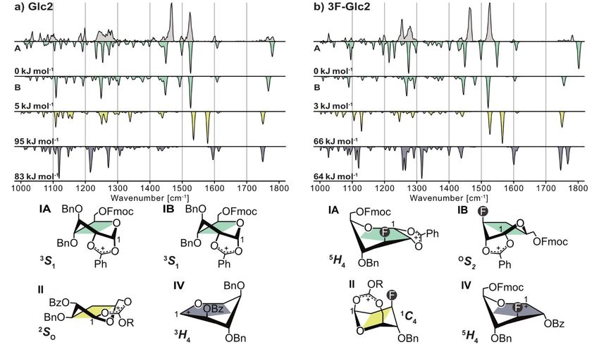
Research Article
doi.org/10.1002/ejoc.202200255
Results and Discussion
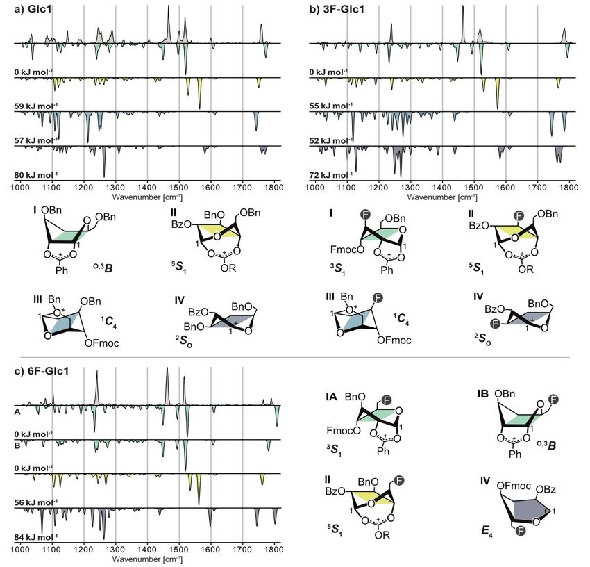
First, the IR signature of the non-fluorinated glycosyl cation
Glc1 is shown (Figure 1a). The functional group region (1450–
1800 cm 1) shows five resolved absorption bands that clearly
match the computed spectrum of the lowest-energy structure I
(Glc1), with an O,3B ring pucker, exhibiting neighboring group
participation (NGP) of the C2-benzoyl group with a covalent
bond (1.51 Å) between the carbonyl oxygen and the anomeric
carbon. The signals at 1466 and 1500 cm 1 originate from the
symmetric and antisymmetric dioxolenium stretches ν(O C O)
of the participating Bz PG, while the signal at 1759 cm 1 stems
from a carbonyl stretch ν(C=O) within the non-participating
Fmoc PG. Interestingly, the vibrations at 1519 and 1600 cm 1
are due to ν(C=C) stretches connected to resonance stabiliza-
Scheme 1. Modes of participation in glycosyl cations. tion of the positive charge by the phenyl ring of the Bz PG in
the dioxolenium motif (Scheme 3). The strong absorption at
1519 cm 1 is caused by the vibration of the formed C=C double
bond, while the weak absorption at 1600 cm 1 can be
the anomeric carbon to yield a bicyclic dioxolenium intermedi- attributed to the ν(C=C) stretches within the phenyl ring. The
ate. The studies also revealed that the gas-phase structures of increased partial double bond character is also visible in the
the investigated glycosyl cations correlate with the experimen- length of the C C bond that decreases from 1.47 to 1.43 Å
tal stereoselectivity observed in solution-phase studies of their compared to the lowest-energy oxocarbenium structure where
precursors. the PGs do not participate. Thus, the charge of the glycosyl
Interestingly, despite being formally known as non-partic- cation is not only delocalized within the dioxolenium motif, but
ipating PGs, benzyl ether oxygens can also stabilize the positive also within the phenyl ring, leading to further stabilization. A
charge at the anomeric carbon, resulting in the formation of similar stabilization by resonance effects in cations was
oxonium ions (III).[15b] previously reported for 4-aminobenzoic acid in gas-phase IR
Here, we combine cryogenic infrared spectroscopy with experiments.[16]
density functional theory (DFT) to probe glycosyl cations of The fingerprint region (1000–1450 cm 1) contains a unique
functionalized glucose building blocks that are commonly used signature for each species, however, it is rather difficult to
in glycan synthesis. The C2 position is always benzoylated (Bz), derive a structural assignment solely based on this region.
while the other hydroxyl groups are either protected with Computational methods often fail to accurately model the
fluorenylmethoxycarbonyl (Fmoc) or benzyl (Bn) groups. In fingerprint region in more complex systems, also due to
selected building blocks fluorine is introduced at the C3 or C6 anharmonicities.[17] The vibrations observed herein are mainly
position to study its impact on the structure of the glycosyl originating from C C and C O stretching vibrations (1000–
cation (Scheme 2). Further, the gas-phase structures are corre- 1350 cm 1) as well as C H bends (1350–1450 cm 1). The
lated to the experimentally observed β-stereoselectivity. spectral signature corresponds the best to the lowest-energy
structure I (Glc1). Other structural motifs, such as remote
participation of the Fmoc PG II (Glc1) (+ 61 kJ mol 1), remote
benzyl ether participation III (Glc1) (+ 57 kJ mol 1) or oxocarbe-
nium structures IV (Glc1) (+ 80 kJ mol 1), can be clearly ruled
out due to two reasons: 1) their free energies at 90 K are
significantly higher than those of structures exhibiting NGP; 2)
Scheme 3. Resonance stabilization of the positive charge by the phenyl ring
Scheme 2. Differentially protected monosaccharide building blocks used in in benzoyl neighboring group participation. Glycosyl cations with this mode
this study to generate glycosyl cations, which are subsequently probed by of participation are further stabilized by increased delocalization of the
cryogenic infrared spectroscopy. positive charge.
Eur. J. Org. Chem. 2022, e202200255 (2 of 6) © 2022 The Authors. European Journal of Organic Chemistry published by Wiley-VCH GmbH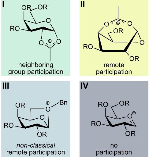
Research Article
                                                  doi.org/10.1002/ejoc.202200255

Figure 1. Infrared spectra of (a) Glc1, (b) 3F Glc1, and (c) 6F Glc1 glycosyl cations generated from β-thiotolyl (a) and β-thioethyl (b,c) precursors.
Experimental IR spectra are shown as light gray traces. Computed spectra of lowest-energy dioxolenium structures, exhibiting neighboring group (green) and
remote participation (yellow), oxonium (blue), and oxocarbenium structures (dark gray) are shown as inverted traces in respective colors. Relative free energies
at 90 K are indicated. The lowest-energy structures are shown in a simplified representation below the spectra, with their ring pucker annotated. For clarity,
some protecting groups have been omitted and R used as abbreviation for fluorenylmethyl. 3D-representation of the structures and xyz-coordinates can be
found in the SI.

their computed infrared spectra do not agree with the                             adopt dioxolenium-type structures I exhibiting benzoyl NGP.
experimental spectrum (Figure 1a).                                                Although all three experimental spectra share some similarities,
    The IR spectra of the C3- and C6-fluorinated glycosyl cations                 the absorption bands differ in shape and exact position. Thus,
3F Glc1 and 6F Glc1 are shown in Figure 1b and Figure 1c.                         each spectrum is a unique pattern for the probed glycosyl
Compared to Glc1, the spectral signature of the fluorinated                       cation. Further evidence for a C2-dioxolenium motif is provided
counterparts is less crowded in the fingerprint region. Here,                     by the computed spectra of structures exhibiting benzoyl NGP
mainly one intense absorption band can be observed at                             that also possess the lowest free energy of all sampled
1234 cm 1 associated with a ν(C O) stretch within the Fmoc PG.                    structures. In both cases, a 3S1 pucker is adopted with a bond
Otherwise, the spectral signature resembles that of Glc1. As a                    distance of 1.50 Å between the carbonyl oxygen of the Bz PG
consequence, the glycosyl cations 3F Glc1 and 6F Glc1 mainly                      and the anomeric carbon. The vibrations associated with the

Eur. J. Org. Chem. 2022, e202200255 (3 of 6)                              © 2022 The Authors. European Journal of Organic Chemistry published by Wiley-VCH GmbH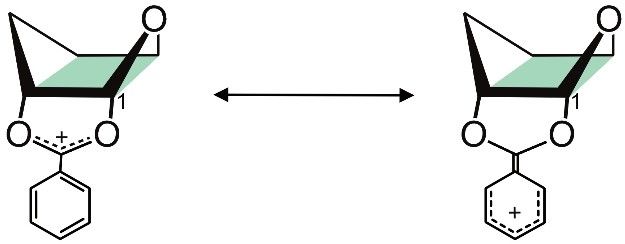
Research Article
                                                   doi.org/10.1002/ejoc.202200255

dioxolenium motif and the + M effect within the benzoyl group                      group region, the spectra look similar to those previously
clearly correspond to the experimental signature. The carbonyl                     shown, being diagnostic for C2-dioxolenium structures exhibit-
absorption band in I (3F Glc1) corresponds to the experiment,                      ing NGP. For Glc2, the lowest-energy structure IA exhibits
while the experimental spectrum of 6F Glc1 exhibits two                            benzoyl NGP, with a 3S1 pucker and a bond distance of 1.51 Å
carbonyl bands, which is diagnostic for a second low-energy                        between the carbonyl oxygen of the benzoyl group and the
conformer (IB) simultaneously present in the ion trap. Like for                    anomeric carbon. A second low-energy conformer IB (+
Glc1, other structural motifs can be excluded based on their                       5 kJ mol 1) was sampled, in which the Fmoc, the C4-Bn and the
computed spectral signatures and unfavorable free energies.                        participating Bz PG are stacked. The differently orientated Fmoc
    Although the substitution of a benzyl group by fluorine                        PG leads to a shift of the position of the carbonyl band. The
changes the ring pucker from O,3B in Glc1 to 3S1 in 3/6F Glc1, it                  population of these two low-energy conformers might explain
does not have an influence on the participation of the                             the presence of two carbonyl bands and the wealth of
neighboring benzoyl group. The changes in ring pucker could                        absorption bands in the fingerprint region in the experimental
be attributed to a decreased steric hindrance of fluorine                          spectrum. For 3F Glc2, the lowest-energy conformer IA exhib-
compared to the bulkier benzyl PG. In all three cases, the α-side                  its a 5H4 pucker, however, its IR signature matches the experi-
of the glycosyl cation is efficiently shielded, leading to β-                      ment slightly less well than that of a second low-energy
stereoselectivity. This selectivity was observed in the AGA of                     structure IB (+ 3 kJ mol 1) with a OS2 pucker and a 1.50 Å bond
deoxyfluorinated β(1,4) hexaglucoside analogues (employing                         distance. Again, other structural motifs are unlikely, considering
building blocks Glc1, 3F Glc1, and 6F Glc1, see the Support-                       their spectral signature and energetics. Here, fluorine has an
ing Information).[7b,18]                                                           influence on the ring pucker, but not on the overall structural
    In a second set of glycosyl cations, Glc2 and 3F Glc2, the                     motif, strongly correlated to the experimental β-stereoselectiv-
C4 and C6 PGs are permuted, compared to Glc1 analogues. The                        ity. Formation of β-linkages was observed in the AGA of
IR spectra are shown in Figure 2. Generally, the spectral                          deoxyfluorinated glucosides (employing building blocks Glc2
signature is slightly more congested than the corresponding                        and 3F Glc2).[19]
Glc1 species, which is attributed to the population of multiple
low-energy conformers enabled by the increased flexibility of
the Fmoc PG now located at the C6 position. In the functional

Figure 2. Infrared spectra of (a) Glc2 and (b) 3F Glc2 glycosyl cations generated from β-thioethyl precursors. Experimental IR spectra are shown as light gray
traces. Computed spectra of lowest-energy dioxolenium structures, exhibiting neighboring group (green) and remote participation (yellow), and
oxocarbenium structures (dark gray) are shown as inverted traces in respective colors. Relative free energies at 90 K are indicated. The lowest-energy
structures are shown in a simplified representation below the spectra, with their ring pucker annotated (for Glc2, IA and IB the differences in structures are
too subtle to represent them in the simplified representation, therefore, the reader is referred to the 3D-structure in Figure S12). For clarity, some protecting
groups have been omitted and R used as abbreviation for fluorenylmethyl. 3D-representation of the structures and xyz-coordinates can be found in the SI.

Eur. J. Org. Chem. 2022, e202200255 (4 of 6)                               © 2022 The Authors. European Journal of Organic Chemistry published by Wiley-VCH GmbH
Research Article
doi.org/10.1002/ejoc.202200255
Conclusion DFT functionals were chosen because they showed chemical
accuracy in a benchmark study on carbohydrates.[28] Details of the
To conclude, we have shown that it is possible to generate and reoptimized structures, such as energetics, ring puckers, and
coordinates can be found in the Supporting Information.
probe glycosyl cations and their fluorinated analogues from
precursors readily used in glycan synthesis. In each case, the
underlying structural motif can be clearly identified as neigh-
boring group participation of C2-benzoyl protecting groups. Acknowledgements
Interestingly, participation of the Bz protecting groups is
connected to resonance effects involving the phenyl ring, which The authors gratefully acknowledge the expertise of Dr. Wieland
can be directly monitored due to vibrations associated with the Schöllkopf and Sandy Gewinner for running the FHI FEL. K.G.
delocalized electrons. The permutation of the protecting groups thanks the Fonds National de la Recherche (FNR), Luxembourg, for
as well as their substitution by the less bulky fluorine leads to a funding the project GlycoCat (13549747). C.K. is grateful for
change in the conformation of the ring pucker. However, the financial support by Fonds der Chemischen Industrie. R.C. and K.P.
structure of the active dioxolenium site remains unchanged and thank the Deutsche Forschungsgemeinschaft (DFG) for support
the stereoselectivity observed for these building blocks in under project number 387284271-SFB 1349. K.P. acknowledges
glycosylation reactions is therefore not affected. Further experi- generous funding by the European Research Council, ERC-2019-
ments are needed to explore the effects of a C2- and C4- CoG-863934-GlycoSpec. M.D., G.F., and P.H.S. thank the MPG-FhG
fluorination, which are expected to have a much more Cooperation Project Glyco3Dysplay and the German Federal
significant impact on the structure of the reactive glycosyl- Ministry of Education and Research (BMBF, grant number
cation intermediate. 13XP5114) for financial support. P.H.S. thanks the Max Planck
Society for generous financial support. Open Access funding
enabled and organized by Projekt DEAL.
Experimental Section
Cryogenic infrared spectroscopy Conflict of Interest
A detailed description of the experimental setup can be found in
the SI (Figure S1) and in previous publications.[20] Briefly, thioglyco- The authors declare no conflict of interest.
side precursors were transferred into the gas phase via nano-
electrospray ionization (nESI). The leaving group is cleaved by in-
source fragmentation leading to glycosyl cations. Mass spectra can Data Availability Statement
be found in the SI (Figures S2–S6). The ions of interest are mass-to-
charge selected by a quadrupole mass filter and accumulated in a
hexapole ion trap, which is cooled to approximately 90 K by liquid The data that support the findings of this study are available
nitrogen. Superfluid helium nanodroplets (0.4 K) are generated by from the corresponding author upon reasonable request.
an Even-Lavie valve and traverse the ion trap, picking up ions, and
guide them to a detection region, where the embedded ions are
Keywords: Carbohydrates · Fluorine · Glycosylation · IR
excited by IR photons generated by the free-electron laser of the
Fritz Haber Institute (FHI FEL[21]). Upon absorption of resonant Spectroscopy · Mass spectrometry
photons, ions are eventually released from the droplets and
afterwards detected by a time-of-flight detector. Monitoring the ion
signal as a function of the IR photon wavenumber leads to a high-
resolution IR signature of the probed ion. [1] A. Varki, Glycobiology 2017, 27, 3.
[2] P. H. Seeberger, R. D. Cummings, in: Essentials of Glycobiology, 3rd ed.
(Eds.: rd, A. Varki, R. D. Cummings, J. D. Esko, P. Stanley, G. W. Hart, M.
Aebi, A. G. Darvill, T. Kinoshita, N. H. Packer, J. H. Prestegard, R. L.
Computational methods Schnaar, P. H. Seeberger), Cold Spring Harbor (NY), 2015, pp. 729.
[3] a) R. Hevey, Chem. Eur. J. 2021, 27, 2240; b) B. Linclau, A. Ardá, N. C.
To model the IR spectra of the probed ions, candidate structures
Reichardt, M. Sollogoub, L. Unione, S. P. Vincent, J. Jiménez-Barbero,
were sampled using the genetic algorithm (GA) FAFOOM.[22] The GA Chem. Soc. Rev. 2020, 49, 3863.
allows sampling flexible bonds and ring puckers and sends each [4] a) A. Geissner, L. Baumann, T. J. Morley, A. K. O. Wong, L. Sim, J. R. Rich,
sampled geometry to an external software (ORCA 4.1.1)[23] for DFT P. P. L. So, E. M. Dullaghan, E. Lessard, U. Iqbal, M. Moreno, W. W.
optimization at the PBE/def2-SVP[24] level of theory. This conforma- Wakarchuk, S. G. Withers, ACS Cent. Sci. 2021, 7, 345; b) A. Axer, R. P.
tional search mainly yielded dioxolenium-type structures I, in which Jumde, S. Adam, A. Faust, M. Schäfers, M. Fobker, J. Koehnke, A. K. H.
the benzoyl group shields the anomeric carbon from the α-side, Hirsch, R. Gilmour, Chem. Sci. 2021, 12, 1286; c) H. J. Lo, L. Krasnova, S.
and oxocarbenium-type structures IV, in which no participation Dey, T. Cheng, H. Liu, T. I. Tsai, K. B. Wu, C. Y. Wu, C. H. Wong, J. Am.
Chem. Soc. 2019, 141, 6484.
takes place. Furthermore, the algorithm also generated structures
[5] a) J. St-Gelais, E. Côté, D. Lainé, P. A. Johnson, D. Giguère, Chem. Eur. J.
in which either the remote Fmoc (C4 and C6) or Bn PGs (C6 only) 2020, 26, 13499; b) J. St-Gelais, M. Bouchard, V. Denavit, D. Giguère, J.
interact with the anomeric carbon (dioxolenium II and oxonium Org. Chem. 2019, 84, 8509; c) D. Lainé, O. Lessard, J. St-Gelais, D.
structures III). A subset of structures of each type was reoptimized Giguère, Chem. Eur. J. 2021, 27, 3799.
and their harmonic frequencies computed at the PBE0 + D3/6-311 [6] J. Vaugenot, A. El Harras, O. Tasseau, R. Marchal, L. Legentil, B.
+ G(d,p)[25] level of theory using Gaussian 16.[26] Each computed IR Le Guennic, T. Benvegnu, V. Ferrières, Org. Biomol. Chem. 2020, 18,
spectrum was normalized and scaled by 0.965. Ring puckers were 1462.
[7] a) M. Delbianco, P. H. Seeberger, Mater. Horiz. 2020, 7, 963; b) Y. Yu, T.
assigned according to Cremer-Pople coordinates.[27] The employed
Tyrikos-Ergas, Y. Zhu, G. Fittolani, V. Bordoni, A. Singhal, R. J. Fair, A.
Eur. J. Org. Chem. 2022, e202200255 (5 of 6) © 2022 The Authors. European Journal of Organic Chemistry published by Wiley-VCH GmbHResearch Article
doi.org/10.1002/ejoc.202200255
Grafmuller, P. H. Seeberger, M. Delbianco, Angew. Chem. Int. Ed. 2019, Fittolani, Y. Yu, Y. Zhu, P. H. Seeberger, Y. Ogawa, M. Delbianco, Chem.
58, 13127. Eur. J. 2021, 27, 13139; c) A. Poveda, G. Fittolani, P. H. Seeberger, M.
[8] O. J. Plante, E. R. Palmacci, P. H. Seeberger, Science 2001, 291, 1523. Delbianco, J. Jiménez-Barbero, Front. Mol. Biosci. 2021, 8, 784318.
[9] A. A. Joseph, A. Pardo-Vargas, P. H. Seeberger, J. Am. Chem. Soc. 2020, [20] a) D. A. Thomas, E. Mucha, M. Lettow, G. Meijer, M. Rossi, G. von Helden,
142, 8561. J. Am. Chem. Soc. 2019, 141, 5815; b) D. A. Thomas, R. Chang, E. Mucha,
[10] a) P. O. Adero, H. Amarasekara, P. Wen, L. Bohe, D. Crich, Chem. Rev. M. Lettow, K. Greis, S. Gewinner, W. Schöllkopf, G. Meijer, G. von Helden,
2018, 118, 8242; b) S. Chatterjee, S. Moon, F. Hentschel, K. Gilmore, P. H. Phys. Chem. Chem. Phys. 2020, 22, 18400; c) M. Lettow, M. Grabarics, K.
Seeberger, J. Am. Chem. Soc. 2018, 140, 11942. Greis, E. Mucha, D. A. Thomas, P. Chopra, G. J. Boons, R. Karlsson, J. E.
[11] a) E. Mucha, M. Marianski, F.-F. Xu, D. A. Thomas, G. Meijer, G. Turnbull, G. Meijer, R. L. Miller, G. von Helden, K. Pagel, Anal. Chem.
von Helden, P. H. Seeberger, K. Pagel, Nat. Commun. 2018, 9, 4174; b) H. 2020, 92, 10228.
Elferink, M. E. Severijnen, J. Martens, R. A. Mensink, G. Berden, J. [21] W. Schöllkopf, S. Gewinner, H. Junkes, A. Paarmann, G. von Helden, H. P.
Oomens, F. Rutjes, A. M. Rijs, T. J. Boltje, J. Am. Chem. Soc. 2018, 140, Bluem, A. M. M. Todd, Proc. SPIE-Int. Soc. Opt. Eng. 2015, 9512, 95121 L.
6034; c) A. A. Hettikankanamalage, R. Lassfolk, F. S. Ekholm, R. Leino, D. [22] A. Supady, V. Blum, C. Baldauf, J. Chem. Inf. Model. 2015, 55, 2338.
Crich, Chem. Rev. 2020, 120, 7104. [23] F. Neese, WIREs Comput. Mol. Sci. 2012, 2, 73.
[12] H. S. Hahm, M. Hurevich, P. H. Seeberger, Nat. Commun. 2016, 7, 12482. [24] a) J. P. Perdew, K. Burke, M. Ernzerhof, Phys. Rev. Lett. 1996, 77, 3865;
[13] L. Lebedel, A. Ardá, A. Martin, J. Désiré, A. Mingot, M. Aufiero, N. b) F. Weigend, R. Ahlrichs, Phys. Chem. Chem. Phys. 2005, 7, 3297.
Aiguabella Font, R. Gilmour, J. Jiménez-Barbero, Y. Blériot, S. Thibau- [25] a) C. Adamo, V. Barone, J. Chem. Phys. 1999, 110, 6158; b) S. Grimme, J.
deau, Angew. Chem. Int. Ed. 2019, 58, 13758. Antony, S. Ehrlich, H. Krieg, J. Chem. Phys. 2010, 132, 154104.
[14] K. Greis, C. Kirschbaum, S. Leichnitz, S. Gewinner, W. Schöllkopf, G. [26] M. J. Frisch, G. W. Trucks, H. B. Schlegel, G. E. Scuseria, M. A. Robb, J. R.
von Helden, G. Meijer, P. H. Seeberger, K. Pagel, Org. Lett. 2020, 22, Cheeseman, G. Scalmani, V. Barone, G. A. Petersson, H. Nakatsuji, X. Li,
8916. M. Caricato, A. V. Marenich, J. Bloino, B. G. Janesko, R. Gomperts, B.
[15] a) H. Elferink, R. A. Mensink, W. W. A. Castelijns, O. Jansen, J. P. J. Mennucci, H. P. Hratchian, J. V. Ortiz, A. F. Izmaylov, J. L. Sonnenberg,
Bruekers, J. Martens, J. Oomens, A. M. Rijs, T. J. Boltje, Angew. Chem. Int. Williams, F. Ding, F. Lipparini, F. Egidi, J. Goings, B. Peng, A. Petrone, T.
Ed. 2019, 58, 8746; b) M. Marianski, E. Mucha, K. Greis, S. Moon, A. Pardo, Henderson, D. Ranasinghe, V. G. Zakrzewski, J. Gao, N. Rega, G. Zheng,
C. Kirschbaum, D. A. Thomas, G. Meijer, G. von Helden, K. Gilmore, P. H. W. Liang, M. Hada, M. Ehara, K. Toyota, R. Fukuda, J. Hasegawa, M.
Seeberger, K. Pagel, Angew. Chem. Int. Ed. 2020, 59, 6166; c) T. Hansen, Ishida, T. Nakajima, Y. Honda, O. Kitao, H. Nakai, T. Vreven, K. Throssell,
H. Elferink, J. M. A. van Hengst, K. J. Houthuijs, W. A. Remmerswaal, A. J. A. Montgomery Jr., J. E. Peralta, F. Ogliaro, M. J. Bearpark, J. J. Heyd,
Kromm, G. Berden, S. van der Vorm, A. M. Rijs, H. S. Overkleeft, D. V. E. N. Brothers, K. N. Kudin, V. N. Staroverov, T. A. Keith, R. Kobayashi, J.
Filippov, F. Rutjes, G. A. van der Marel, J. Martens, J. Oomens, J. D. C. Normand, K. Raghavachari, A. P. Rendell, J. C. Burant, S. S. Iyengar, J.
Codee, T. J. Boltje, Nat. Commun. 2020, 11, 2664; d) K. Greis, E. Mucha, Tomasi, M. Cossi, J. M. Millam, M. Klene, C. Adamo, R. Cammi, J. W.
M. Lettow, D. A. Thomas, C. Kirschbaum, S. Moon, A. Pardo-Vargas, G. Ochterski, R. L. Martin, K. Morokuma, O. Farkas, J. B. Foresman, D. J. Fox,
von Helden, G. Meijer, K. Gilmore, P. H. Seeberger, K. Pagel, Wallingford, CT, 2016.
ChemPhysChem 2020, 21, 1905. [27] D. Cremer, J. A. Pople, J. Am. Chem. Soc. 1975, 97, 1354.
[16] a) J. Seo, S. Warnke, S. Gewinner, W. Schollkopf, M. T. Bowers, K. Pagel, [28] M. Marianski, A. Supady, T. Ingram, M. Schneider, C. Baldauf, J. Chem.
G. von Helden, Phys. Chem. Chem. Phys. 2016, 18, 25474; b) T. Khuu, N. Theory Comput. 2016, 12, 6157.
Yang, M. A. Johnson, Int. J. Mass Spectrom. 2020, 457.
[17] a) B. Brauer, M. Pincu, V. Buch, I. Bar, J. P. Simons, R. B. Gerber, J. Phys.
Chem. A 2011, 115, 5859; b) E. Mucha, A. Stuckmann, M. Marianski, W. B.
Struwe, G. Meijer, K. Pagel, Chem. Sci. 2019, 10, 1272; c) M. Grabarics, M.
Lettow, C. Kirschbaum, K. Greis, C. Manz, K. Pagel, Chem. Rev. 2021.
[18] G. Fittolani, E. Shanina, M. Guberman, P. H. Seeberger, C. Rademacher,
M. Delbianco, Angew. Chem. Int. Ed. 2021, 60, 13302. Manuscript received: March 2, 2022
[19] a) M. Delbianco, A. Kononov, A. Poveda, Y. Yu, T. Diercks, J. Jiménez- Revised manuscript received: March 23, 2022
Barbero, P. H. Seeberger, J. Am. Chem. Soc. 2018, 140, 5421; b) S. Gim, G. Accepted manuscript online: March 24, 2022
Eur. J. Org. Chem. 2022, e202200255 (6 of 6) © 2022 The Authors. European Journal of Organic Chemistry published by Wiley-VCH GmbHYou can also read