Muscle atrophy induced by overexpression of ALAS2 is related to muscle mitochondrial dysfunction
←
→
Page content transcription
If your browser does not render page correctly, please read the page content below
Peng et al. Skeletal Muscle     (2021) 11:9
https://doi.org/10.1186/s13395-021-00263-8

 RESEARCH                                                                                                                                            Open Access

Muscle atrophy induced by overexpression
of ALAS2 is related to muscle
mitochondrial dysfunction
Yahui Peng1,3,4†, Jihong Li1,3,4†, Dixian Luo2, Shuai Zhang1,3,4, Sijia Li1,3,4, Dayong Wang1,3,4, Xidi Wang1,3,4,
Zhujun Zhang1,3,4, Xue Wang1,3,4, Changhui Sun1,3,4, Xu Gao1,3,4, Yang Hui1,3,4* and Rongzhang He2,5*

  Abstract
  Background: ALAS2 (delta-aminolevulinate synthase 2) is one of the two isoenzymes catalyzing the synthesis of
  delta-aminolevulinic acid (ALA), which is the first precursor of heme synthesis. ALAS2-overexpressing transgenic
  mice (Tg mice) showed syndrome of porphyria, a series of diseases related to the heme anabolism deficiency. Tg
  mice showed an obvious decrease in muscle size. Muscle atrophy results from a decrease in protein synthesis and
  an increase in protein degradation, which ultimately leads to a decrease in myofiber size due to loss of contractile
  proteins, organelles, nuclei, and cytoplasm.
  Methods: The forelimb muscle grip strength of age-matched ALAS-2 transgenic mice (Tg mice) and wild-type mice
  (WT mice) were measured with an automated grip strength meter. The activities of serum LDH and CK-MB were
  measured by Modular DPP. The histology of skeletal muscle (quadriceps femoris and gastrocnemius) was observed
  by hematoxylin and eosin (HE) staining, immunohistochemistry, and transmission electron microscope. Real-time
  PCR was used to detect mtDNA content and UCP3 mRNA expression. Evans blue dye staining was used to detect
  the membrane damage of the muscle fiber. Single skeletal muscle fiber diameter was measured by single-fiber
  analyses. Muscle adenosine triphosphate (ATP) levels were detected by a luminometric assay with an ATP assay kit.
  Results: Compared with WT mice, the strength of forelimb muscle and mass of gastrocnemius were decreased in
  Tg mice. The activities of serum CK-MB and LDH, the number of central nuclei fibers, and Evans blue positive fibers
  were more than those in WT mice, while the diameter of single fibers was smaller, which were associated with
  suppressed expression levels of MHC, myoD1, dystrophin, atrogin1, and MuRF1. Re-expression of eMyHC was only
  showed in the quadriceps of Tg mice, but not in WT mice. Muscle mitochondria in Tg mice showed dysfunction
  with descented ATP production and mtDNA content, downregulated UCP3 mRNA expression, and swelling of
  mitochondria.
  (Continued on next page)

* Correspondence: huiyang79@126.com; rongzhang412@163.com
†
 Yahui Peng and Jihong Li contributed equally to this work.
1
 Department of Biochemistry and Molecular Biology, Harbin Medical
University, Harbin 150086, China
2
 Institute of Translational Medicine, National and Local Joint Engineering
Laboratory of High-through Molecular Diagnostic Technology, the First
People’s Hospital of Chenzhou, The First Affiliated Hospital of Xiangnan
University, Chenzhou 423000, China
Full list of author information is available at the end of the article

                                         © The Author(s). 2021 Open Access This article is licensed under a Creative Commons Attribution 4.0 International License,
                                         which permits use, sharing, adaptation, distribution and reproduction in any medium or format, as long as you give
                                         appropriate credit to the original author(s) and the source, provide a link to the Creative Commons licence, and indicate if
                                         changes were made. The images or other third party material in this article are included in the article's Creative Commons
                                         licence, unless indicated otherwise in a credit line to the material. If material is not included in the article's Creative Commons
                                         licence and your intended use is not permitted by statutory regulation or exceeds the permitted use, you will need to obtain
                                         permission directly from the copyright holder. To view a copy of this licence, visit http://creativecommons.org/licenses/by/4.0/.
                                         The Creative Commons Public Domain Dedication waiver (http://creativecommons.org/publicdomain/zero/1.0/) applies to the
                                         data made available in this article, unless otherwise stated in a credit line to the data.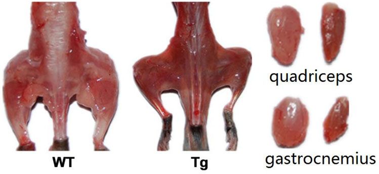
Peng et al. Skeletal Muscle    (2021) 11:9                                                                       Page 2 of 11

  (Continued from previous page)
  Conclusion: ALAS2 overexpressing-transgenic mice (Tg mice) showed muscle dystrophy, which was associated
  with decreased atrogin-1 and MuRF-1, and closely related to mitochondrial dysfunction.
  Keywords: Muscle atrophy, Mitochondrial dysfunction, Transgenic mice, Delta-aminolevulinate synthase 2

Background                                                        of ALAS2 mRNA and protein among patients with
The heme biosynthetic pathway begins from delta-                  erythropoietic protoporphyria (EPP) [14]. Additionally,
aminolevulinic acid synthase (ALAS) catalyzing the con-           four recurrent gain-of-function mutations in the
densation of glycine and succinyl-CoA to delta-                   catalytic domain of the ALAS2 enzyme resulting in an
aminolevulinic acid (ALA) in the mitochondria [1]. ALAS           increased ALAS2 activity have been described as being
is coded by two genes: ALAS1 and ALAS2 [2]. ALAS1 is              responsible for X-linked protoporphyria (XLPP) [15].
ubiquitously expressed in all cells, and the negative feed-       To-Figueras et al. presented convincing evidence that
back is regulated by the heme pool [3, 4]; however, ALAS2         ALAS2 acts as a modifier gene in patients with congeni-
is specifically expressed only in erythroid cells [2, 5] and is   tal erythropoietic porphyria [16]. In this study, we
not inhibited by heme [6]. Bechara reported that reactive         reported that ALAS2 overexpressing-transgenic mice
oxygen species (ROS) is formed by the metal-catalyzed aer-        (Tg mice) showed muscle atrophy, which was associated
obic oxidation of ALA at abnormally high levels [6, 7];           with decreased atrogin-1 and MuRF-1, and closely
mitochondria are the main source of ROS and are also the          related to mitochondrial dysfunction.
primary target of oxidant-induced damage. ALA, as a puta-
tive endogenous source of ROS, induces mitochondrial              Materials and methods
swelling and transmembrane potential collapse [7], and            Animals
ALA-treated rats under swimming training experienced fa-          ALAS2 transgenic mice were generated by the standard
tigue earlier [8]. Defective mitochondrial function has been      pronuclear injection technique using C57BL/6 mice.
shown to cause muscle weakness [9]. The loss of mitochon-         Briefly, mouse ALAS2 cDNA was cloned into the
dria has also been shown to result in muscle wasting [10].        pCAGGS plasmid with the chicken β-actin promoter,
Notably, studies have reported abnormalities in the mito-         which drove the mouse ALAS2 cDNA expression, and
chondria during sarcopenia, muscle wasting, associated            terminated with the poly (A) signal. All animals were
with chronic illness (cachexia), and disuse atrophy [11, 12].     identified by analysis of tail DNA by PCR. Sequences of
   The mitochondria produce adenosine triphosphate                primers were as follows (5′−3′): forward primer was
(ATP) as a source of chemical energy, and skeletal                GCCTTCTTCTTTTTCCTACAGCTC; reverse primer
muscle contains abundant mitochondria. Increased                  was GCACAATCTTGCTCTTCCTGTCTTGG. Both
mitochondrial ROS production can promote disuse                   the ALAS2 transgenic mice and wild type mice were
muscle atrophy by increasing proteolysis and depressing           housed in a temperature-controlled room (22 °C) with a
protein synthesis, and ROS can contribute to mitochon-            12-h light to 12-h dark cycle. Unless otherwise noted, 6-
drial damage and impaired the ability to produce ATP,             to 12-month-old male mice were used in the experi-
which results in energy stress [10]. Theoretically,               ments. All procedures were approved by the Institutional
mitochondrial damage could decrease the level of cellu-           Animal Care and Use Committee of Harbin Medical
lar energy available for protein synthesis, and energy            University.
stress could promote proteolysis via the AMPK-FoxO3
axis [10].                                                        Measurement of forelimb muscle grip strength
   Disorder of the heme biosynthesis pathway could in-            The forelimb muscle grip strength was measured by an
duce porphyria. Each enzymatic alteration of the heme             automated grip strength meter (Jinan, China) as
biosynthesis system can cause a specific porphyria [13].          described previously [17]. Briefly, mice were lifted by
The clinical manifestations include acute neurovisceral           their tails and made to hold a horizontal bar with their
attacks, skin lesions, and muscle atrophy, which are as-          forelimbs. Next, they were pulled slowly backwards until
sociated with the accumulation of porphyrin precursors            they could no longer hold the grip. The maximal force
(5-aminolevulinic acid, porphobilinogen) and porphy-              was recorded during consecutive attempts (at least 20 at-
rins. Muscle weakness due to porphyria can progress               tempts per mouse), and the average was set as the result.
and lead to tetraplegia, with respiratory and bulbar par-
alysis [13]. ALAS2-overexpressing transgenic mice (Tg             Biochemical assays
mice) showed obvious muscle atrophy. Jasmin Barman-               Serum samples were separated from whole blood by
Aksözen et al. found a significant increase in the amount         centrifugation at 1,000×g for 10 minutes after the blood
Peng et al. Skeletal Muscle   (2021) 11:9                                                                    Page 3 of 11

was allowed to clot at room temperature for 30 minutes.        were captured with the Olympus Bx51 microscope, and
The activities of serum lactate dehydrogenase (LDH),           fiber diameter was measured by Olympus Element
creatine kinase (CK), and creatine kinase-MB (CK-MB)           software.
were measured by Modular DPP (Roche).
                                                               Muscle ATP level
Processing of tissues for histology                            For the muscle ATP level, we used a luminometric assay
The gastrocnemius and/or quadriceps muscles were               with ATP Assay kit (Beyotime) according to the manu-
fixed with 4% formalin for at least 36 h. The tissues were     facturer’s instructions.
embedded in paraffin and cut into 4 μm thick sections
in the transverse myofilament direction. Then, the sec-
                                                               Real-time PCR
tions were stained with hematoxylin and eosin (HE), and
                                                               The muscles were harvested from ALAS2 transgenic and
the images were visualized and captured with the
                                                               wild-type (WT) mice and were frozen immediately in
Olympus Bx51 microscope.
                                                               liquid nitrogen, and then they were stored frozen at – 80
                                                               °C. For RNA isolation, the tissue was homogenized in
Immunohistochemistry
                                                               Trizol reagent (Invitrogen) and total RNA was prepared
The sections of paraffin-embedded muscle tissue were
                                                               according to the manufacturer’s protocol. The RNA was
deparaffinized in xylene and rehydrated in ethyl alcohol.
                                                               reverse transcribed into cDNA by High-Capacity cDNA
Then, the sections were blocked with 1% hydrogen
                                                               Reverse Transcription kit (Applied Biosystems). Real-
peroxide (H2O2) in distilled water for 10 min, and the
                                                               time PCR (RT-PCR) analyses were performed by the
non-specific sites were blocked with bovine serum albu-
                                                               ABI 7500 real-time PCR system (Applied Biosystems).
min (BSA, DAKO) for 20 min at room temperature. For
                                                               The glyceraldehyde-3-phosphate dehydrogenase (GAPD
detecting eMyHC, heat-induced antigen retrieval was
                                                               H) expression was used to normalize the expression
performed (Tris/EDTA buffer, pH 8, DAKO) prior to
                                                               levels. The relative expression values gained were used
staining the muscle samples. The sections were then
                                                               to calculate fold change. The primer sequences are listed
incubated overnight at 4 °C with primary antibody of
                                                               in Table 1 (5′−3′).
anti-eMyHC (clone BF-45, mouse, 1:400). The BF-45
monoclonal antibody was obtained from DSHB at the
University of Iowa in USA. After thorough washing in           Mitochondrial DNA (mtDNA) content assay
PBS, the sections were incubated with biotin-conjugated        The muscles were digested and the DNA was isolated
secondary antibodies (DAKO) at 37 °C for 20 min. We            using the DNeasy Blood and Tissue kit (QIAGEN). The
used a standard peroxidase-based method with DAB               mtDNA content was quantified by qRT-PCR using a
(DAKO) to detect the antibody. The sections were dehy-         SYBR Green-based detection system by the ABI 7500
drated with ethyl alcohol and coverslipped with mount-         real-time PCR system (Applied Biosystems) according to
ing medium. The stained sections were imaged using an          the manufacturer’s protocol in a similar way as the pre-
Olympus BX51 microscope.                                       vious description [20]. The qRT-PCR primer sequences
                                                               of mtDNA and nucleus DNA were as follows (5′-3′),
Evans blue assay                                               mtDNA: Forward primer was AAGTCGTAACAAGG
Evans blue assay was performed as described previously         TAAGCA, and Reverse primer was ATATTTGTGT
[18]. Evans blue dye (10 mg/ml) was dissolved in phos-         AGGGCTAGGG; Nuc.DNA: Forward primer was
phate buffered saline (PBS). Then, it was filtered sterilely   GGGTATATTTTTGATACCTTCAATGAGTTA, and
by a 0.2-μm pore filter. The Evans blue dye was intraper-      Reverse primer was TCTGAAACAGTAGGTAGAGAC-
itoneally injected into the mouse (0.1 ml/10 g body            CAAAGC.
weight). The mice were killed 24 h after injection. The
quadriceps muscle of these killed mice was prepared and        Transmission electron microscopy (TEM)
observed under the Olympus Bx51microscope.                     The muscle blocks were prepared and soaked immedi-
                                                               ately in 2.5% glutaraldehyde. After 6-8 h at 4 °C, they
Single-fiber analyses                                          were cut into 1mm thick coronal slices. Next, the sam-
Single fibers were isolated and fiber size was measured        ples were rinsed with PBS (0.1 M) before being post-
as described previously [19]. The quadriceps muscles           fixed by osmium tetroxide for 1-2 h. The muscle blocks
were fixed with 4% paraformaldehyde (PFA) for more             were dehydrated through a graded series of alcohol and
than 2 days. Dissected small bundles of fibers were incu-      acetone. Subsequently, we used epoxy resin for embed-
bated in 40% NaOH for 2-3 h and vigorously shaken.             ding prior to slicing of the ultra-thin sections. Then,
Isolated myofibers were washed in PBS and stained with         double staining by uranyl acetate and lead citrate was
10 μM DAPI. Images of 40-60 single fibers per animal           performed. Finally, the images were acquired by a
Peng et al. Skeletal Muscle (2021) 11:9 Page 4 of 11
Table 1 Primers table
transmission electron microscope (JEM-1220, JEOL Ltd, Results
Tokyo, Japan). Reduction of forelimb muscle grip strength in ALAS2
transgenic mice
Western blotting As muscle weakness is a clinical manifestation of
About 20 mg muscle tissue was lysed in RIPA Lysis porphyria [18, 21, 22], we measured the forelimb muscle
Buffer (Beyotime) for 10 minutes on ice. RIPA Lysis grip strength by the automated grip strength meter.
Buffer is configured with 20 mM Tris PH7.5, 150 mM Interestingly, we found that ALAS2 transgenic (Tg) mice
NaCl, 1% Triton X-100, 2.5 mM sodium pyrophosphate, had reduced forelimb muscle strength compared with
1 mM EDTA, 1%Na3VO4, 0.5 μg/ml leupeptin, and 1 the age-matched WT littermates (Fig. 1). The data
mM phenyl methane sulfonyl fluoride (PMSF). The strongly suggest that muscle weakness is present in
lysate homogenate was centrifuged at 12,000×g at 4 °C ALAS2 transgenic (Tg) mice.
for 5 min. The protein concentration was measured with
the DC Protein Assay kit (Bio-Rad Laboratories). Protein Loss of muscle mass in ALAS2 transgenic mice
samples were boiled for 10 min in the presence of 4× Tg mice were smaller and thinner than the age-matched
Loading Dye. Equal amounts of total proteins (25 μg) WT littermates (data not shown). On visual analysis,
were loaded on a 12% SDS-polyacrylamide gel for elec- overall loss of hindlimb muscle mass was clearly evident
trophoresis followed by a transfer to PVDF membranes in Tg mice (Fig. 2a). The wet weight of the quadriceps
(Millipore) at 70 V for 1 h. The membranes were femoris was approximately half of that in the age-
blocked with 5% non-fat powdered milk in PBS (10 mM, matched WT littermates, and this finding was similar to
pH 7.4) for 1 h at 4 °C. The blot was incubated with the that in the gastrocnemius muscle (Fig. 2b). As Tg mice
primary antibody (GAPDH, 1:2000, Cell Signaling were smaller and thinner, we normalize the wet weight
Technology, HO-1, Santa Cruz Biotechnology) overnight of the quadriceps femoris mass by the body weight. The
at 4 °C. The membrane was washed three times by results showed that the muscle mass percentage in Tg
PBST, followed by incubation with the appropriate mice was lower than that in WT mice (Fig. 2c). The my-
secondary antibody. The signal was detected by an osin heavy chain (MHC) mRNA expression was also
Enzymatic Chemiluminescence (ECL) kit (Applygen). measured. We found the MHC mRNA level in Tg mice
was decreased compared with that in WT mice (Fig. 2d).
Statistics The data indicated muscle mass loss in Tg mice.
All quantitative data are expressed as means ± SD.
Statistical analysis was performed using either Student’s Muscle atrophy in ALAS2 transgenic mice
t test (two groups) or one-way analysis of variance (more To determine the cause of loss of muscle mass, HE-
than two groups), followed by Bonferroni post hoc test. stained transverse sections of the quadriceps femoris
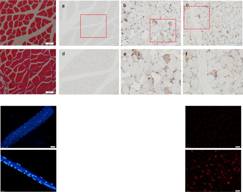
Differences were considered significant at P < 0.05. from age-matched Tg mice and WT mice werePeng et al. Skeletal Muscle (2021) 11:9 Page 5 of 11 Fig. 1 Reduced forelimb muscle grip strength in ALAS2 transgenic mice. Grip strength in ALAS2 transgenic mice (n = 9) and WT mice (n = 8). Values are means ± SD. *P < 0.05;**P < 0.01;***P < 0.001 compared. A high number of muscle fibers with were compared by immunostaining for quadriceps cross centrally located nuclei was found in Tg mice, a primary section from age-matched Tg mice and WT mice at 6 pathological sign of muscular dystrophy [23], but not in months old. Re-expression of eMyHC was only showed in WT mice (Fig. 3a). Moreover, to detect the regeneration the quadriceps of Tg mice, but not in WT mice (Fig. 3b), of the quadriceps in Tg mice, re-expression of eMyHC indicating that the central nucleation determined in Tg Fig. 2 Loss of muscle mass in the ALAS2 transgenic mice. a Left panel: gross morphology of skinned hindlimb muscles of ALAS2 transgenic mice and WT mice. Right panel: comparison of individual muscles. b Comparison of changes in wet weight of individual muscles mass (n = 6–7). c Comparison of in wet weight of individual muscles mass normalized to body weight (n = 6–7). d The MHC mRNA expression (n = 6–7). Values are means ± SD.*P < 0.05; **P < 0.01; ***P < 0.001
Peng et al. Skeletal Muscle    (2021) 11:9   Page 6 of 11

 Fig. 3 (See legend on next page.)
Peng et al. Skeletal Muscle     (2021) 11:9                                                                                          Page 7 of 11

 (See figure on previous page.)
 Fig. 3 Muscle atrophy in the ALAS2 transgenic mice. a Quadriceps cross section from hematoxylin and eosin stain. (bar, 50 μm). b Expression of
 eMyHC in WT and ALAS2 transgenic mice (Tg) muscle. (Bar of a-c is 50 μm, and bar of d-e 20 μm). c Single-fiber size comparison of WT and
 ALAS2 transgenic mice quadriceps muscle. d Quantification of average fiber diameter across single fibers in quadriceps muscle from WT (n = 4)
 and ALAS2 transgenic mice (n = 4). e Evans blue dye was intraperitoneal injected and mice sacked 24 h later. Transverse cross-sections of
 quadriceps muscle were observed with OLYMPUS BX51 equipped with green activation filters. Evans blue-positive fibers are denoted by red
 staining. Bar, 50 μm. f Serum LDH levels of WT (n = 7) and ALAS2 transgenic mice (n = 6). g Serum CK-MB levels of WT (n = 7) and ALAS2
 transgenic mice (n = 6). h The relative mRNA expression of myod1, myogenin, and S6K1 (n = 5–8). i The relative mRNA expression of utrophin,
 dystrophin, Atrogin-1, and MuRF1. Values are means ± SD.*P < 0.05; **P < 0.01; ***P < 0.001

mice muscles resulted from muscle regeneration [24, 25].                   energetic failure played an important role in the expres-
With respect to single-fiber analyses [19, 26], the average                sion of acute intermittent porphyria (AIP) [28].
diameter of single fibers isolated from the muscle of Tg
mice was smaller than that isolated from the muscle of                     Discussion
WT mice (Fig. 3c, d). In addition, to examine leakage into                 ALAS2-overexpressing Tg mice were developed to in-
the muscle fiber, Evans blue assay was employed [18, 27].                  vestigate the mechanism of porphyria. The expression of
The fluorescent dye accumulated in the interior of dystro-                 ALAS2 was increased in Tg mice [29]. Tg mice showed
phied muscle fibers in Tg mice (Fig. 3e). We showed that                   obvious muscular atrophy, which is also a clinical char-
the myocyte membrane was damaged in dystrophied                            acteristic of porphyria. Therefore, we explored the
muscle of Tg mice. Moreover, we detected the activities of                 mechanism of muscular atrophy in Tg mice. Firstly, we
serum CK, CK-MB, and LDH, and we found the elevation                       found that Tg mice experienced a decrease in muscle
of the activity of serum CK-MB and LDH in Tg mice (Fig.                    mass and grip strength of the forelimb muscles. Sec-
3f, g). We analyzed the expression of the genes, and we                    ondly, increased activities of serum CK-MB and LDH,
found that the expression levels of MyoD1, dystrophin,                     increased central nuclear fiber and Evans blue positive
Atrogin-1, and MuRF1 were decreased, but the expression                    fiber and decreased single-fiber diameter confirmed
level of utrophin was increased. There was no difference                   muscle atrophy in Tg mice. In addition, Re-expression
in myogenin and S6K1 in Tg mice compared with WT                           of eMyHC was only showed in the quadriceps of Tg
mice (Fig. 3h, i). Albertyn CH et al. also described that                  mice, but not in WT mice, indicating that the central
acute intermittent porphyria presenting as progressive                     nucleation determined in Tg mice muscles resulted from
muscular atrophy in a 23-year-old black South African                      muscle regeneration. Furthermore, the expression levels
man [12].                                                                  of MyoD1, S6K1 (anabolic factor), atrogin1, and MuRF1
                                                                           (catabolic factor) were determined. Finally, muscle mito-
Mitochondrial dysfunction in the muscle of ALAS2                           chondrial dysfunction in ALAS2 Tg mice was detected
transgenic mice                                                            based on mitochondrial swelling, decline in ATP pro-
To test whether muscle dystrophy in Tg mice was                            duction and mtDNA content, and downregulation of
affected by mitochondrial damage, we examined the                          UCP3 mRNA expression.
ultrastructure of muscle fibers using a transmission elec-                    We found that the muscle grip strength of forelimbs
tron microscope. Mitochondrial swelling was found in                       of Tg mice was decreased. Since muscle mass deter-
muscles of Tg mice (Fig. 4a, right), but not in muscles of                 mines the skeletal muscle strength [30], the loss of
WT mice (Fig. 4a, left). Then we compared the mtDNA                        muscle strength in Tg mice may be caused by the loss of
content, uncoupling protein 3 (UCP-3) mRNA expres-                         muscle mass. The diameter of single fibers of Tg mice
sion, and ATP production in the hind limb muscles of                       was smaller than that of WT mice, and thinner fiber in-
Tg mice and age-matched WT mice. The mtDNA con-                            dicated less muscle mass. MHC is an important part of
tent quantified by qRT-PCR [20] was significantly re-                      the sarcomere [31]. We found that the mRNA level of
duced in the gastrocnemius muscle of Tg mice (Fig. 4b).                    MHC in Tg mice was decreased, which meant that the
The muscle UCP-3 mRNA expression was decreased in                          loss of muscle mass may be caused by the decrease in
Tg mice (Fig. 4c). ATP production in the gastrocnemius                     MHC content. In addition to the decreased muscle mass
muscle of Tg mice was decreased to 21% of the level in                     of atrophic muscles, a large number of muscle fibers
age-matched WT mice (Fig. 4d). Interestingly, increased                    with centrally located nucleus were observed in Tg mice,
expression levels of SOD1 mRNA, HO-1 mRNA, and                             which is a sign of muscle fiber regeneration [32, 33]. We
HO-1 protein showed that SOD1 and HO-1 were in-                            also found that the re-expression of eMyHC was only
duced in the muscle of Tg mice (Fig. 4e, f). Above all,                    showed in the quadriceps of Tg mice, but not in WT
mitochondrial dysfunction and loss were present in the                     mice, confirming that the central nucleation determined
muscle of Tg mice. It was reported that mitochondrial                      in Tg mice muscles resulted from muscle regeneration.
Peng et al. Skeletal Muscle (2021) 11:9 Page 8 of 11 Fig. 4 Mitochondrial dysfunction in the ALAS2 transgenic mice muscle. a TEM images of gastrocnemius of WT and ALAS2 transgenic mice. Bars, 2 μm. b mtDNA content in gastrocnemius of WT (n = 8) and ALAS2 transgenic mice (n = 7). c UCP3 expression in quadriceps of WT (n = 8) and ALAS2 transgenic mice (n = 7). d ATP production in gastrocnemius of WT (n = 8) and ALAS2 transgenic mice (n = 8). e The SOD1 and HO-1 mRNA expression(n = 5–8). f The HO-1 protein expression (n = 4). Values are means ± SD. *P < 0.05; **P < 0.01; ***P < 0.001 The Evans blue dye could enter into the myocyte myocyte of Tg mice suggested that the myocyte through the damaged cytomembrane and get accu- membrane was damaged in Tg mice. Also, increased mulated in the myocyte, and thus, Evans blue dye activities of serum LDH and CK-MB indicated that was used to identify damaged skeletal myofibers [27, the muscular membrane of Tg mice was damaged 34, 35]. Accumulation of Evans blue dye in the [36–38]. The expression of MyoD1 was decreased in
Peng et al. Skeletal Muscle   (2021) 11:9                                                                                      Page 9 of 11

Tg mice compared with WT mice. Since muscle                    mitochondrial damage. A decrease in the ATP production
regeneration has been reported to be delayed in                was observed in Tg mice, which was probably induced by
MyoD (−/−) mice [39], decreased MyoD1 might                    mitochondrial damage and loss. The pCAGGS expression
cause a disturbance in the regeneration in Tg mice.            vector can drive EGFP expression in all tissues, except
   The expression of utrophin was increased and that of        erythrocytes and hair in mice, particularly higher in the
dystrophin was decreased in Tg mice compared with              muscle [53]. Also, Tg mice have ubiquitous overexpres-
WT mice, which was similar to that in other dystrophy          sion of ALAS-2 in all tissues and higher expression of
reports [33, 40]. MAFbx and MuRF1 belong to the ubi-           ALAS-2 in the muscle. Moreover, the accumulation of
quitin proteasome pathway, which plays a critical role in      ALA is much higher in Tg mice than in WT mice. Be-
the intracellular protein degradation of skeletal muscle       cause ALA is synthesized in the mitochondria and ALA is
[41]. Upregulation of atrophy-related genes atrogin-1          a putative endogenous source of ROS [7], ALA might
(MAFbX) and MuRF1 in skeletal muscle atrophy has               damage the mitochondria of muscle in Tg mice.
been reported previously [42, 43]. However, atrogin-1            Porphyrias are a group of eight metabolic disorders of
and MuRF1 were downregulated in aging-related loss of          the heme biosynthesis pathway [7]. Every porphyria is
skeletal muscle [44] and in mTOR-mice [33], and here,          caused by abnormal function of a separate enzymatic
we also found that atrogin-1 and MuRF1 levels were             step, resulting in a specific accumulation of heme pre-
decreased in Tg mice. Inhibition of MuRF1 is sufficient        cursors, including ALA, PBG, and porphyrins. In some
to maintain the MHC [44]. However, MHC in Tg mice              cases, muscle atrophy was present in porphyria; how-
was decreased, which indicated that the loss of muscle         ever, the underlying mechanism is still unknown.
mass in Tg mice was not related to activation of the ubi-      ALAS2-overexpressing Tg mice also show accumulation
quitin proteasome pathway. A previous study showed             of ALA, thus it may be a new model of porphyria. In the
that chronic spinal cord-injured patients with severe at-      future, we will further verify whether ALAS2-
rophy of the quadriceps muscles showed a reduction in          overexpressing Tg mice can be used as a porphyria
atrogin-1 and MuRF1, which suggested an internal               model, and we will use this model to investigate the rela-
mechanism aimed at reducing the further loss of muscle         tionship of mitochondrial dysfunction and porphyria-
proteins [45, 46]. The reduction of atrogin-1 and MuRF1        related muscle weakness.
in Tg mice may also be a protective attempt to reduce
further muscle wasting in muscle atrophy.                      Conclusion
   Mitochondrial dysfunction is a hallmark trait that oc-      Muscle weakness in ALAS2-overexpressing mice is re-
curs during prolonged muscle inactivity in both animals        lated to muscle mitochondrial dysfunction induced by
and humans. Mitochondrial fission and remodeling con-          the accumulation of ALA.
tribute to muscle atrophy [47]. Increased superoxide
in vivo accelerates age-associated muscle atrophy through      Abbreviations
mitochondrial dysfunction [19]. Mitochondria play an im-       ALAS2: Delta-aminolevulinate synthase 2; ALA: Delta-aminolevulinic acid; Tg
                                                               mice: Transgenic mice; WT mice: Wild-type mice; ATP: Adenosine
portant role in muscle atrophy [19, 47, 48]. It has been re-   triphosphate; HE: Hematoxylin and eosin; ROS: Reactive oxygen species;
ported that ALA-generated oxidant promotes dysfunction         EPP: Erythropoietic protoporphyria; XLPP: X-linked protoporphyria;
and swelling of the isolated rat liver mitochondria [7].       mtDNA: Mitochondrial DNA; LDH: Lactate dehydrogenase ; CK: Creatine
                                                               kinase; CK-MB: Creatine kinase-MB; GAPDH: Glyceraldehyde-3-phosphate
Similarly, mitochondrial swelling and mitochondrial cris-      dehydrogenase; TEM: Transmission electron microscopy
tae reduction were shown in muscles of Tg mice. As Tg
mice have high expression of ALAS2 [29] and accumula-
tion of ALA in the muscles (data not shown), mitochon-         Supplementary Information
                                                               The online version contains supplementary material available at https://doi.
drial damage in the muscle of Tg mice is most likely to be     org/10.1186/s13395-021-00263-8.
induced by ALA. There is an increase of SOD-1 in the
brain, muscle, and liver of chronic ALA-treated rats [8],       Additional file 1 : FigS. 1 Overexpresion ALAS-2 in in mouse myoblasts
and thus, we speculate that SOD-1 and HO-1 are induced          (C2C12). A, The relative mRNA expression of ALAS-2. B, The relative mRNA
                                                                expression of myod1, myogein and S6K1. C, The relative mRNA expres-
by ALA in Tg mice. Increasing of anti-oxidant enzymes           sion of utrophin, dystrophin, Atrogin-1 and MuRF1. Values are means ±
in Tg mice indicated oxidant existence. As excessive free       SD.*P
Peng et al. Skeletal Muscle (2021) 11:9 Page 10 of 11
Funding 12. Albertyn CH, Sonderup M, Bryer A, Corrigall A, Meissner P, Heckmann JM.
This work was supported by Natural Science Foundation of China (81773165, Acute intermittent porphyria presenting as progressive muscular atrophy in
81671255), Hunan Province Science Fund for Distinguished Young Scholars a young black man. S Afr Med J. 2014;104(4):283–5.
(2018JJ1021), the Natural Science Foundation of Hunan Province 13. Puy H, Gouya L, Deybach JC. Porphyrias. Lancet. 2010;375(9718):924–37.
(2020JJ5013), and Chinese Scholarship Council Fund for the Visiting Scholars 14. Barman-Aksozen J, Minder EI, Schubiger C, Biolcati G, Schneider-Yin X. In
(No. 201908230108). ferrochelatase-deficient protoporphyria patients, ALAS2 expression is
enhanced and erythrocytic protoporphyrin concentration correlates with
Availability of data and materials iron availability. Blood Cells Mol Dis. 2015;54(1):71–7.
All the data and material could be traced from the paper or can be 15. Manceau H, Gouya L, Puy H. Acute hepatic and erythropoietic porphyrias:
requested from the corresponding author. from ALA synthases 1 and 2 to new molecular bases and treatments. Curr
Opin Hematol. 2017;24(3):198–207.
16. To-Figueras J, Ducamp S, Clayton J, Badenas C, Delaby C, Ged C, Lyoumi S,
Declarations Gouya L, de Verneuil H, Beaumont C, et al. ALAS2 acts as a modifier gene in
patients with congenital erythropoietic porphyria. Blood. 2011;118(6):1443–
Ethics approval and consent to participate 51.
The study was approved by the local Ethic Committee. 17. Windahl SH, Andersson N, Borjesson AE, Swanson C, Svensson J, Moverare-
Skrtic S, Sjogren K, Shao R, Lagerquist MK, Ohlsson C. Reduced bone mass
Competing interests and muscle strength in male 5alpha-reductase type 1 inactivated mice.
The authors declare that they have no competing interests. PLoS One. 2011;6(6):e21402.
18. Crosbie RH, Straub V, Yun HY, Lee JC, Rafael JA, Chamberlain JS, Dawson VL,
Author details Dawson TM. Campbell KP: mdx muscle pathology is independent of nNOS
1
Department of Biochemistry and Molecular Biology, Harbin Medical perturbation. Hum Mol Genet. 1998;7(5):823–9.
University, Harbin 150086, China. 2Institute of Translational Medicine, National 19. Jang YC, Lustgarten MS, Liu Y, Muller FL, Bhattacharya A, Liang H, Salmon
and Local Joint Engineering Laboratory of High-through Molecular AB, Brooks SV, Larkin L, Hayworth CR, et al. Increased superoxide in vivo
Diagnostic Technology, the First People’s Hospital of Chenzhou, The First accelerates age-associated muscle atrophy through mitochondrial
Affiliated Hospital of Xiangnan University, Chenzhou 423000, China. dysfunction and neuromuscular junction degeneration. FASEB J. 2010;24(5):
3
Heilongjiang Academy of Medical Sciences, Harbin 150086, China. 4Key 1376–90.
Laboratory of Preservation of Human Genetic Resources and Disease Control 20. Viader A, Golden JP, Baloh RH, Schmidt RE, Hunter DA, Milbrandt J.
in China (Harbin Medical University), Ministry of Education, Beijing 150086, Schwann cell mitochondrial metabolism supports long-term axonal survival
China. 5Department of Clinical Pharmacology, Xiangya Hospital, Central and peripheral nerve function. J Neurosci. 2011;31(28):10128–40.
South University, Changsha 410078, China. 21. Cavanagh JB, Mellick RS. On the Nature of the Peripheral Nerve Lesions
Associated with Acute Intermittent Porphyria. J Neurol Neurosurg
Received: 15 January 2020 Accepted: 2 March 2021 Psychiatry. 1965;28:320–7.
22. Moorhead PJ, Cooper DJ, Timperley WR. Progressive peripheral neuropathy
in patient with primary hyperoxaluria. Br Med J. 1975;2(5966):312–3.
References 23. Wang B, Li J, Xiao X. Adeno-associated virus vector carrying human
1. Tchaikovskii V, Desnick RJ, Bishop DF. Molecular expression, characterization minidystrophin genes effectively ameliorates muscular dystrophy in mdx
and mechanism of ALAS2 gain-of-function mutants. Mol Med. 2019;25(1):4. mouse model. Proc Natl Acad Sci U S A. 2000;97(25):13714–9.
2. Riddle RD, Yamamoto M, Engel JD. Expression of delta-aminolevulinate 24. Schiaffino S, Rossi AC, Smerdu V, Leinwand LA, Reggiani C. Developmental
synthase in avian cells: separate genes encode erythroid-specific and myosins: expression patterns and functional significance. Skelet Muscle.
nonspecific isozymes. Proc Natl Acad Sci U S A. 1989;86(3):792–6. 2015;5:22.
3. Yamamoto M, Kure S, Engel JD, Hiraga K. Structure, turnover, and heme- 25. Silva WJ, Graca FA, Cruz A, Silvestre JG, Labeit S, Miyabara EH, Yan CYI,
mediated suppression of the level of mRNA encoding rat liver delta- Wang DZ. Moriscot AS: miR-29c improves skeletal muscle mass and
aminolevulinate synthase. J Biol Chem. 1988;263(31):15973–9. function throughout myocyte proliferation and differentiation and by
4. Kolluri S, Sadlon TJ, May BK, Bonkovsky HL. Haem repression of the repressing atrophy-related genes. Acta Physiol. 2019;226(4):e13278.
housekeeping 5-aminolaevulinic acid synthase gene in the hepatoma cell 26. Wada KI, Takahashi H, Katsuta S, Soya H. No decrease in myonuclear
line LMH. Biochem J. 2005;392(Pt 1):173–80. number after long-term denervation in mature mice. Am J Physiol Cell
5. Peoc'h K, Nicolas G, Schmitt C, Mirmiran A, Daher R, Lefebvre T, Gouya L, Physiol. 2002;283(2):C484–8.
Karim Z, Puy H. Regulation and tissue-specific expression of delta- 27. Matsuda R, Nishikawa A, Tanaka H. Visualization of dystrophic muscle fibers
aminolevulinic acid synthases in non-syndromic sideroblastic anemias and in mdx mouse by vital staining with Evans blue: evidence of apoptosis in
porphyrias. Mol Genet Metab. 2019;128(3):190–7. dystrophin-deficient muscle. J Biochem. 1995;118(5):959–64.
6. Smith SJ, Cox TM. Translational control of erythroid delta-aminolevulinate 28. Homedan C, Schmitt C, Laafi J, Gueguen N, Desquiret-Dumas V, Lenglet H,
synthase in immature human erythroid cells by heme. Cell Mol Biol. 1997; Karim Z, Gouya L, Deybach JC, Simard G, et al. Mitochondrial energetic
43(1):103–14. defects in muscle and brain of a Hmbs-/- mouse model of acute
7. Hermes-Lima M, Castilho RF, Valle VG, Bechara EJ, Vercesi AE. Calcium- intermittent porphyria. Hum Mol Genet. 2015;24(17):5015–23.
dependent mitochondrial oxidative damage promoted by 5-aminolevulinic 29. Huang H, Wang W, Zou J, Liu Z, Zhou Z, Nakajima O, Zhang L, Luo J, Li M,
acid. Biochim Biophys Acta. 1992;1180(2):201–6. He Q, et al. Over-expression 5-aminolevulinic acid synthase 2 in
8. Pereira B, Curi R, Kokubun E, Bechara EJ. 5-aminolevulinic acid-induced nonerythroid cell may causes protoporphyrin IX accumulation.
alterations of oxidative metabolism in sedentary and exercise-trained rats. J Photodiagnosis Photodyn Ther. 2017;17:22–8.
Appl Physiol. 1992;72(1):226–30. 30. Frontera WR, Hughes VA, Lutz KJ, Evans WJ. A cross-sectional study of
9. Yamada T, Ivarsson N, Hernandez A, Fahlstrom A, Cheng AJ, Zhang SJ, muscle strength and mass in 45- to 78-yr-old men and women. J Appl
Bruton JD, Ulfhake B, Westerblad H. Impaired mitochondrial respiration Physiol. 1991;71(2):644–50.
and decreased fatigue resistance followed by severe muscle weakness 31. Derbre F, Ferrando B, Gomez-Cabrera MC, Sanchis-Gomar F, Martinez-Bello
in skeletal muscle of mitochondrial DNA mutator mice. J Physiol. 2012; VE, Olaso-Gonzalez G, Diaz A, Gratas-Delamarche A, Cerda M, Vina J.
590(23):6187–97. Inhibition of xanthine oxidase by allopurinol prevents skeletal muscle
10. Powers SK, Wiggs MP, Duarte JA, Zergeroglu AM, Demirel HA. Mitochondrial atrophy: role of p38 MAPKinase and E3 ubiquitin ligases. PLoS One. 2012;
signaling contributes to disuse muscle atrophy. Am J Physiol Endocrinol 7(10):e46668.
Metab. 2012;303(1):E31–9. 32. Handschin C, Chin S, Li P, Liu F, Maratos-Flier E, Lebrasseur NK, Yan Z,
11. Peker N, Donipadi V, Sharma M, McFarlane C, Kambadur R. Loss of Parkin Spiegelman BM. Skeletal muscle fiber-type switching, exercise intolerance,
impairs mitochondrial function and leads to muscle atrophy. Am J Physiol and myopathy in PGC-1alpha muscle-specific knock-out animals. J Biol
Cell Physiol. 2018;315(2):C164–85. Chem. 2007;282(41):30014–21.Peng et al. Skeletal Muscle (2021) 11:9 Page 11 of 11
33. Risson V, Mazelin L, Roceri M, Sanchez H, Moncollin V, Corneloup C, Richard-
Bulteau H, Vignaud A, Baas D, Defour A, et al. Muscle inactivation of mTOR
causes metabolic and dystrophin defects leading to severe myopathy. J Cell
Biol. 2009;187(6):859–74.
34. Brussee V, Tardif F, Tremblay JP. Muscle fibers of mdx mice are more
vulnerable to exercise than those of normal mice. Neuromuscul Disord.
1997;7(8):487–92.
35. Hamer PW, McGeachie JM, Davies MJ, Grounds MD. Evans Blue Dye as an
in vivo marker of myofibre damage: optimising parameters for detecting
initial myofibre membrane permeability. J Anat. 2002;200(Pt 1):69–79.
36. Keshgegian AA, Feinberg NV. Serum creatine kinase MB isoenzyme in
chronic muscle disease. Clin Chem. 1984;30(4):575–8.
37. Apple FS, McGue MK. Serum enzyme changes during marathon training.
Am J Clin Pathol. 1983;79(6):716–9.
38. Siegel AJ, Silverman LM, Evans WJ. Elevated skeletal muscle creatine kinase
MB isoenzyme levels in marathon runners. Jama. 1983;250(20):2835–7.
39. White JD, Scaffidi A, Davies M, McGeachie J, Rudnicki MA, Grounds MD.
Myotube formation is delayed but not prevented in MyoD-deficient skeletal
muscle: studies in regenerating whole muscle grafts of adult mice. J
Histochem Cytochem. 2000;48(11):1531–44.
40. Cifuentes-Diaz C, Frugier T, Tiziano FD, Lacene E, Roblot N, Joshi V, Moreau
MH, Melki J. Deletion of murine SMN exon 7 directed to skeletal muscle
leads to severe muscular dystrophy. J Cell Biol. 2001;152(5):1107–14.
41. Lecker SH, Solomon V, Mitch WE, Goldberg AL. Muscle protein breakdown
and the critical role of the ubiquitin-proteasome pathway in normal and
disease states. J Nutr. 1999;129(1S Suppl):227S–37S.
42. Lagirand-Cantaloube J, Offner N, Csibi A, Leibovitch MP, Batonnet-Pichon S,
Tintignac LA, Segura CT, Leibovitch SA. The initiation factor eIF3-f is a major
target for atrogin1/MAFbx function in skeletal muscle atrophy. EMBO J.
2008;27(8):1266–76.
43. Stitt TN, Drujan D, Clarke BA, Panaro F, Timofeyva Y, Kline WO, Gonzalez M,
Yancopoulos GD, Glass DJ. The IGF-1/PI3K/Akt pathway prevents expression
of muscle atrophy-induced ubiquitin ligases by inhibiting FOXO
transcription factors. Mol Cell. 2004;14(3):395–403.
44. Edstrom E, Altun M, Hagglund M, Ulfhake B. Atrogin-1/MAFbx and MuRF1
are downregulated in aging-related loss of skeletal muscle. J Gerontol A Biol
Sci Med Sci. 2006;61(7):663–74.
45. Foletta VC, White LJ, Larsen AE, Leger B, Russell AP. The role and regulation
of MAFbx/atrogin-1 and MuRF1 in skeletal muscle atrophy. Pflugers Arch.
2011;461(3):325–35.
46. Leger B, Senese R, Al-Khodairy AW, Deriaz O, Gobelet C, Giacobino JP,
Russell AP. Atrogin-1, MuRF1, and FoXO, as well as phosphorylated GSK-
3beta and 4E-BP1 are reduced in skeletal muscle of chronic spinal cord-
injured patients. Muscle Nerve. 2009;40(1):69–78.
47. Romanello V, Guadagnin E, Gomes L, Roder I, Sandri C, Petersen Y, Milan G,
Masiero E, Del Piccolo P, Foretz M, et al. Mitochondrial fission and
remodelling contributes to muscle atrophy. EMBO J. 2010;29(10):1774–85.
48. Chen H, Vermulst M, Wang YE, Chomyn A, Prolla TA, McCaffery JM, Chan
DC. Mitochondrial fusion is required for mtDNA stability in skeletal muscle
and tolerance of mtDNA mutations. Cell. 2010;141(2):280–9.
49. Romanello V, Sandri M. Mitochondrial quality control and muscle mass
maintenance. Front Physiol. 2015;6:422.
50. Bechara EJ, Dutra F, Cardoso VE, Sartori A, Olympio KP, Penatti CA, Adhikari
A, Assuncao NA. The dual face of endogenous alpha-aminoketones: pro-
oxidizing metabolic weapons. Comp Biochem Physiol Toxicol Pharmacol.
2007;146(1-2):88–110.
51. Jones TE, Baar K, Ojuka E, Chen M, Holloszy JO. Exercise induces an increase
in muscle UCP3 as a component of the increase in mitochondrial
biogenesis. Am J Physiol Endocrinol Metab. 2003;284(1):E96–101.
52. Tsuboyama-Kasaoka N, Tsunoda N, Maruyama K, Takahashi M, Kim H,
Ikemoto S, Ezaki O. Up-regulation of uncoupling protein 3 (UCP3) mRNA by
exercise training and down-regulation of UCP3 by denervation in skeletal
muscles. Biochem Biophys Res Commun. 1998;247(2):498–503.
53. Okabe M, Ikawa M, Kominami K, Nakanishi T. Nishimune Y: ‘Green mice’ as a
source of ubiquitous green cells. FEBS Lett. 1997;407(3):313–9.
Publisher’s Note
Springer Nature remains neutral with regard to jurisdictional claims in
published maps and institutional affiliations.You can also read