Multifocus confocal Raman microspectroscopy for rapid single-particle analysis
←
→
Page content transcription
If your browser does not render page correctly, please read the page content below
Multifocus confocal Raman
                 microspectroscopy for rapid
                 single-particle analysis

                 Lingbo Kong
                 Pengfei Zhang
                 Peter Setlow
                 Yong-qing Li

Downloaded From: https://www.spiedigitallibrary.org/journals/Journal-of-Biomedical-Optics on 03 Jan 2022
Terms of Use: https://www.spiedigitallibrary.org/terms-of-use
JBO Letters

                Multifocus confocal Raman                                                          tens of seconds to obtain a Raman spectrum of an unknown par-
                                                                                                   ticle, especially biological particles, and single-focus excitation
                microspectroscopy for                                                              can only analyze one particle within a Raman acquisition time.
                                                                                                   Consequently, this technique becomes time-consuming when
                rapid single-particle                                                              the analysis of large numbers of single particles is desired, and
                                                                                                   the laser power shining on each particle is constrained, such as
                analysis                                                                           when monitoring physiological dynamics of multiple individual
                                                                                                   microbial cells in a population4 and when attempting to quantify
                                                                                                   individual airborne particles impacted at random positions on a
                Lingbo Kong,a Pengfei Zhang,a Peter Setlow,b and                                   cover slip.6, 7
                Yong-qing Lia                                                                          In this paper, we report the development of a multifocus
                a EastCarolina University, Department of Physics, Greenville,                      confocal Raman microspectroscopy system that allows parallel
                North Carolina 27858–4353                                                          analysis of multiple individual particles in random positions on a
                b University of Connecticut Health Center, Department
                of Molecular, Microbial and Structural Biology, Farmington,
                                                                                                   cover slip based on a precise image-guided technique. The mul-
                Connecticut 06030–3305                                                             tifocus excitation technique has been used in laser scanning flu-
                                                                                                   orescence microscopy,8 coherent anti-stokes Raman scattering
                Abstract. We have developed a multifocus confocal Raman                            microscopy,9 Raman imaging,10 and recently in multiple-trap
                microspectroscopy system that allows simultaneous analy-                           laser tweezers Raman spectroscopy.4 Here, we show that this
                ses of ∼80 individual biological or airborne microparticles                        technique can be used to rapidly measure relative concentration
                based on a precise image-guided technique. Multiple indi-                          of microparticles, to rapidly identify individual aerosol particles
                vidual particles adhered in random positions on a coverslip                        collected in indoor air and monitor biological dynamics of single
                were illuminated by a multifocus excitation pattern formed                         bacterial spores during their germination. As a result, it allows
                by rapidly steering a single laser beam with a pair of galvo-                      parallel monitoring of dynamic process of 80 individual cells
                mirrors, and their Raman scatterings were synchronously                            in 1 h, which otherwise would take at least 80 h to obtain the
                projected with another galvo-mirror to different rows of a
                                                                                                   statistical data by using single-focus Raman microspectroscopy.
                CCD chip for parallel spectroscopic analyses. We show that
                this technique can be used to rapidly identify single air-                             The experimental schematic is shown in Fig. 1(a). A laser
                borne particles or bacteria collected on a slide and to mon-                       beam at 780 nm was introduced into an inverted microscope
                itor germination dynamics of multiple bacterial spores in                          (Nikon TiS) equipped with an objective (Plan Apo 60×,
                real-time. C 2011 Society of Photo-Optical Instrumentation Engineers (SPIE).      NA = 1.4) and a green-filtered halogen lamp was used for
                [DOI: 10.1117/1.3662456]                                                           illumination.4, 5 An imaging camera was used to record a bright-
                                                                                                   field or phase-contrast image which was analyzed by a MATLAB
                Keywords: Raman microspectroscopy; single-cell analysis.
                                                                                                   program to locate the centroid positions (xi , yi ) of 80 parti-
                Paper 11532LR received Sep. 20, 2011; revised manuscript received                  cles in a field of view. These coordinates were used to drive a
                Oct. 24, 2011; accepted for publication Oct. 27, 2011; published
                online Nov. 23, 2011.                                                              pair of galvo-mirrors GM1 and GM2 (Cambridge Technology,
                                                                                                   6220H) to rapidly steer a single laser beam with step-function
                                                                                                   waveforms to form multifocus excitation on 80 individual parti-
                                                                                                   cles. Another galvo-mirror GM3 (Thorlabs, GVS001) in front of
                    Raman microspectroscopy combines Raman spectroscopy                            the spectrograph (Princeton Instruments, LS785) synchronously
                with optical microscopy1 and has been widely applied in                            steered the backward Raman scattering from each focus onto dif-
                biomedicine, materials, and environmental science.1, 2 It uses a                   ferent vertical positions of a multichannel CCD chip (Princeton
                focused laser beam as a single-point illumination on the sample                    Instruments, PIXIS 400BR) so that full Raman spectra from
                by an objective, and the Raman-scattered signal from a tiny vol-                   each focus can be collected simultaneously.10 The time that the
                ume (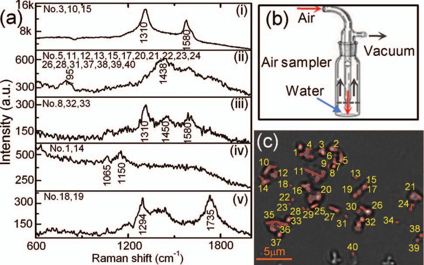
JBO Letters
Fig. 2 Relative concentration measurements of mixed B. megaterium
and B. subtilis spores. Raman spectra of (a) B. megaterium and (b)
B. subtilis spores. (c) Phase-contrast image of the mixed spores of B.
megaterium (numbered in red) and B. subtilis (numbered in black).
(d) Measured ratios of B. megaterium and B. subtilis spores versus the
relative concentration of B. megaterium spores in the mixture. The red
curve is the fitted line.
Due to the rapid identification capability noted above, it is
possible to rapidly measure the relative percentage of specific
microparticles in a mixture. As a demonstration, we mixed B.
megaterium and B. subtilis spores with relative ratios of 1:3,
1:2, 1:1, 2:1, and 3:1, and ∼10 μl of each mixture was dried
on a coverslip. For each mixture, we randomly measured 20
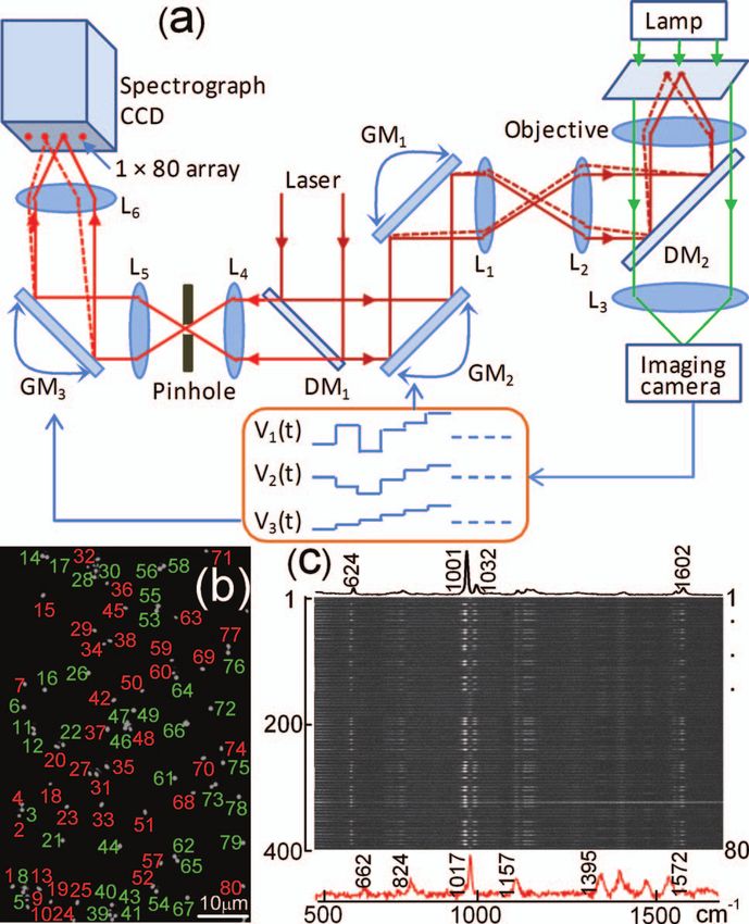
Fig. 1 Experimental setup. (a) Schematic of multifocus confocal fields of view that each contained ∼40 spores, as shown in
Raman microscopy. GM: galvo-mirrors; DM: dichroic mirrors; L: lens. Fig. 2(c), and ∼800 particles were analyzed for each mixture.
(b) Phase-contrast image of a mixture of polystyrene beads and B.
Figures 2(a) and 2(b) show Raman spectra of B. megaterium and
megaterium spores. Beads (numbered in green) and spores (numbered
in red) have been identified by multifocus confocal Raman microscopy B. subtilis spores, respectively. They can be easily distinguished
as shown in (c). (c) Individual Raman spectral images of 80 particles in because of the two carotenoid-specific bands at 1157 and 1520
(b) measured by the spectrograph’s CCD chip with one exposure time. cm − 1 uniquely from B. megaterium spores.10 Therefore, the
Typical Raman spectra of a polystyrene bead and a B. megaterium ratio of the numbers of B. megaterium and B. subtilis spores
spore are shown at the top and bottom, respectively.
in a mixture can be determined for each condition simply by
measuring the carotenoid-specific bands. Figure 2(d) shows
the ratios as the function of the relative concentrations of the
independently changed by the third galvo-mirror so that cross sample mixtures and a linear relationship was observed.
talk between different spectral channels can effectively be Multifocus confocal Raman microspectroscopy can also be
avoided.4 The difference between current work and multifoci- used to rapidly identify airborne particles. Aerosol particles in
scan Raman imaging10 is that the 80 laser foci were only a laboratory room were collected into deionized water (10 ml)
switched to each individual particle’s position here, while in with an AGI-30 impinger at a flow rate of 15 l/min for 30 min, as
Raman imaging the laser foci are scanned across the entire area shown in Fig. 3(b). A small drop of the collected particles were
of the sample. This switching scheme effectively avoids useless deposited and dried on a quartz coverslip. Figure 3(c) shows a
Raman acquisition from the positions without particle occupa- bright field image of 40 individual unknown particles. Raman
tion and thus increases the speed of multiple particle analysis. spectra of these particles were simultaneously collected with a
Figure 1(b) shows a phase-contrast image of 80 particles of laser power of 2.5 mW per particle and an exposure time of
an unknown mixture of 1-μm polystyrene beads and dormant 5 s. These spectra can be classified into five different types, as
Bacillus megaterium spores. Apparently, these particles cannot shown in Fig. 3(a). These particles were identified as i. carbon
be distinguished by their image sizes and shapes. Figure 1(c) (with 1300 and 1600 cm − 1 bands); ii. glassy silicates; iii. the
shows the Raman spectral image of the 80 particles on the mixed compounds of carbonates and silicates; iv. and v. silicate
CCD chip with typical Raman spectra of a polystyrene bead particles with and without fluorescence background.6, 7
(top spectrum) and a B. megaterium spore (bottom spectrum) The most effective application of multifocus confocal
recorded with an incident power of 1.0 mW per focus (with Raman microspectroscopy could be simultaneous monitoring
total power of 80 mW for the 80 foci) and a 5 s exposure of dynamic physiological processes of multiple living cells ad-
time. From the single-particle Raman spectra, these particles hered at random positions on a coverslip. As an example, we
can be easily distinguished, based on characteristic bands at monitored the germination of 20 B. megaterium spores using
1001 cm − 1 for polystyrene beads and 1017 cm − 1 for B. Raman microspectroscopy and phase-contrast microscopy. The
megaterium spores. The latter band is due to the presence of spores were first heat-activated at 60 ◦ C for 15 min in a water
(∼10% of dry wt) 1:1 chelate of Ca2 + with pyridine-2,6- bath and were then adhered firmly on the surface of a quartz
dicarboxylic acid (dipicolinic acid) (CaDPA) in spores.4 coverslip. The germination solution of 0.5 mm D-glucose with
Journal of Biomedical Optics 120503-2 December 2011 r Vol. 16(12)
Downloaded From: https://www.spiedigitallibrary.org/journals/Journal-of-Biomedical-Optics on 03 Jan 2022
Terms of Use: https://www.spiedigitallibrary.org/terms-of-use
JBO Letters

                                                                                                   intensity and the rapid CaDPA release process normally took 1
                                                                                                   to 2 min.5 Up to 80 spores can be monitored in parallel by using
                                                                                                   this multifocus Raman microspectroscopy.
                                                                                                       In summary, we have developed a multifocus confocal
                                                                                                   Raman microspectroscopy system based on an image-guided
                                                                                                   synchronous galvo-mirror scheme, allowing ∼80 individual par-
                                                                                                   ticles in random positions to be analyzed simultaneously. We
                                                                                                   further demonstrated the utility of this analyzer for rapid iden-
                                                                                                   tification of single bacteria and airborne particles collected on a
                                                                                                   cover slip, accurate concentration measurements, and the moni-
                                                                                                   toring of physiological dynamics of multiple individual bacterial
                                                                                                   spores during their germination. This technique can further be
                                                                                                   integrated with differential inference-contrast and fluorescence
                                                                                                   microscopy,4 and may find broad applications in microbiology
                Fig. 3 Identification of airborne particles in the atmosphere. (a)                 and environmental science.
                Raman spectra of five typical aerosol particles with particle numbers
                as labeled in (c). (b) Air sampler that collected the aerosol particles
                into water for spectroscopic analysis. (c) Selected aerosol particles that         Acknowledgments
                were recognized with a MATLAB program.
                                                                                                   This work was supported by a Multi-University Research Initia-
                                                                                                   tive (MURI) award through the U.S. Army Research Laboratory
                25 mm KPO4 buffer (pH 7.4) was added at t = 0 min to initiate                      and the U.S. Army Research Office (PS/YQL) via contract Grant
                germination at 37 ◦ C.4, 5 The phase-contrast images were cap-                     No. W911NF-09-1-0286 and by a grant from the Army Research
                tured at a rate of 10 s per frame and the first frame was used                     Office (YQL/PS) via contract Grant No. W911NF-08-1-0431.
                to locate the positions of the 20 spores, as shown in Fig. 4(a).
                During the germination process of 60 min, the microscope stage
                may have slight movements in the horizontal direction, caus-                       References
                ing the displacement of spores from the laser foci. Therefore,                      1. G. J. Puppels, F. F. M. de Mul, C. Otto, J. Greve, M. Robert-Nicoud, D.
                a new frame of phase-contrast images was used to recalculate                           J. Arndt-Jovin, and T. M. Jovin, “Studying single living cells and chro-
                                                                                                       mosomes by confocal Raman microspectroscopy,” Nature 347(6290),
                the coordinates of the 20 spores every 2 min to precisely guide                        301–303 (1990).
                the laser foci to the spores. The Raman spectra of these spores                     2. G. Turrell and J. Corset, Raman Microscopy: Developments and Appli-
                were acquired with an exposure time of 20 s and a laser power                          cations, Academic Press, London (1996).
                of 5 mW per focus. Figures 4(b) and 4(c) show the time-lapse                        3. K. Kneipp, Y. Wang, H. Kneipp, L. T. Perelman, I. Itzkan, R.
                changes in the CaDPA level (intensity of the 1017 cm − 1 band)                         Dasari, and M. S. Feld, “Single molecule detection using surface-
                                                                                                       enhanced Raman scattering (SERS),” Phys. Rev. Lett. 78(9), 1667–1670
                and refractility (intensity of the phase-contrast image) of seven                      (1997).
                germinating spores, respectively. The data showed that for each                     4. L. B. Kong, P. F. Zhang, G. W. Wang, P. Setlow, and Y. Q. Li, “Character-
                germinating spore, the completion of CaDPA release precisely                           ization of bacterial spore germination using phase contrast microscopy,
                corresponded to the end of the rapid fall in phase-contrast image                      fluorescence microscopy, Raman spectroscopy and optical tweezers,”
                                                                                                       Nat. Protoc. 6(5), 625–639 (2011).
                                                                                                    5. L. B. Kong, P. F. Zhang, P. Setlow, and Y. Q. Li, “Characterization of
                                                                                                       bacterial spore germination using integrated phase contrast microscopy,
                                                                                                       Raman spectroscopy and optical tweezers,” Anal. Chem. 82(9),
                                                                                                       3840–3847 (2010).
                                                                                                    6. N. P. Ivleva, U. McKeon, R. Niessner, and U. Pöschl, “Raman mi-
                                                                                                       crospectroscopic analysis of size-resolved atmospheric aerosol par-
                                                                                                       ticle samples collected with an ELPI: soot, humic-like substances,
                                                                                                       and inorganic compounds,” Aerosol Sci. Technol. 41(7), 655–671
                                                                                                       (2007).
                                                                                                    7. S. Mertes, B. Dippel, and A. Schwarzenbock, “Quantification
                                                                                                       of graphitic carbon in atmospheric aerosol particles by Raman
                                                                                                       spectroscopy and first application for the determination of mass ab-
                                                                                                       sorption efficiencies,” Aerosol Sci. 35(3), 347–361 (2004).
                                                                                                    8. J. Bewersdorf, R. Pick, and S. W. Hell, “Multifocal multiphoton mi-
                                                                                                       croscopy,” Opt. Lett. 23(9), 655–657 (1998).
                                                                                                    9. T. Minamikawa, M. Hashimoto, K. Fujita, S. Kawata, and T. Araki,
                                                                                                       “Multi-focus excitation coherent anti-Stokes Raman scattering (CARS)
                                                                                                       microscopy and its applications for real-time imaging,” Opt. Express
                Fig. 4 Simultaneous monitoring of the germination of multiple individ-                 17(12), 9526–9536 (2009).
                ual B. megaterium spores. (a) Phase-contrast image of 20 B. megaterium             10. L. B. Kong, P. F. Zhang, J. Yu, P. Setlow, and Y. Q. Li “Rapid confocal
                spores before the addition of germinant. (b) Intensities of the CaDPA-                 Raman imaging using a synchro multifoci-scan scheme for dynamic
                specific band at 1017 cm − 1 and (c) intensities of phase-contrast images              monitoring of single living cells,” Appl. Phys. Lett. 98(21), 213703
                of seven germinating spores as a function of the germination time.                     (2011).

                Journal of Biomedical Optics                                              120503-3                                           December 2011 r Vol. 16(12)

Downloaded From: https://www.spiedigitallibrary.org/journals/Journal-of-Biomedical-Optics on 03 Jan 2022
Terms of Use: https://www.spiedigitallibrary.org/terms-of-use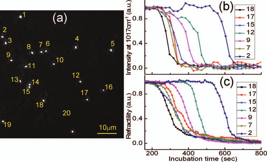
You can also read