Management of Traumatic Herniation of Buccal Fat Pad in a 22-month-old Child: A Case Report
←
→
Page content transcription
If your browser does not render page correctly, please read the page content below
Case Report J Nepal Assoc Pediatr Dent. 2020;1(1):17-9
Management of Traumatic Herniation of Buccal Fat Pad in a
22-month-old Child: A Case Report
Bhawana Sigdel,1 Sneha Shrestha,2 Mamta Dali,3 Amita Rai,4 Ashish Shrestha,5 Bandana Koirala6
1
Post-graduate Resident, 2Assistant Professor, 3Associate Professor, 4Consultant, 5,6Professor
1-3, 6
Department of Pedodontics and Preventive Dentistry, B.P. Koirala Institute of Health Sciences, Dharan, Nepal,
4
Kanti Children’s Hospital, Kathmandu, Nepal, 5Department of Oral Pathology B.P. Koirala Institute of Health Sciences, Dharan, Nepal
ABSTRACT
The buccal fat pad is an encapsulated mass located within the buccal facial spaces. It is relatively large in neonate and infants. A tiny
perforation on buccal mucosa, fascia or buccinator muscle can lead to the herniation of buccal fat pad in young children. Trauma
is the most common etiology. Such situation demands careful examination of oral cavity and thorough history taking to avoid any
misdiagnosis. This paper reports a clinical presentation and management of herniation of buccal fat pad on a 22-month-old-girl
following trauma to her left cheek region with sugarcane.
Keywords: Buccal fat pad, excision, herniation.
INTRODUCTION Traumatic herniation is a rare entity commonly seen in
infants and young children from five months to12 years.5
Buccal fat pad is a specialized type of adipose tissue that
Perforation of buccinator muscle due to blunt injury of
serves to line the masticatory space, separating muscles
buccal mucosa by any foreign objects is the cause of large
of mastication from each other and from the zygomatic
portion of buccal fat pad to herniate into the oral cavity. At
arch and the ramus of mandible. It has got four processes:
the same time, sucking activity of infants further promotes
buccal, pterygoid, superficial, and deep temporal processes.
its herniation through the mucosal defect.6
The pterygoid and temporal processes are situated deep
whereas the buccal extension is located superficially within CASE REPORT
the cheek.1 Buccal fat pad is prominent in young children
A 22-month-old female child reported to the Department
and is responsible for the fullness of the cheeks. It aids in
of Pedodontics and Preventive Dentistry, BPKIHS,
cushioning and sucking functions and is often referred to
Dharan with the chief complaint of swelling on left cheek
as sucking or suctorial pad.2 Bichat (1802) first described
region. There was a history of traumatic injury while
the true fatty nature of buccal fat pad.3 Earlier, it was
eating sugarcane twenty-four hours back. Parents noted
accidentally encountered during various operations in the
an extruding mass with minimal bleeding from the site of
maxillofacial region and had little importance.2 However,
injury though it did not cause any difficulty in eating or
these days it is considered as an ideal flap to reconstruct
breathing. The medical history was not significant.
oral defect due to its high vascularity, ease of mobilization
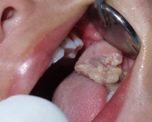
and minimal donor site morbidity.4 On examination, extraoral swelling was present on
the left cheek region. Intraorally, reddish yellow, soft
Correspondence pedunculated mass measuring 2x1x1 cm size was seen
Dr. Bhawana Sigdel protruding inferior and distal to the left parotid papilla
Post Graduate Student, Department of Pedodontics and (Figure 1), with no active bleeding. Henceforth, the clinical
Preventive Dentistry, BPKIHS, Dharan, Nepal.
diagnosis of herniation of buccal fat pad was made.
E-mail: bhawana.sigdel@gmail.com
Citation The necrotic mass was surgically excised under local
Sigdel B, Shrestha S, Dali M, Rai A, Shrestha A, Koirala B.
anesthesia and healthy tissue was repositioned back
Management of Traumatic Herniation of Buccal Fat Pad in a
22-month-old Child: A Case Report. J Nepalese Assoc Pediatr Dent. followed by primary closure using 4-0 vicryl resorbable
2020;1(1):17-9.
suture (Figure 2). The excised mass was sent for
Journal of Nepalese Association of Pediatric Dentistry : Vol. 1, No. 1, Jan-Dec, 2020 17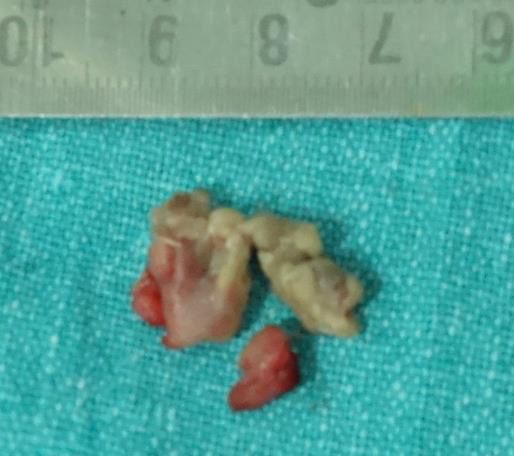
Sigdel et al. Management of Traumatic Herniation of Buccal Fat Pad in a 22-month-old Child: A Case Report
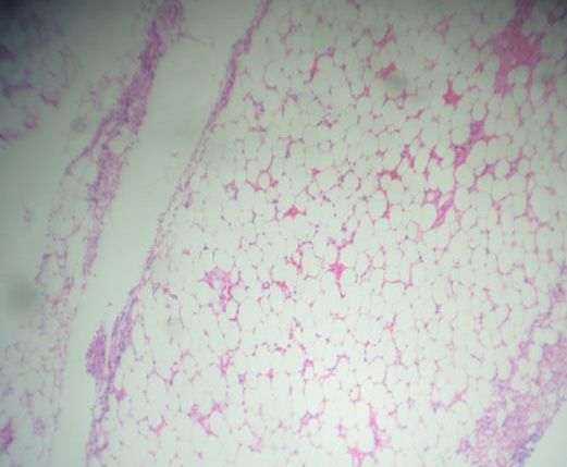
Figure 1. Intraoral herniated buccal Figure 2. Excision and primary closure Figure 3. Excised mass from
fat pad. (vicryl suture). buccal fat pad.
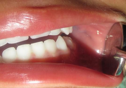
Figure 4. Satisfactory healing after one Figure 5. Four months follow up Figure 6. Photomicrograph showing
week. showing complete healing. numerous fat cells in the loose
fibrousstroma and inflammatory cells
(H&E; 4X).
histopathological examination (Figure 3). Antibiotics, anti- prone to injury due to their active and adventurous nature
inflammatory drugs and antiseptic mouthwashes were as compared to girls. But in our case, it was a girl child
prescribed and satisfactory healing was observed one with her left buccal mucosa involved.
week postoperatively (Figure 4). The patient came for
Depending upon the situation, herniation of buccal fat
follow up after four months and it showed normal healed
pad is managed by different approaches. If the herniated
area (Figure 5).
buccal fat pad mass is too large and necrosed, excision
Histopathologically it showed aggregates of numerous is treatment of choice. Alternative to excision, in cases
normal adipocytes with peripherally placed nuclei and where there is early evaluation and the protruded mass is
central vacuolization in a fibrous connective tissue along small with minimal inflammatory changes, repositioning
with numerous endothelial cells lined blood vessels and of the herniated mass to its anatomic position followed by
dispersed inflammatory cells, predominantly lymphocytes primary closure can be done. Some cases reported that
(Figure 6). the approximate period between the injury and the first
visit was less than four hours.8,9
DISCUSSION
Another better option for management could be the
Intraoral buccal fat pad herniation is seen frequently in
partial conservative excision, as an en-masse removal of
young children. This is due to children’s propensity to
the buccal fat pad may reduce cheek fullness and change
place every reachable object (sharp, pointed sticks, toys,
facial contour.6 Wolford et al.10 surgically excised the
lead pencil, comb, toothbrushes etc.) in their mouth
lesion under general anesthesia in an 8-month-old girl.
making them extremely vulnerable to oral injuries. An
However in our case, we opted for the partial conservative
interesting characteristic of this lesion is that the size of the
excision which was carried out under local anesthesia.
extruded mass is greater than the site of injury.5 According
to a report by Abdulai et al.,7 male children are more Close proximity of buccal fat pad to the parotid gland
18 Journal of Nepalese Association of Pediatric Dentistry : Vol. 1, No. 1, Jan-Dec, 2020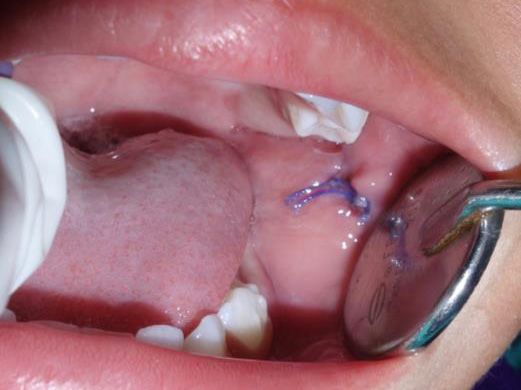
Sigdel et al. Management of Traumatic Herniation of Buccal Fat Pad in a 22-month-old Child: A Case Report

and facial nerve demands for precise and careful wound                                    confirm the diagnosis are history of trauma, site of injury,
management during treatment. Likewise, exploration for                                    fatty appearance of lesion and histopathology.
foreign body and proper irrigation is of utmost importance
                                                                                          CONCLUSIONS
before closing the wound. The situation can be complicated
by factors like occlusal trauma, inflammation, infection,                                 Traumatic herniation of buccal fat pad though rare,
salivary contamination, and necrosis of the tissue.6 Most                                 presents with a challenging scenario to manage. In the
of the cases reported had occurred at occlusal level near                                 present case, the herniated buccal fat pad did not directly
parotid papilla where prevention of damage to the parotid                                 involve any of the vital structures. The excision and
duct is very important.9 Timely management of the                                         repositioning back of the healthy tissues with primary
lacerated wound with proper oral hygiene maintenance                                      closure should be taken with care to avoid injury to vital
gives satisfactory healing in terms of both physical and                                  structures and asymmetry or facial dysmorphosim.
psychological aspects.
                                                                                          Conflict of Interest: None
The differential diagnosis of buccal fat pad herniation
includes tumors like lipoma; salivary gland neoplasm,                                                                            JNAPD
hemangioma, and traumatic fibroma.3 Features that

 REFERENCES
 1.    Zhang HM, Yan YP, Qi KM, Wang JQ, Liu ZF. Anatomical structure of the buccal fat pad and its clinical adaptations. Plastic and Reconstructive Surgery. 2002
       Jun;109(7): 2509-18. [Pub Med | DOI]
 2.    Singhal M, Sagar S. Post Traumatic Buccal Fat Pad Injury in a Child: A missed entity in ER. Oman Medical Journal. 2010 Jul;25(3):e002. [PubMed | DOI]
 3.    Zipfel TE, Street DF, Gibson WS, Wood WE. Traumatic herniation of the buccal fat pad: a report of two cases and a review of the literature. Int
       J Pediatr Otorhinolaryngol. 1996 Dec 20;38(2):175-9. [PubMed | DOI]
 4.    Baumann A, Ewers R. Application of the buccal fat pad in oral reconstruction. J Oral Maxillofac Surg. 2000 Apr;58(4):389-92; discussion 392-3. [PubMed | DOI]
 5.    Gadipelly S, Sudheer MV, Neshangi S, Harsha G, Reddy V. Traumatic herniation of buccal fat pad in 1 year old child: case report and review of literature.
       J Maxillofac Oral Surg. 2015 Mar;14(Suppl 1):435-7. [PubMed | DOI]
 6.    Patil R, Singh S, Subba Reddy VV. Herniation of the buccal fat pad into the oral cavity: a case report. J Indian Soc Pedod Prev Dent. 2003 Dec;21(4):152-4. [PubMed]
 7.    Abdulai AE, Avogo D. Traumatic herniation of the buccal fat pad: A case report. Ghana Medical Journal. 2004 Sep;38(3):120-2. [Full Text | DOI]
 8.    Fleming P. Traumatic herniation of buccal fat pad: a report of two cases. Br J Oral Maxillofac Surg. 1986 Aug;24(4):265-8. [PubMed | DOI]
 9.    Horie N, Shimoyama T, Kaneko T, Ide F. Traumatic herniation of the buccal fat pad. Pediatr Dent. 2001 May-Jun;23(3):249-52. [PubMed]
 10.   Wolford DG, Stapleford RG, Forte RA, Heath M. Traumatic herniation of the buccal fat pad: report of case. J Am Dent Assoc. 1981 Oct;103(4):593-4. [PubMed |DOI]

                                  Journal of Nepalese Association of Pediatric Dentistry : Vol. 1, No. 1, Jan-Dec, 2020                                                   19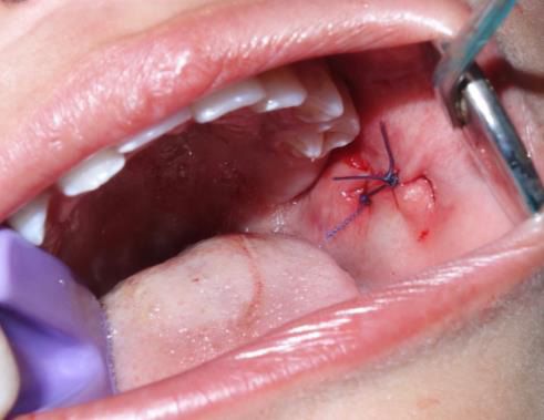
You can also read