Leiomyoma of the uterine round ligament: A case report
←
→
Page content transcription
If your browser does not render page correctly, please read the page content below
EXPERIMENTAL AND THERAPEUTIC MEDICINE 22: 1285, 2021

           Leiomyoma of the uterine round ligament: A case report
                    ALIKI TYMPA1, CHARALAMPOS GRIGORIADIS2, EMMANOUIL TERZAKIS2,
                             CHRISTINA GOUDELI2 and AIKATERINI MELEMENI1

       1
        First Department of Anaesthesiology, Aretaieion Hospital, University of Athens, Medical School, Athens 11528;
                 2
                   Department of Obstetrics and Gynaecology, Leto Maternity Hospital, Athens 11524, Greece

                                       Received January 28, 2021; Accepted July 6, 2021

                                                  DOI: 10.3892/etm.2021.10720

Abstract. Leiomyomas are common benign tumours that                is attached to the superior and lateral aspect of the uterus at
can arise in any anatomical structure containing smooth            the anatomical location of the uterine cornu. Arising from
muscle. Their localization in the uterine round ligament is        the cornu of the uterus, the round ligament is covered by
rare, although leiomyomas are the most frequent tumour             peritoneum over a length of 10‑12 cm, crosses the pelvis via
of this structure. Leiomyomas present as inguinal, labial          the deep inguinal ring, then traverses the inguinal canal and
or intra‑abdominal masses, and are often misdiagnosed as           finally enters the labia majora, where it terminates with its
hernias or enlarged lymph nodes. The aim of the present            fibres blending into the mons pubis (1).
study was to describe a rare case of a large intra‑abdominal           Leiomyomas are benign tumours that may arise in any
mesenchymal neoplasm arising from the right round liga‑            anatomical structure containing smooth muscle (2). However,
ment of the uterus. A 51‑year‑old asymptomatic female              in contrast to typical leiomyomas, which are a relatively
patient (gravida 3, para 3) presented herself for a routine        common gynaecological problem, leiomyomas of the uterine
gynaecological examination. A transvaginal ultrasound              round ligament are rare (2). Tumours of the round ligament are
examination revealed a solid heterogeneous mass with a             exceedingly rare, with roughly 300 cases previously published
maximum diameter of 9 cm localized at the right parame‑            in the literature (3). Although rare, leiomyomas are the most
trial space. Further preoperative evaluation using magnetic        frequent tumour of the uterine round ligament (3). Other
resonance imaging revealed that the mass contained solid           tumours encountered at the uterine round ligament according
and cystic components, which was suggestive of a mesen‑            to histological type include adenomyomas, mesothelial cysts,
chymal neoplasm with possible involvement of the right             endometriotic cysts, leiomyosarcomas and other, even rarer
ovary. Complete excision of the tumour and total abdominal         entities (4).
hysterectomy with bilateral salpingo‑oophorectomy was                  Leiomyomas of the uterine round ligament may appear
performed via laparotomy under general anaesthesia. The            in multiple different anatomical locations at any point along
intraoperative findings, frozen section biopsies and final         the length of this structure, and are commonly classified as
histological examination of the tumour established the             intra‑ or extra‑abdominal (inguinal or labial). Intra‑abdominal
diagnosis of an intra‑abdominal myoma of the right uterine         leiomyomas of the uterine round ligament typically remain
round ligament. The majority of abdominal round ligament           asymptomatic. On pelvic and ultrasound examination, the
myomas are initially asymptomatic. The role of synchro‑            differential diagnosis of leiomyomas from pedunculated
nous imaging examinations, such as ultrasonography and             subserosal myomas or solid ovarian neoplasms is difficult (4).
magnetic resonance imaging, in the diagnosis of these              Even following computed tomography and magnetic reso‑
lesions is crucial.                                                nance imaging examinations, the lack of specific findings, as
                                                                   leiomyomas appear as encapsulated heterogeneous tumours,
Introduction                                                       hinders final diagnosis prior to surgical intervention and histo‑
                                                                   logical examination (5). On the other hand, extra‑abdominal
The uterine round ligament is a round, rope‑like band of           leiomyomas are frequently detected by the patient as a mass
fibromuscular connective tissue (1). One side of the ligament      lesion with synchronous symptoms, including pain, and
                                                                   differential diagnosis from hernias or enlarged lymph nodes
                                                                   is necessary (4,6).
                                                                       In the present study, a rare case of a large leiomyoma
Correspondence to: Dr Charalampos Grigoriadis, Department          of the right uterine round ligament detected during routine
of Obstetrics and Gynaecology, Leto        Maternity   Hospital,   gynaecological examination is described, in line with the
7‑13 Mouson Street, Athens 11524, Greece                           Surgical CAse REport (SCARE) criteria (7). The aim was
E‑mail: xarisgrigoriadis@yahoo.gr                                  to underline the role of routine gynaecological examination
                                                                   and the importance of complete preoperative diagnostic
Key words: leiomyoma, uterus, round ligament, ultrasonography      evaluation in cases of large intra‑abdominal lesions to
                                                                   plan the appropriate surgical approach and ensure optimal
                                                                   management.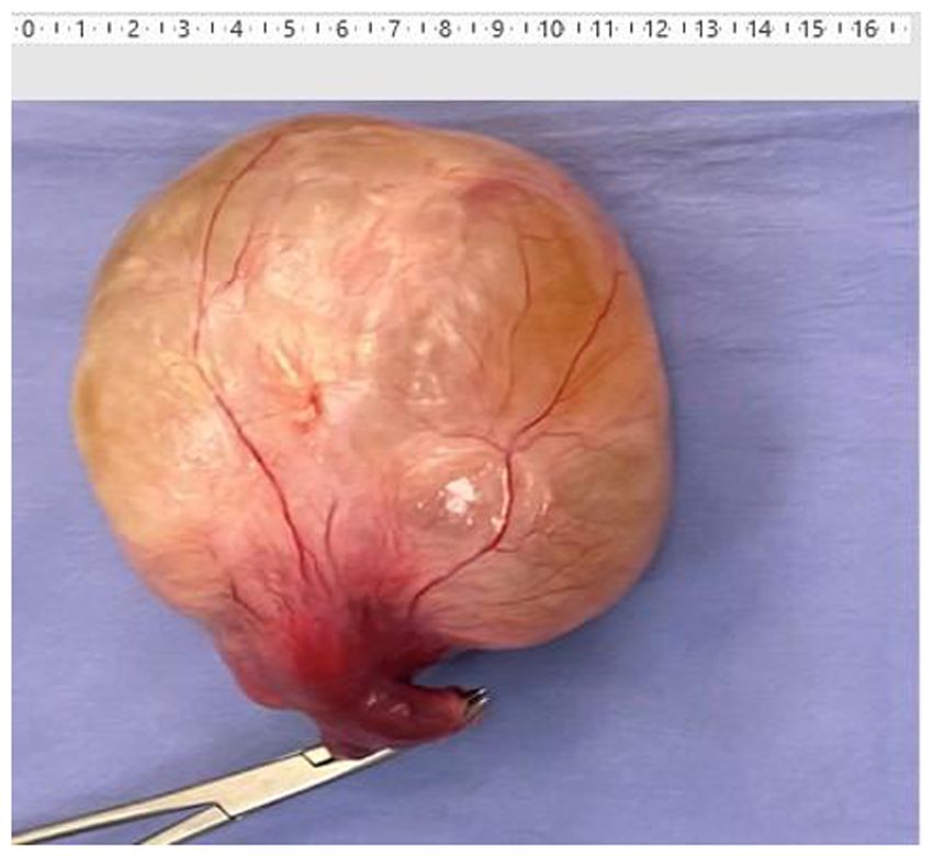
2 TYMPA et al: LARGE INTRA-ABDOMINAL LEIOMYOMA ARISING FROM THE UTERINE ROUND LIGAMENT
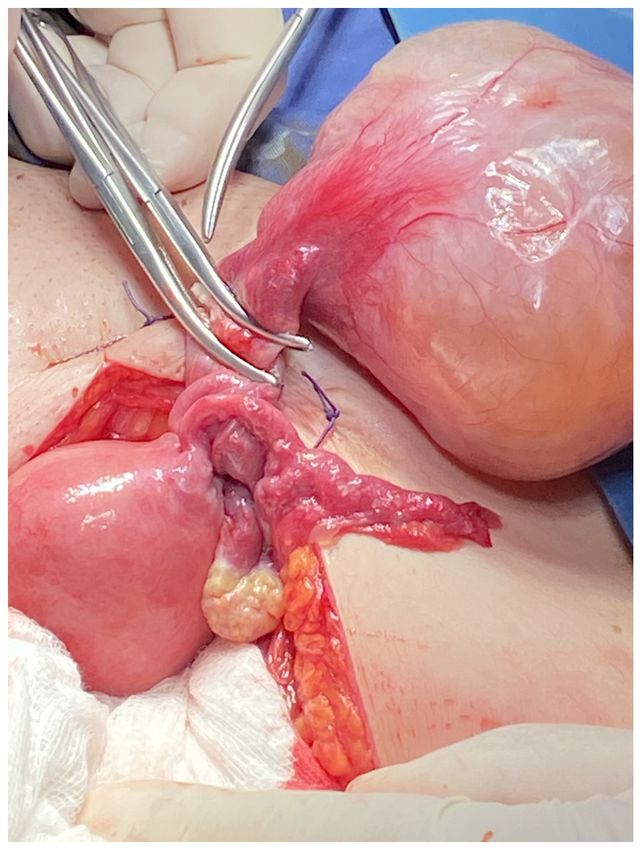
Figure 1. Transvaginal ultrasonographic appearance of a large solid tumour Figure 2. Surgical excision of the tumour following dissection from the right
of the right parametrium attached to the uterus (maximum tumour diameter, uterine round ligament.
9.2‑cm; thickness of the endometrium, 8.5 mm).
of necrosis, with morphological characteristics suggestive
Case report of a benign leiomyoma of the uterine round ligament. Total
abdominal hysterectomy and bilateral salpingo‑oophorectomy
A 51‑year‑old asymptomatic female patient (gravida 3, para 3) were subsequently performed.
presented herself on October 2020 for a routine gynaecolog‑ Histological examination revealed a benign leiomyoma
ical examination. The patient had obstetrical history of three with a maximum diameter of 10.5 cm, with signs of fibrosis,
full‑term vaginal deliveries and no previous surgical interven‑ hyalinosis and oedema. On immunohistological examination,
tions, while her medical history was unremarkable. the neoplastic cells were positive for desmin and smooth muscle
A transvaginal ultrasound examination revealed a solid actin, as expected. High density of mononuclear inflammatory
heterogeneous mass localized at the right parametrial space cells, mainly small lymphocytes with perivascular location,
with a maximum diameter of 9 cm, which was sugges‑ were detected. Among them, rare eosinophil leukocytes and
tive of a large pelvic lesion with possible involvement of mastocytes were recognised (data not shown). There were
the right ovary (Fig. 1). Routine blood investigations and no malignant findings from the uterus, cervix oradnexae.
tumour marker levels were within the normal range. Further The postoperative period was uneventful and the patient was
preoperative evaluation using magnetic resonance imaging discharged on postoperative day 2 without complications.
revealed that the mass (maximum diameter, 9‑cm) contained No recurrence was identified until her last follow‑up visit on
solid as well as cystic components, which was suggestive June 2021.
of a mesenchymal neoplasm, possibly originating from the
right ovary. There were no enlarged pelvic lymph nodes or Discussion
ascites.
After obtaining informed consent from the patient, Leiomyomas of the uterine round ligament are rare, and
a laparotomy was performed at Leto Maternity Hospital may present with multiple clinical manifestations according
(Athens, Greece) on November 2020, under general anaes‑ to the location along the anatomical course of the ligament.
thesia via a midline vertical subumbilical incision. A large Symptoms associated with leiomyomas largely depend upon
mobile solid mass arising from the right uterine round liga‑ location, size and rate of growth (4). In the vast majority
ment was detected (Fig. 2). Complete excision of the lesion of cases, intra‑abdominal leiomyomas of the round liga‑
was performed (Fig. 3). The frozen section biopsy revealed ment remain asymptomatic, and diagnosis is often made
a mesenchymal neoplasm without mitotic activity or signs incidentally during routine ultrasound examination or surgical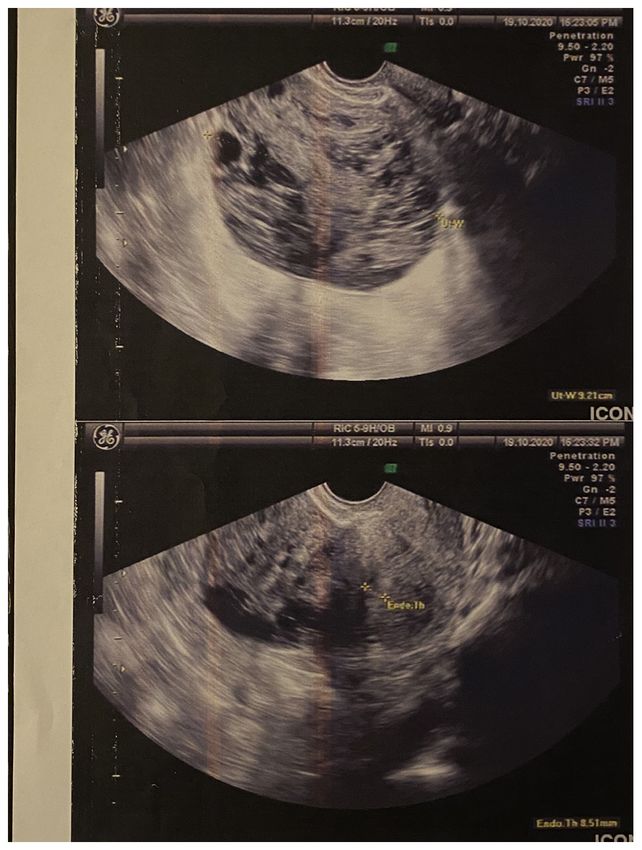
EXPERIMENTAL AND THERAPEUTIC MEDICINE 22: 1285, 2021                                                  3

                           Figure 3. Surgical specimen of neoplasm arising from the right round ligament of the uterus.

intervention for other reasons (3,4). These literature data                    Leiomyomas of the uterine round ligament are primarily
agree with the history of the patient in the present case, as              solitary and unilateral, although they have also been reported
she was asymptomatic and the tumour was detected on routine                to be multiple and bilateral (4). A number of studies have
gynaecological examination. The patient's last transvaginal                reported that leiomyomas appear more frequently on the
ultrasound gynaecological examination had been performed                   right compared with the left uterine round ligament (4,8,9).
4 years earlier, without signs of pathology at the right para‑             Although the aforementioned studies support this predilection
metrium, which suggested that the solid heterogeneous mass                 for the right round ligament, which was also the case in our
identified in the present study was a pelvic tumour with a                 patient, there appears to be no medical explanation for this
high rate of growth. Surgical intervention with oncological                observation.
parameters was recommended as a safe therapeutic strategy                      Surgical intervention is currently considered as the most
for a tumour with a potentially aggressive biological behav‑               appropriate therapeutic strategy in cases of large pelvic tumours
iour. This was the reason for which laparotomy was suggested               with uncertain differential diagnosis. Complete excision of the
instead of laparoscopic excision of the tumour in order to                 lesions is necessary in cases of uterine leiomyomas, without
avoid morcelation. In addition, the tumour was sent for frozen             the requirement for hysterectomy in women of reproductive
section biopsy so as to have a first histological view and decide          age (2‑4). The patient presented herein was a perimenopausal
if further pelvic/paraaortic lymphadenectomy was necessary                 51‑year‑old woman, and the total abdominal hysterectomy
or not.                                                                    with bilateral salpingo‑oophorectomy that followed the exci‑
    Preoperative investigation via computed tomography or                  sion of the leiomyoma of the right uterine round ligament was
magnetic resonance imaging often fails to distinguish the origin           performed as a preventive measure.
of solid pelvic tumours. Leiomyomas appear as encapsulated                     Routine gynaecological examination is necessary to
heterogeneous tumours on spontaneous contrast‑enhanced                     prevent women from experiencing life‑threatening patho‑
computed tomography and post‑gadolinium contrast magnetic                  logical conditions that affect the reproductive system. The
resonance imaging (5). The definitive differential diagnosis               vast majority of patients may be asymptomatic, as abdominal
from subserosal myomas or solid ovarian neoplasms often                    tumours arising from the ovaries or the uterus may not give
requires surgical intervention. In the present case, magnetic              rise to symptoms, even when they reach a large size. Surgical
resonance imaging was unable to identify the origin of the                 intervention remains the optimal therapeutic strategy (3,4),
pelvic mass, and final diagnosis was made intraoperatively.                following appropriate preoperative evaluation, as a means of
4 TYMPA et al: LARGE INTRA-ABDOMINAL LEIOMYOMA ARISING FROM THE UTERINE ROUND LIGAMENT
definitive diagnosis, as well as curative treatment in cases of Competing interests
large intra‑abdominal tumours with a challenging differential
diagnosis. The authors declare that they have no competing interests.
Acknowledgements References
Not applicable. 1. Chaudhry SR and Chaudhry K: Anatomy, abdomen and pelvis,
uterus round ligament. In: StatPearls [Internet]. StatPearls
Publishing, Treasure Island, FL, 2021. https://www.ncbi.nlm.nih.
Funding gov/books/NBK499970/. Accessed July 26, 2021.
2. Kirkham JC, Nero CJ, Tambouret RH and Yoon SS: Leiomyoma
No funding was received. and leiomyosarcoma arising from the round ligament of the
uterus. J Am Coll Surg 207: 452, 2008.
3. Klingbeil KD, Polcari AM, Azab B and Franceschi D: Large,
Availability of data and materials extra‑abdominal leiomyoma of the round ligament with carneous
degeneration. BMJ Case Rep 2017: bcr2017222454, 2017.
4. Breen JL and Neubecker RD: Tumors of the round ligament: A
The datasets used and/or analyzed during the current study review of the literature and report of 25 cases. Obstet Gynecol 19:
are available from the corresponding author on reasonable 771‑780, 1962.
request. 5. Michel P and Viola D: Abdomino‑pelvic leiomyoma of the round
ligament: Contribution of computed tomography and magnetic
resonance imaging. J Gynecol Obstet Biol Reprod (Paris) 32:
Authors' contributions 571‑574, 2003 (In French).
6. Christodoulou IM, Angelopoulos A, Siaperas P, Ioannidis A,
Skarpas A, Tellos A, Velimezis G and Karanikas I: Leiomyoma
AT was involved in the study conception and design, data of the round ligament of the uterus mimicking inguinal hernia.
collection and analysis and the writing of the manuscript. CG Case Rep Surg 2018: 6702494, 2018.
participated in the writing of the manuscript, data collection 7. Agha RA, Borrelli MR, Farwana R, Koshy K, Fowler A and
Orgill DP; SCARE Group: The SCARE 2018 statement:
and data analysis. ET was involved in the study design and Updating consensus Surgical CAse REport (SCARE) guidelines.
data analysis. CG was involved in data analysis. AM was Int J Surg 60: 132‑136, 2018.
involved in study conception and design. All authors have read 8. Emanuel R: Veber tumoren des ligamentum rotundum uteri.
Ztschr f Geburtsh u Gynak Stuttgart 49: 383, 1903.
and approved the final manuscript. CG, ET and CG confirm 9. Taussig FG: Sarcoma of the round ligament of the uterus. Surg
the authenticity of the raw data. Gynec Obst 19: 218, 1914.
Ethics approval and consent to participate
This work is licensed under a Creative Commons
The present study was approved by the Ethics Committee of Attribution-NonCommercial-NoDerivatives 4.0
Leto Maternity Hospital (approval no. 38/2021). International (CC BY-NC-ND 4.0) License.
Patient consent for publication
The patient provided written informed consent for the publica‑
tion of the case details and any associated images.You can also read