Case Report: Protein-Losing Enteropathy in Association With Tuberculosis-Related Constrictive Pericarditis
←
→
Page content transcription
If your browser does not render page correctly, please read the page content below
CASE REPORT
                                                                                                                                                     published: 30 May 2022
                                                                                                                                             doi: 10.3389/fped.2022.875032

                                                Case Report: Protein-Losing
                                                Enteropathy in Association With
                                                Tuberculosis-Related Constrictive
                                                Pericarditis
                                                Yue Xi 1 , Zhi Chen 1 , Kun Hao 2 and Xiaorong Liu 1*
                                                1
                                                 Department of Nephrology, Beijing Children’s Hospital, Capital Medical University, National Center for Children’s Health,
                                                Beijing, China, 2 Department of Lymphatic Surgery, Beijing Shijitan Hospital, Capital Medical University, Beijing, China

                                                Protein-losing enteropathy (PLE) is a clinical disorder in which an excessive amount of
                                                serum protein is lost into the gastrointestinal tract, resulting in hypoproteinemia and
                           Edited by:           malnutrition. PLE is associated with a wide range of gastrointestinal disorders and the
                       Andrew S. Day,
     University of Otago, New Zealand
                                                rare complication of constrictive pericarditis. We report a case in which pericardiectomy
                       Reviewed by:
                                                achieved marked improvement of extremely severe hypoalbuminemia caused by PLE
                Hisayoshi Kawahara,             associated with tuberculosis-related constrictive pericarditis. The formation of diarrhea
          Naramachi Hospital, Japan             and edema was aggravated by PLE, resulting in hypoalbuminemia. Cardiac computed
                         Ashish Garg,
          Washington State University           tomography showed a calcified pericardium. Echocardiography showed decreased
             Tri-Cities, United States          cardiac function underlying PLE. Functional imaging with technetium-99m serum albumin
                   *Correspondence:             identified the region of protein leakage as the intestine. After pericardiectomy, the diarrhea
                          Xiaorong Liu
                     lxrbch@sina.com
                                                ceased completely. Serum albumin concentrations were increased (3.3–3.7 g/dL), which
                                                indicated resolution of the PLE.
                    Specialty section:          Keywords: protein-losing enteropathy, hypoalbuminemia, intestinal lymphangiectasia, hematuria, constrictive
          This article was submitted to         pericarditis
           Pediatric Gastroenterology,
             Hepatology and Nutrition,
                a section of the journal
                  Frontiers in Pediatrics
                                                CASE REPORT
         Received: 13 February 2022             A 14-year-old boy was admitted to our hospital because of diarrhea, hypoalbuminemia, and gross
            Accepted: 04 May 2022               hematuria. He had been well until 5 years earlier, when he developed edema in the eyelid and
            Published: 30 May 2022
                                                lower extremities. At that time, he had a serum albumin concentration of 2.63 g/dL and 24-h
                               Citation:        urinary protein quantitation of 4,400 mg. Stool cultures were sterile, and a stool examination
 Xi Y, Chen Z, Hao K and Liu X (2022)
                                                showed no ova or parasites. A tuberculin skin test (purified protein derivative, 5 TU) was strongly
           Case Report: Protein-Losing
       Enteropathy in Association With
                                                positive at the 72-h timepoint. The TSPOT.TB assay qualitative results were positive. A patchy
     Tuberculosis-Related Constrictive          shadow was present in a computed tomography (CT) scan of the chest. The clinical diagnosis was
                             Pericarditis.      pulmonary tuberculosis. The patient was treated with a combination of isoniazid, rifampin, and
             Front. Pediatr. 10:875032.         pyrazinamide for 6 months. However, his serum albumin concentration remained at 2.5–3.6 g/dL
       doi: 10.3389/fped.2022.875032            after the therapy.

Frontiers in Pediatrics | www.frontiersin.org                                          1                                              May 2022 | Volume 10 | Article 875032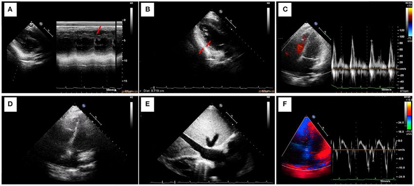
Xi et al. Case Report: PLE Associated With Constrictive Pericarditis
The sediment contained 100–200 red cells/high-powered
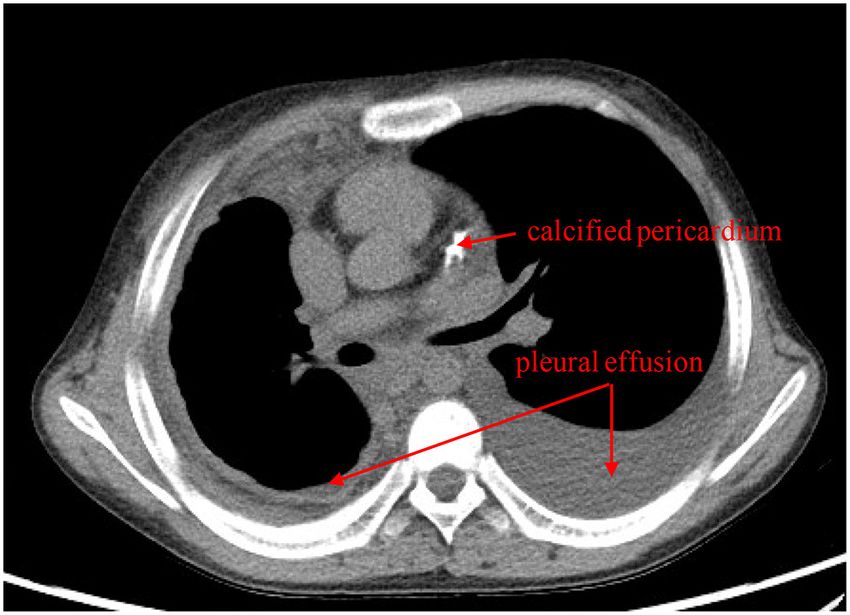
field. The patient had pulmonary tuberculosis. Cardiac CT
showed a calcified pericardium (Figure 1). Echocardiography
showed decreased cardiac function underlying protein-
losing enteropathy (PLE; Figure 2). The inferior vena cava
was widened. Ultrasonographic examination findings of
the urinary tract were normal. Functional imaging with
technetium-99m serum albumin identified the region of
protein leakage as the intestine (Figure 3). A biopsy of
the liver showed fibrotic changes. On the basis of these
findings, the patient was diagnosed with tuberculosis-related
constrictive pericarditis, which was considered to have caused
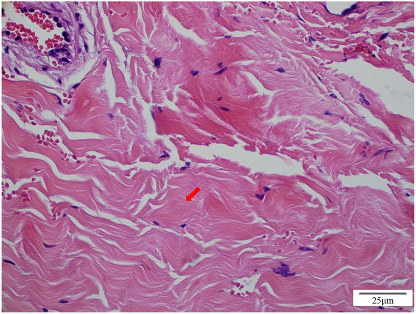
the PLE. Pericardiectomy was then performed. A pathological
examination of the pericardium showed hyaline degeneration
(Figure 4).
The diarrhea ceased completely soon after the
pericardiectomy. The patient’s cardiac condition then improved.
FIGURE 1 | Cardiac computed tomography shows bilateral pleural effusion
and a calcified pericardium (red arrows).
During a 2-year follow-up, the patient reported no symptoms,
edema of the extremities, or pleural effusion. His serum albumin
concentrations were increased (3.3–3.7 g/dL), which indicated
resolution of the PLE (Figure 5).
Two years before the present admission, he developed
diarrhea with watery stools, but had no fever or vomiting. A DISCUSSION
physical examination showed hepatic and splenic enlargement.
Laboratory tests showed that his serum albumin concentration PLE is a rare, but well-known, complication of constrictive
was low at 1.75 g/dL. The qualitative results of the TSPOT.TB pericarditis (1). Considerable intestinal loss of protein leads
assay were positive. A dense consolidation was present in the to hypoalbuminemia, which aggravates the extravascular fluid
right lobe with enlarged mediastinal lymph nodes, ascites, and overload. The exact pathophysiological relationship between
pleural and pericardial effusions on chest CT. Cardiac CT showed constrictive pericarditis and PLE remains obscure. In the present
a calcified cardiac base and pericardium. Echocardiography case, pulmonary tuberculosis led to constrictive pericarditis. A
showed moderate right ventricular and auricular dilatation with high venous pressure has been hypothesized to increase the
pulmonary arterial hypertension. The clinical diagnoses were hydrostatic pressure in the thoracic duct and presumably leads
pulmonary tuberculosis, hypoproteinemia, and diarrhea. The to diminished lymphatic drainage in the intestine. Notably,
patient was treated with a combination of isoniazid, rifampin, however, most patients with elevated venous pressure do not
ethambutol, and pyrazinamide for 8 months. Albumin infusions develop PLE (2). Most reports describe the resolution of PLE
were also administered. However, the albumin infusions had after pericardiectomy (1, 3). However, more research is required
only short-term effects and could be used only as a bridging to determine the relationship between constrictive pericarditis
intervention. Serum albumin concentrations remained at 1.5–2.5 and PLE.
g/dL. The diarrhea was still present. Previous case reports have described postoperative
Four months before the present admission, the patient improvements in the serum albumin concentration (4).
visited a local hospital because of hematuria. Persistent However, the decision to operate requires a risk/benefit analysis
splenomegaly and hepatomegaly were found. The serum albumin regarding the expected outcome of pericardiectomy in the
concentration was 2.17 g/dL. Urinalysis showed a protein presence of hypoalbuminemia. This analysis is required because
concentration of 220 mg/24 h and a red cell count of 8,634/µL. hypoalbuminemia has also been identified as an independent
Cardiac CT showed a calcified pericardium. Chest CT showed predictor of mortality after pericardiectomy (5). Although
right-lobe pneumonia with pleural effusion. The patient began our patient had extremely severe hypoalbuminemia (1.75
treatment with captopril (an angiotensin-converting enzyme g/dL) compared with previously reported cases, his serum
inhibitor) and dipyridamole. The diarrhea persisted. albumin concentration increased to almost the normal range
On admission to our hospital, the patient had a normal after pericardiectomy. Our results and previous data suggest
temperature, pulse rate, respiratory rate, and blood pressure. that patients with constrictive pericarditis can be successfully
A physical examination showed eyelid edema and jugular treated with pericardiectomy, even if PLE has caused extremely
vein engorgement. The lungs were clear. The heart sounds severe hypoalbuminemia.
were slightly weak without murmurs. Palpable splenomegaly Besides the loss of serum protein into the gastrointestinal
and hepatomegaly were found. The urine was positive for tract, other mechanisms that induce hypoalbuminemia should
protein (+), with a protein concentration of 160 mg/24 h. be considered. These mechanisms include hemodilution, liver
Frontiers in Pediatrics | www.frontiersin.org 2 May 2022 | Volume 10 | Article 875032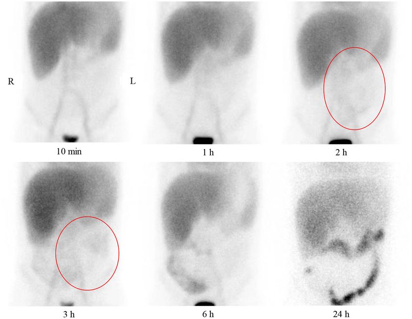
Xi et al.                                                                                                          Case Report: PLE Associated With Constrictive Pericarditis

  FIGURE 2 | Ultrasonography shows decreased cardiac function underlying protein-losing enteropathy. (A) M-mode ultrasonography shows thickening of the posterior
  pericardium of the posterior wall of the left ventricle and signs of a septal notch. The left ventricular ejection fraction is 77%. (B) The posterior pericardium of the
  posterior wall of the left ventricle is thickened (thickness, 7 mm). (C) Pulse Doppler shows limited ventricular diastolic activity. The high peak E and low peak A result in
  an E/A of >2. (D) A four-chamber view of the heart shows marked thickening of the pericardium and atrial enlargement. (E) The inferior vena cava is greatly widened
  (width, 26 mm). (F) Myocardial relaxation is normal.

  FIGURE 3 | Anterior plane abdominal scintigraphy at 10 min and 1, 2, 3, 6, and 24 h after intravenous administration of technetium-99m-labeled human serum
  albumin. Tracer accumulation in large vessels, heart blood pool, liver, and kidney is present at 10 min and 1 h. Tracer accumulation in the abdomen is not present at
  10 min and 1 h. Tracer accumulation in the lower abdomen is vaguely observed at 2 and 3 h (red circles). A large amount of tracer accumulation is present in the right
  lower abdomen at 6 h, and the radionuclide shows the bowel. Clear colonic tracer accumulation is present at 24 h.

Frontiers in Pediatrics | www.frontiersin.org                                          3                                                May 2022 | Volume 10 | Article 875032
Xi et al.                                                                                                       Case Report: PLE Associated With Constrictive Pericarditis

                                                                                         dysfunction (liver cirrhosis), inflammation, malnutrition, and
                                                                                         cachexia resulting from heart failure or non-heart failure-related
                                                                                         illness. Pericardiectomy is effective in patients with constrictive
                                                                                         pericarditis complicated by severe hypoalbuminemia because
                                                                                         of PLE. However, the ultimate decision to operate should be
                                                                                         made cautiously by the whole healthcare team comprising
                                                                                         gastroenterologists, hepatologists, and cardiovascular surgeons.

                                                                                         DATA AVAILABILITY STATEMENT
                                                                                         The original contributions presented in the study are included
                                                                                         in the article/supplementary material, further inquiries can be
                                                                                         directed to the corresponding author.

                                                                                         AUTHOR CONTRIBUTIONS
                                                                                         YX and XL examined the patient, analyzed the laboratory and
  FIGURE 4 | Pathological examination of the pericardium shows hyaline                   histopathology data, and discussed the case with the relevant
  degeneration (red arrow).                                                              consulting physicians. YX was the major contributor to drafting
                                                                                         of the manuscript. ZC provided a supervising and editorial
                                                                                         role. KH provided the functional imaging with technetium-99m
                                                                                         serum albumin with its analysis. All authors read and approved
                                                                                         the final manuscript.

                                                                                         FUNDING
                                                                                         This work was supported by the Capital Health Research and
                                                                                         Development of Special Grant (No. 2016-2-2094), the Research
                                                                                         on the Application of Capital Clinical Characteristics Program
                                                                                         of Beijing Municipal Science and Technology Commission
                                                                                         (No. Z161100000516106), the Project of Beijing Science and
                                                                                         Technology Commission (No. D181100000118006), and the
  FIGURE 5 | Clinical course of the patient. Serum albumin concentrations are            Cultivation Fund of Beijing Children’s Hospital, Capital Medical
  improved after pericardiectomy.                                                        University (GPQN202007).

REFERENCES                                                                               Conflict of Interest: The authors declare that the research was conducted in the
                                                                                         absence of any commercial or financial relationships that could be construed as a
1. Mller C H, Globits S, Glogar D, Klepetko W, Knoflach P, Constrictive                  potential conflict of interest.
   pericarditis without typical haemodynamic changes as a cause of oedema
   formation due to protein-losing enteropathy. Eur Heart J. (1991) 12:1140–             Publisher’s Note: All claims expressed in this article are solely those of the authors
   3. doi: 10.1093/oxfordjournals.eurheartj.a059848                                      and do not necessarily represent those of their affiliated organizations, or those of
2. Güneri S, Nazli C, Kinay O, Mermut C, Hazan E. Chylous                                the publisher, the editors and the reviewers. Any product that may be evaluated in
   ascites due to constrictive pericarditis. Int J Cardiac Imag. (2000)
                                                                                         this article, or claim that may be made by its manufacturer, is not guaranteed or
   16:49. doi: 10.1023/a:1006379625554
                                                                                         endorsed by the publisher.
3. Nikolaidis N, Tziomalos K, Giouleme O, Gkisakis D, Kokkinomagoulou
   A, Karatzas N, et al. Protein-losing enteropathy as the principal
   manifestation of constrictive pericarditis. J General Intern Med. (2005)
   20:C5–7. doi: 10.1111/j.1525-1497.2005.0202.x                                         Copyright © 2022 Xi, Chen, Hao and Liu. This is an open-access article distributed
4. Meijers B, Schalla S, Eerens F, Van Suylen RJ, Broers B, Cheriex EM, et al.           under the terms of the Creative Commons Attribution License (CC BY). The
   Protein-losing enteropathy in association with constrictive pericarditis. Int J       use, distribution or reproduction in other forums is permitted, provided the
   Cardiovasc Imag. (2006) 22:389–92. doi: 10.1007/s10554-005-9067-2                     original author(s) and the copyright owner(s) are credited and that the original
5. Moriyama H, Kohno T, Nishiyama T, Hattori O, Maekawa Y, Yoshida K, et al.             publication in this journal is cited, in accordance with accepted academic practice.
   Constrictive pericarditis and protein-losing enteropathy. Circul Heart Fail.          No use, distribution or reproduction is permitted which does not comply with these
   (2016) 9:e003666. doi: 10.1161/CIRCHEARTFAILURE.116.003666                            terms.

Frontiers in Pediatrics | www.frontiersin.org                                        4                                                May 2022 | Volume 10 | Article 875032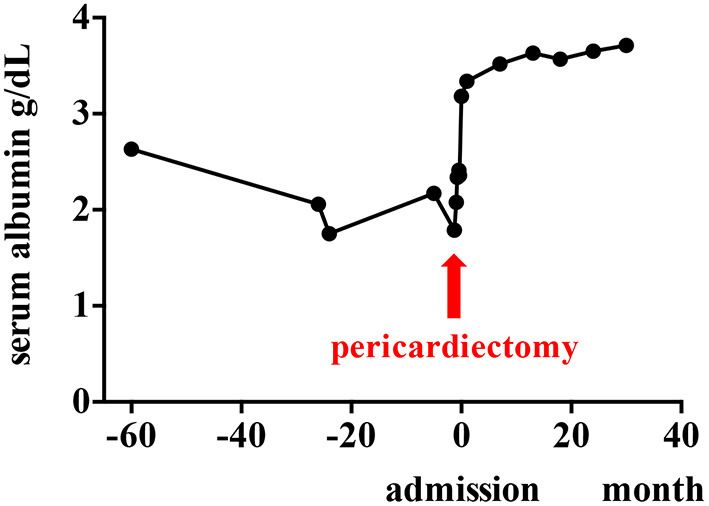
You can also read