Hip Adductor Muscle Abscess Descending From Septic Symphysitis
←
→
Page content transcription
If your browser does not render page correctly, please read the page content below
Open Access Case
Report DOI: 10.7759/cureus.21138
Hip Adductor Muscle Abscess Descending From
Septic Symphysitis
Review began 01/02/2022
Benjamin Kraler 1 , Eldaras Gotovski-Getman 1 , Henk Eijer 1
Review ended 01/08/2022
Published 01/11/2022 1. Orthopaedics and Traumatology, Emmental Hospital, Burgdorf, CHE
© Copyright 2022
Kraler et al. This is an open access article Corresponding author: Benjamin Kraler, b.kraler@protonmail.com
distributed under the terms of the Creative
Commons Attribution License CC-BY 4.0.,
which permits unrestricted use, distribution,
and reproduction in any medium, provided
the original author and source are credited. Abstract
Hip adductor muscle abscesses that descend from an infected symphysis pubis are rare but cause serious
morbidity. We present a case of a 73-year-old male patient with unilateral hip adductor muscle abscess that
descended from septic symphysitis caused by Staphylococcus aureus. Surgical debridement of the adductor
compartment could not clear the infection and secondary debridement of the symphysis was necessary to
eradicate S. aureus. Additionally, we review another four cases with similarities to our case comparing their
investigation, treatment, and outcome.
Categories: Infectious Disease, Orthopedics, Trauma
Keywords: symphysis pubis, symphysitis, staphylococcus aureus bacteremia, adductor muscle abscess, pubic
osteomyelitis, septic symphysitis
Introduction
Hip adductor muscle abscesses that descend from an infected symphysis are rare but cause serious illness in
the affected individual. In a comprehensive review of septic symphysitis including 100 cases, only one
patient was diagnosed with concomitant hip adductor muscle abscess [1]. Nonetheless, awareness of the
disease is important because the treatment has to be initiated early and address the adductor compartment
as well as the symphysis. Differential diagnoses include septic hip arthritis and the non-infectious condition
of osteitis pubis frequently seen in athletes [2].
We report a case of hip adductor muscle abscess secondary to septic symphysitis and present a review of four
other cases that were identified in PubMed (US national library of medicine) [3-6]. We discuss their
laboratory, radiology, microbiology, treatment, and outcome. Written informed consent was obtained for the
publication of this case report and accompanying images.
Case Presentation
A 73-year-old male patient with a history of sigma resection for colorectal cancer 16 years ago and diabetes
presented at the emergency department with acute right groin pain without previous trauma. Vital signs
showed a blood pressure of 138 over 65 mmHg, heart rate of 73 bpm, and body temperature of 37.2°C. Local
examination of the right adductors showed no swelling or rubor, but tenderness over the proximal adductor
compartment. Right hip flexion/extension was limited to 30/0/10 degrees and abduction was restricted to 10
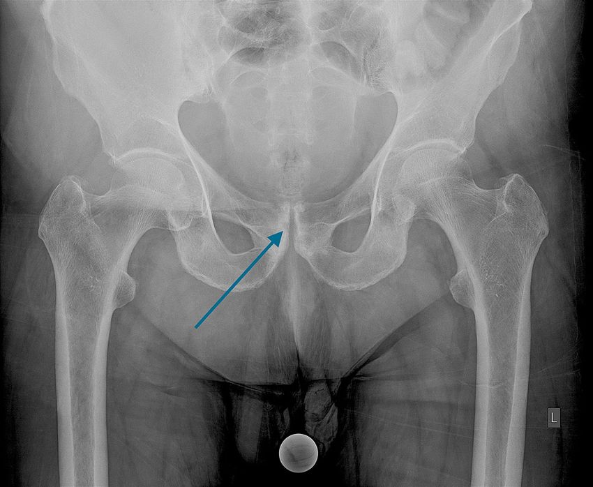
degrees. Pelvic X-ray revealed subtle erosive changes of the symphysis (Figure 1).
How to cite this article
Kraler B, Gotovski-Getman E, Eijer H (January 11, 2022) Hip Adductor Muscle Abscess Descending From Septic Symphysitis. Cureus 14(1):
e21138. DOI 10.7759/cureus.21138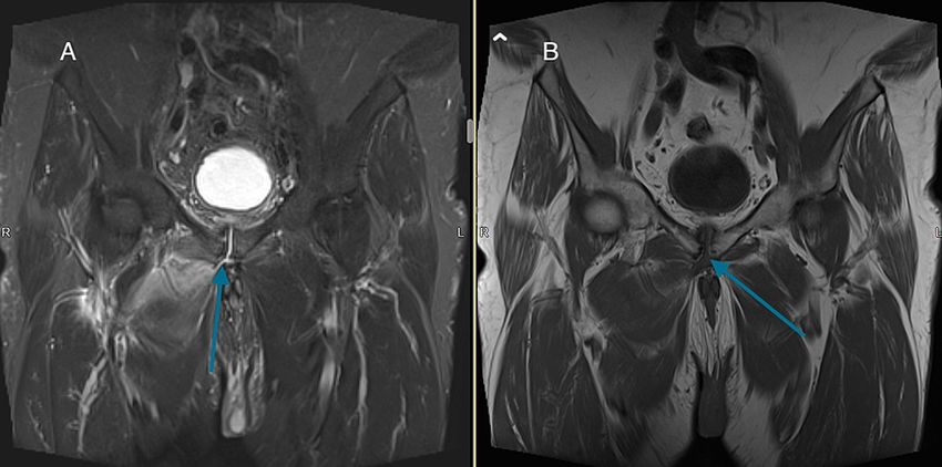
FIGURE 1: Anteroposterior radiograph of the pelvis at presentation with
                                                    subtle erosive changes of the symphysis (arrow).

                                                  The patient was afebrile, but C-reactive protein (CRP) was 149 mg/l with a normal white blood cell (WBC)
                                                  count. Adductor strain was presumed and he was discharged with analgesic treatment and crutches. Two
                                                  days later he came back with increasing adductor pain. Laboratory revealed a CRP of 298 mg/l, WBC count of
                                                  14.7 × 10 9/l, blood glucose levels of 8.46 mmol/l, and creatinine of 183 μmol/l. More laboratory results are
                                                  summarized in Table 1.

2022 Kraler et al. Cureus 14(1): e21138. DOI 10.7759/cureus.21138                                                                                                 2 of 7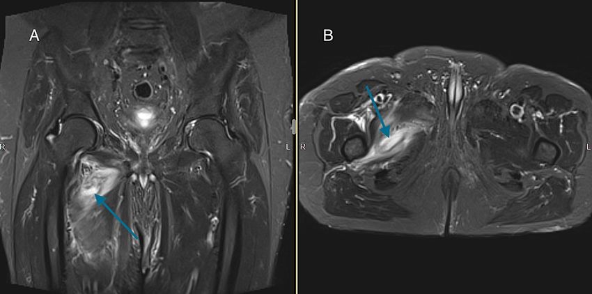
Laboratory value (reference range)                                                                   Patient's results

               ASAT (0-50 U/l)                                                                                      21

               ALAT (0-50 U/l)                                                                                      18

               Alkaline phosphatase (40-130 U/l)                                                                    78

               y-GT (
FIGURE 3: Coronal views: (A) turbo inversion recovery magnitude
(TIRM) and (B) T1-weighted image with fluid descending from the
symphysis to the right adductor compartment (arrows).
Open debridement of the adductors through a medial approach drained pus. Biopsies and blood cultures
identified Staphylococcus aureus (methicillin-sensitive). Intravenous antibiotic treatment was started with
amoxicillin-clavulanic acid 2200 mg followed one day later by intravenous flucloxacillin 2000 mg four times
a day. Blood cultures taken 3 and 5 days postoperatively remained positive, which prompted a follow-up
MRI that showed residual fluid collection within the right adductor compartment. Revision adductor
debridement and additional debridement of the symphysis through a Pfannenstiel incision were performed.
Biopsies identified S. aureus at the symphysis and adductors. Due to persisting wound drainage, a third look
surgery of the adductor compartment followed 2 weeks later and subcutaneous seroma was drained. Four
weeks after the first surgery, CRP was 23 mg/l, WBC count was normal, the wound was bland and the patient
was able to ambulate with crutches. Before leaving the hospital the visual analog scale for pain was zero at
rest and also when walking with crutches. He was discharged home with a 9-week course of oral clindamycin
600 mg three times daily. At four months follow-up there was no evidence of infection locally or systemically
with a normal CRP level. There was no pain over the symphysis or the right adductor compartment and the
patient used a cane but was able to walk without aid.
Discussion
We identified four cases of septic symphysitis with descending adductor muscle abscesses in PubMED that
were supported by MRI or CT imaging and had microbiologic confirmation (table 2). Including our case,
three out of five patients with adductor muscle abscesses and symphysitis had a surgically treated urologic
or abdominal malignancy. Possible other risk factors included diabetes, drug-induced immunosuppression
and preexisting osteitis pubis. Although osteitis pubis is an aseptic condition, some authors suggest that the
susceptibility to develop septic symphysitis is increased due to degenerative changes of the symphysis [2,7].
Complaints at admission include groin pain, pubic, thigh and/or gluteal pain as well as painful gait. Septic
hip arthritis can mimic similar symptoms and needs to be excluded. Fever was present in four and CRP was
elevated in all cases with documented CRP levels. Although CRP is unspecific, a normal CRP level makes hip
adductor muscle abscesses or septic symphysitis highly unlikely.
2022 Kraler et al. Cureus 14(1): e21138. DOI 10.7759/cureus.21138 4 of 7Laboratory
Age
(WBC in Imaging
Author (in Sex Risk factors Complaints Imaging findings
109 /l, CRP modality
years)
in mg/l)
Juvenile
Groin pain,
Alqahtani idiopathic WBC 19.1, Fluid collection in symphysis and bilateral
17 M painful gait, MRI
et al. [3] arthritis under CRP 232 adductor compartments
fever
methotrexate
Cardoso Pubic pain, Fluid collection in symphysis and right adductor
57 F osteitis pubis -- MRI
et al. [4] fever compartment
Diabetes,
radical Pubic pain, Symphyseal erosion, pubic bone enhancement,
Degheili WBC 11.6,
68 M prostatectomy painful gait, MRI and fluid collection in bilateral adductor
et al. [5] CRP 179
for prostate fever compartments
cancer
X-ray X-ray: unremarkable; CT: symphyseal erosion,
Transurethral Thigh, groin,
Trubiano pelvis, fluid collection in bilateral adductor compartments;
78 M resection for gluteal pain, CRP 83
et al. [6] CT, CT- CT-cystogram: cysto-symphyseal-adductor
prostate cancer afebrile
cystogram fistulas
Diabetes, sigma Groin and
X-ray
Current resection for pubic pain, WBC 14.7, X-ray: symphyseal erosion; MRI: fluid collection in
73 M pelvis,
case colorectal painful gait, CRP 298 symphysis and right adductor compartment
MRI
cancer fever
TABLE 2: Age, sex, risk factors, complaints, laboratory results, imaging modality, and imaging
findings in septic symphysitis with adductor muscle abscess
MRI with gadolinium contrast detected symphyseal and adductor fluid collections in four patients.
Interestingly, in one case CT was the imaging modality of choice and CT-cystogram could confirm fistulas
from the bladder to the symphysis and bilateral adductor compartments. However, MRI has shown the
highest sensitivity and specificity to highlight symphyseal fluid and pubic bone marrow edema in adjacent
osteomyelitis as well as adductor muscle abscesses [8]. Pelvic x-ray can delineate symphyseal erosive
changes, but it cannot help discern between early stage septic symphysitis and osteitis pubis since both can
show symphyseal erosions [2]. Microbiology was secured by adductor and/or symphyseal biopsies or blood
cultures and pathogens isolated are summarized in table 3. As one would expect, pathogens found in hip
adductor muscle abscesses resemble the spectrum of pathogens in septic symphysitis [1,4-6] with
Staphylococcus aureus being the most frequently identified pathogen.
2022 Kraler et al. Cureus 14(1): e21138. DOI 10.7759/cureus.21138 5 of 7Follow-
Author Microbiology Pathogen Management Antimicrobial agent (duration in weeks) up Outcome
(months)
No
infectious
Alqahtani Streptococcus Open debridement
Biopsy adductors ceftriaxone (6) 6 sequelae
et al. [3] group A adductors
at follow-
up
No
infectious
Cardoso Staphylococcus Percutaneous adductor
Blood cultures vancomycin (8) 36 sequelae
et al. [4] aureus drainage
at follow-
up
No
Open debridement
Aspiration Vancomycin + meropenem (1) , infectious
Degheili Enterococcus symphysis,
adductors CT piperacillin/tazobactam (2) + vancomycin 6 sequelae
et al. [5] spp. percutaneous adductor
guided (1.5), rifampicin + ciprofloxacin (8) at follow-
drainage
up
No
Candida albicans Open debridement infectious
Trubiano Agent not specified, intravenous (6) and
Biopsy symphysis + Pseudomonas symphysis, 3 sequelae
et al. [6] oral (12)
aeruginosa cystoprostatectomy at follow-
up
No
Blood cultures, Open debridement Amoxicillin/clavulanic acid (0.14), infectious
Current Staphylococcus
biopsy adductors, adductors open flucloxacillin (3), amoxicillin/clavulanic acid 4 sequelae
case aureus
biopsy symphysis debridement symphysis (1), clindamycin (9) at follow-
up
TABLE 3: Microbiology, pathogen, management, antimicrobial treatment, follow-up, and outcome
in septic symphysitis with adductor muscle abscess
Management of hip adductor muscle abscesses included percutaneous adductor drainage and open
debridement of the adductor compartment in two cases, respectively. Primary open debridement of the
symphysis was performed in two cases; one was combined with percutaneous adductor drainage [5], the
other with cystoprostatectomy due to fistulas from the bladder to the symphysis and adductors [6]. In our
case, open drainage of the adductor muscle abscess and antibiotic therapy could not clear the infection and
symphyseal debridement revealed S. aureus confirming that the symphysis served as the source of infection.
In contrast to our experience, Cardoso et al. reported a case of a 57-year-old with symphysitis and an
adductor abscess caused by S. aureus that healed without surgically addressing the symphysis and by only
draining the adductor abscess [4]. However, this was an otherwise healthy individual. In contrast, our patient
had undergone sigma resection for colorectal cancer, although this was performed 16 years ago. Likewise, in
a 17-year-old with bilateral adductor abscesses following septic symphysitis, Streptococcus group A infection
was cleared by open debridement of both adductor compartments as a single procedure [3]. This patient
received ceftriaxone for 6 weeks and apart from juvenile idiopathic arthritis did not show other
comorbidities. This suggests that patients without comorbidities might be best treated by open or
percutaneous drainage of adductor muscle abscesses. In patients with medical comorbidities, especially
previous abdominal or urologic malignancy, draining the adductor muscle abscess but also debriding the
symphysis seems necessary to rapidly clear infection and prevent relapse. Despite different treatment
modalities, all patients were infection-free at the final follow-up (range: 3 months to 3 years).
Conclusions
Hip adductor muscle abscesses originating from septic symphysitis are a rare entity but contribute to
significant patient morbidity. Pelvic MRI with gadolinium contrast has the highest sensitivity and specificity
to diagnose not only adductor muscle abscesses but also septic symphysitis and fluid communicating
between the symphysis and adductor compartment. Microbiologic confirmation is best achieved by tissue
samples of the adductors and symphysis. If the patient’s status necessitates immediate empiric antibiotic
treatment, at least blood samples for culture need to be obtained before antibiotic therapy is started. S.
aureus is the most frequently isolated pathogen in septic symphysitis with adductor muscle abscesses but
2022 Kraler et al. Cureus 14(1): e21138. DOI 10.7759/cureus.21138 6 of 7polymicrobial infections can occur. Open or percutaneous adductor drainage in combination with tailored
antibiotic treatment seems adequate care in otherwise healthy individuals. However, in patients with
previous abdominal or urologic malignancies draining the adductor compartment alone might not be
sufficient and debridement of the symphysis is needed to clear the source of infection. Serial CRP and repeat
blood cultures help to monitor the success of surgical and antibiotic treatment.
Additional Information
Disclosures
Human subjects: Consent was obtained or waived by all participants in this study. Conflicts of interest: In
compliance with the ICMJE uniform disclosure form, all authors declare the following: Payment/services
info: All authors have declared that no financial support was received from any organization for the
submitted work. Financial relationships: All authors have declared that they have no financial
relationships at present or within the previous three years with any organizations that might have an
interest in the submitted work. Other relationships: All authors have declared that there are no other
relationships or activities that could appear to have influenced the submitted work.
References
1. Ross JJ, Hu LT: Septic arthritis of the pubic symphysis: review of 100 cases . Medicine. 2003, 82:340-5.
10.1097/01.md.0000091180.93122.1c
2. Choi H, McCartney M, Best TM: Treatment of osteitis pubis and osteomyelitis of the pubic symphysis in
athletes: a systematic review. Br J Sports Med. 2011, 45:57-64. 10.1136/bjsm.2008.050989
3. Alqahtani SM, Jiang F, Barimani B, Gdalevitch M: Symphysis pubis osteomyelitis with bilateral adductor
muscles abscess. Case Rep Orthop. 2014, 2014:1-3. 10.1155/2014/982171
4. Cardoso L, Alves P, Santos F, Ross JJ: Septic arthritis of the pubic symphysis . BMJ Case Rep. 2017, 2017:1.
10.1136/bcr-2016-216784
5. Degheili JA, Mansour MM, Nasr RW: Symphysis pubis osteomyelitis: an uncommon complication after
robotic assisted radical prostatectomy-case description with literature review. Case Rep Urol. 2018, 2018:1-
5. 10.1155/2018/5648970
6. Trubiano JA, Yang N, Mahony AA: Bilateral thigh pain after treatment for prostate cancer . BMJ Case Rep.
2013, 2013:1. 10.1136/bcr-2013-008784
7. Smits FJ, Frima H, Schaeffeler C, Sommer C: Spontaneous septic arthritis of pubic symphysis in an elite
athlete. Case Rep Surg. 2016, 2016:1-4. 10.1155/2016/6384707
8. Sexton SJ, Lavien G, Said N, Eward W, Peterson AC, Gupta RT: Magnetic resonance imaging features of pubic
symphysis urinary fistula with pubic bone osteomyelitis in the treated prostate cancer patient. Abdom
Radiol. 2019, 44:1453-60. 10.1007/s00261-018-1827-2
2022 Kraler et al. Cureus 14(1): e21138. DOI 10.7759/cureus.21138 7 of 7You can also read