Individual Sensory Modality Dominance as an Influential Factor in the Prefrontal Neurofeedback Training for Spatial Processing: A Functional ...
←
→
Page content transcription
If your browser does not render page correctly, please read the page content below

ORIGINAL RESEARCH
                                                                                                                                          published: 10 February 2022
                                                                                                                                     doi: 10.3389/fnsys.2022.774475

                                           Individual Sensory Modality
                                           Dominance as an Influential Factor in
                                           the Prefrontal Neurofeedback
                                           Training for Spatial Processing:
                                           A Functional Near-Infrared
                                           Spectroscopy Study
                                           Takeshi Sakurada 1,2,3* , Mayuko Matsumoto 2,4 and Shin-ichiroh Yamamoto 4
                                           1
                                             Department of Robotics, College of Science and Engineering, Ritsumeikan University, Shiga, Japan, 2 Functional Brain
                                           Science Laboratory, Center for Development of Advanced Medical Technology, Jichi Medical University, Tochigi, Japan,
                                           3
                                             Department of Neurosurgery, Jichi Medical University, Tochigi, Japan, 4 Graduate School of Systems Engineering
                                           and Science, Shibaura Institute of Technology, Saitama, Japan

                                           Neurofeedback is a neuromodulation technique used to improve brain function by
                                           self-regulating brain activity. However, the efficacy of neurofeedback training varies
                                           widely between individuals, and some participants fail to self-regulate brain activity.
                                           To overcome intersubject variation in neurofeedback training efficacy, it is critical to
                       Edited by:          identify the factors that influence this type of neuromodulation. In this study, we
                   Tomohiko Takei,
         Tamagawa University, Japan
                                           considered that individual differences in cognitive ability may influence neurofeedback
                       Reviewed by:
                                           training efficacy and aimed to clarify the effect of individual working memory (WM)
                           Nan Liang,      abilities, as characterized by sensory modality dominance, on neurofeedback training
             Kyoto University, Japan       efficacy in healthy young adults. In particular, we focused on the abilities of individuals
                    Hiroyuki Hamada,
       The University of Tokyo, Japan      to retain internal (tactile or somatosensory) or external (visual) body information in
                 *Correspondence:          their WM. Forty participants performed functional near-infrared spectroscopy-based
                   Takeshi Sakurada        neurofeedback training aimed at producing efficient and lower-level activity in the
           sakurada@fc.ritsumei.ac.jp
                                           bilateral dorsolateral prefrontal cortex and frontopolar cortex. We carried out a
      Received: 12 September 2021          randomized, sham-controlled, double-blind study that compared WM ability before
        Accepted: 07 January 2022          and after neurofeedback training. Individual WM ability was quantified using a target
       Published: 10 February 2022
                                           searching task that required the participants to retain spatial information presented as
                              Citation:
      Sakurada T, Matsumoto M and
                                           vibrotactile or visual stimuli. Participants who received feedback information based on
        Yamamoto S (2022) Individual       their own prefrontal activity showed gradually decreasing activity in the right prefrontal
  Sensory Modality Dominance as an
                                           area during the neurofeedback training and demonstrated superior WM ability during
    Influential Factor in the Prefrontal
   Neurofeedback Training for Spatial      the target searching task with vibrotactile stimuli compared with the participants who
             Processing: A Functional      performed dummy neurofeedback training. In comparison, left prefrontal activity was not
  Near-Infrared Spectroscopy Study.
   Front. Syst. Neurosci. 16:774475.
                                           influenced by the neurofeedback training. Furthermore, the efficacy of neurofeedback
    doi: 10.3389/fnsys.2022.774475         training (i.e., lower right prefrontal activity and better searching task performance) was

Frontiers in Systems Neuroscience | www.frontiersin.org                           1                                        February 2022 | Volume 16 | Article 774475
Sakurada et al.                                                                                                   fNIRS Neurofeedback for Working Memory

                                           higher in participants who exhibited tactile dominance rather than visual dominance in
                                           their WM. These findings indicate that sensory modality dominance in WM may be an
                                           influential neurophysiological factor in determining the efficacy of neurofeedback training.
                                           These results may be useful in the development of neurofeedback training protocols
                                           tailored to individual needs.
                                           Keywords: neurofeedback, functional near-infrared spectroscopy, working memory, prefrontal cortex, individual
                                           differences, sensory modality

INTRODUCTION                                                                         Although various types of neurofeedback training have been
                                                                                 reported to be useful for brain functional training, the efficacy
Neurofeedback is a neuromodulation technique that aims to                        of neurofeedback training varies widely among individuals, and
self-regulate brain activity patterns to improve specific functions              a certain population obtains no benefit from neurofeedback
(Ehlis et al., 2018; Xiang et al., 2018; Jeunet et al., 2019). In healthy        training and fails to self-regulate brain activity patterns (Alkoby
individuals, theta electroencephalography (EEG) power increases                  et al., 2018; Diaz Hernandez et al., 2018; Kadosh and Staunton,
during neurofeedback training have been shown to assist in                       2019). A recent review pointed out that participants who failed
improving motor sequential learning (Rozengurt et al., 2016),                    to self-regulate during neurofeedback training represented about
and high sensorimotor rhythm power has been associated with                      50% of the population (Alkoby et al., 2018). For instance, a
better motor performance in a sporting task (Cheng et al., 2015).                neurofeedback training protocol that aimed to improve WM
Moreover, neurofeedback training has been noted as a useful                      ability by increasing upper alpha power indicated that three
protocol for improving cognitive functions such as attention                     of nine participants failed to control their alpha power after
(Wang and Hsieh, 2013), working memory (WM) (Escolano                            several training sessions (Escolano et al., 2011). Moreover, during
et al., 2011) and mental rotation (Hanslmayr et al., 2005). In                   neurofeedback training to modulate sensorimotor rhythm,
clinical fields, neuromodulation by neurofeedback has been used                  approximately half of the participants did not learn to regulate
successfully to alter brain activity in a wide range of disorders,               their own brain activities even after numerous training sessions
such as attention deficit hyperactivity disorder (Leins et al., 2007)            (Weber et al., 2011).
and Parkinson’s disease (Subramanian et al., 2011).                                  To overcome these individual differences in the efficacy
    Although EEG and fMRI are the imaging techniques most                        of neurofeedback training, we need to identify the predictor
commonly used for neurofeedback training, the use of functional                  (influential factor) of the neuromodulation. Although the factors
near-infrared spectroscopy (fNIRS) has increased over the                        influencing neurofeedback training are not fully understood,
last 10 years (Ehlis et al., 2018; Kohl et al., 2020). fNIRS                     the issue has been discussed recently, and several factors
recording has several advantages. For example, if the probes                     have been pointed out (Alkoby et al., 2018; Diaz Hernandez
are in close contact with the head, we can measure brain                         et al., 2018; Kadosh and Staunton, 2019). First, the effect
activity, regardless of posture or any slight body movement.                     of psychological factors such as mental strategy, motivation,
Furthermore, fNIRS is resistant to electrical noise, an especially               and mood has been discussed frequently. The most successful
useful feature when conducting neuroimaging experiments. For                     mental strategies during neurofeedback training were found
instance, using fNIRS-based neurofeedback, self-regulation of                    to be related to positive thoughts such as those of friends,
the right dorsolateral prefrontal cortex (DLPFC) has been                        love, or family (Nan et al., 2012). Another study demonstrated
shown to contribute to improved emotional regulation (Yu                         that the psychological effects depended on the targeted EEG
et al., 2021), and postural stability has been found to be                       component. Specifically, individuals with no specific mental
affected by greater supplementary motor area activity (Fujimoto                  strategy showed improvements from neurofeedback training
et al., 2017). Moreover, neurofeedback of the premotor area                      for sensorimotor rhythm, and the efficacy of neurofeedback
during a rehabilitation protocol requiring motor imagery of                      training for gamma power did not vary among individuals,
a paretic hand’s movements enhanced the recovery of finger                       regardless of their mental strategies (Kober et al., 2013). The
motor function in patients with hemiplegic stroke. Specifically,                 authors pointed out the disadvantage of overloading cognitive
the motor imagery-related premotor activity changes during                       resource data from the use of a specific mental strategy during
neurofeedback training significantly correlated with functional                  neurofeedback training. Second, in addition to psychological
recovery (Mihara et al., 2013). Altogether, these reports                        factors, neurophysiological factors also influence the ability
demonstrate that fNIRS-based neurofeedback might constitute                      to regulate brain activity using neurofeedback. For instance,
an effective approach to neuromodulation. However, it is worth                   alpha powers in the eyes-closed and eyes-open resting state
noting that acquiring higher brain activity does not always                      before neurofeedback training were significantly correlated with
correlate with improvement in a specific brain function. Lower                   successful EEG learning (Wan et al., 2014). Another study
(i.e., efficient) brain activity can also lead to high brain function            found that an individual’s ability to modulate sensorimotor
performance (Jansma et al., 2001; Ramsey et al., 2004; Koike et al.,             rhythm in early neurofeedback training sessions predicted later
2013). Therefore, the direction of self-regulation of brain activity             neurofeedback training efficacy (Weber et al., 2011). Thus, even
must be appropriately determined in neurofeedback training.                      if the psychological state is similar among trainees, different

Frontiers in Systems Neuroscience | www.frontiersin.org                      2                                   February 2022 | Volume 16 | Article 774475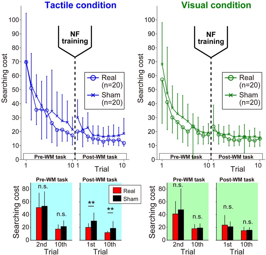
Sakurada et al.                                                                                           fNIRS Neurofeedback for Working Memory

functional brain characteristics might affect the efficacy of             accordance with the Declaration of Helsinki and approved by
neurofeedback training.                                                   the Institutional Review Board at Jichi Medical University.
   Regarding these neurophysiological factors, we should                  All participants provided written informed consent prior
consider the possibility that individual differences in the               to participation. Each participant completed the following
qualitative characteristics of cognitive function determine               experimental protocol in 1 day, including the fNIRS-based
neurofeedback training efficacy. We recently reported a wide              neurofeedback training task that aimed to regulate bilateral
variation in the sensory modalities that individuals are good at          prefrontal activities and a behavioral task that was used to
processing; hereafter, we refer to these individual differences as        evaluate WM ability before and after neurofeedback training.
modality dominance. Specifically, individual cognitive abilities
in motor imagery or attention control can be characterized by             Regulation of the Prefrontal Activity by
the modality dominance, and this can lead to better processing
                                                                          Functional Near-Infrared
of internal body information such as tactile or somatosensory
stimuli or external body information such as visual stimuli               Spectroscopy-Based Neurofeedback
(Sakurada et al., 2016, 2017, 2019a,b). In these studies,                 Training
individuals with visual motor imagery dominance showed better             Experimental Setup
motor performance when using an external focus attentional                Each participant sat on a chair facing an LCD monitor (size:
strategy, which required that the participant’s attention was             H30.5 × W37.7 cm) that presented the visual stimulus and
focused on a body movement outcome. Conversely, individuals               was asked to hold a computer mouse in their right hand. All
with kinesthetic motor imagery dominance demonstrated better              visual stimuli presented on the monitor were programmed in
motor performance when using an internal focus attentional                MATLAB (MathWorks, Inc., Natick, MA, United States). During
strategy, which required that the participant’s attention was             the fNIRS-based neurofeedback training task, the right hand was
focused on the body movement itself. Furthermore, individual              hidden by a small rack (Figure 1A). A multichannel fNIRS system
WM ability that relates to spatial information processing                 (ETG-7100, Hitachi Medical Corporation, Kashiwa, Japan) with
(Robertson et al., 2001) can be characterized by modality                 probes arranged to cover the prefrontal area was used to measure
dominance. When participants were required to retain spatial              prefrontal activity. All of the fNIRS channel inputs were sampled
information, they could be grouped into those who were good at            at 10 Hz. A 3 × 9 multichannel probe holder contained eight
retaining vibrotactile-based or visual-based spatial information          laser sources that emitted at 695 and 830 nm and seven detecting
(Matsumoto et al., 2020). These findings indicate that modality           probes that were alternately arranged with an interprobe distance
dominance is one of the important parameters in characterizing            of 3 cm. The midpoint between an emitter/detector pair was
the qualitative aspects of cognitive function, but whether distinct       defined as the location of a recording channel (the probe set
qualitative differences in cognitive function among individuals           initially had 22 recording channels). Importantly, fNIRS signals
affects neurofeedback training efficacy remains unclear.                  reflect changes in hemoglobin that originate in cortical tissue due
   This study aimed to examine whether modality dominance                 to brain activation and skin blood flow. To eliminate the impact
in WM influences the efficacy of neurofeedback training.                  of skin blood flow on the fNIRS signals, we set eight additional
Based on our previous findings regarding the neural basis of              short detecting probes at an interprobe distance of 1.5 cm and
individual differences in WM modality dominance (Matsumoto                applied multidistance independent component analysis (ICA)
et al., 2020), the neurofeedback protocol used in this study              to the fNIRS analysis (Hirosaka et al., 2004; Morren et al.,
provided feedback of the bilateral DLPFC and frontopolar                  2004; Akgül et al., 2006; Kohno et al., 2007; Funane et al.,
cortex (FPC), which are also critical for WM (Owen et al.,                2014). Signals from recording channels with a 1.5 cm interprobe
2005; Pleger et al., 2006; Slotnick and Moo, 2006; Kaas                   distance primarily included skin blood flow signals in shallow
et al., 2007; Giglia et al., 2014). We hypothesized that the              tissues. Based on these signals, we discriminated between the
individual modality dominance in WM is one of the influential             effects of cortical tissue and skin blood flow on the fNIRS
factors in neurofeedback training. Specifically, we predicted             signals. As it was possible to apply multidistance ICA only
that the degree of neuromodulation in the prefrontal area                 to the recording channels around the short detecting probes,
and cognitive performance after neurofeedback training would              the number of available recording channels was reduced to 15
highlight differences between individuals exhibiting tactile-             after applying multidistance ICA as shown in Figure 1B. All
versus visual dominance.                                                  results presented in this study were recorded after multidistance
                                                                          ICA processing from the fNIRS system (i.e., the effects of
                                                                          skin blood flow have been removed based on fNIRS signals
MATERIALS AND METHODS                                                     from the 1.5-cm interprobe channels). The probe holder was
                                                                          placed on the scalp with the lowest-row center emitter located
Participants                                                              at the participant’s Fpz position according to the standard
Forty healthy participants (mean age ±SD, 22.4 ± 3.2 years; 19            international 10–20 system (Figure 1B). To spatially register
males, 21 females) were recruited from the student population             fNIRS maps onto the Montreal Neurological Institute coordinate
at Jichi Medical University. All participants were right-handed           space, we individually measured scalp landmarks and all fNIRS
as assessed by the Edinburgh Handedness Inventory (laterality             recording channel positions using a 3D magnetic space digitizer
96.4 ± 8.9) (Oldfield, 1971). This study was conducted in                 (FASTRAK, Polhemus, United States). We then used the position

Frontiers in Systems Neuroscience | www.frontiersin.org               3                                  February 2022 | Volume 16 | Article 774475
Sakurada et al.                                                                                                                  fNIRS Neurofeedback for Working Memory

  FIGURE 1 | Experimental setup. (A) Neurofeedback training set up. The monitor presented visual feedback on the status of participants’ bilateral prefrontal cortex in
  real-time. Furthermore, the six red circles on the monitor indicated the visual stimuli that participants were required to remember. Specifically, in each task block, the
  all-red circles were sequentially presented individually in random order at the predetermined fixed positions {with the center of the monitor as the origin, the
  coordinates of the six circles were [x(cm), y(cm)] = (–10, 8), (10, 8), (–10, 0), (10, 0), (–10, –8), and (10, –8)}. The appearance order changed with every task block.
  The participants were required to remember the spatial appearance order of six visual stimuli on the monitor. (B) Configuration of the fNIRS probe. Probes were
  placed over the prefrontal area. The channels, numbered 1–15, indicated the channels that output fNIRS signals that have been removed based on multidistance
  ICA. Ch.7 and ch.11 conveyed signals from the left and right DLPFC/FPC, respectively, as neurofeedback. (C) Spatial registration of the fNIRS maps onto MNI
  coordinate space. The left and right panels show typical profiles of oxy-Hb and deoxy-Hb in the feedback channels. Time zero indicates the onset of the task block.
  After starting the task block, the oxy-Hb signal showed a stronger response than the deoxy-Hb signal.

data from all of the participants’ recording channels to estimate                         deoxy-Hb signals (Toronov et al., 2001; Strangman et al., 2002).
spatial profiling without MRI (Singh et al., 2005). The estimated                         As shown in Figure 1C, we also confirmed that the oxy-Hb signal
spatial profiling of each recording channel is shown in Table 1.                          change was stronger than that of deoxy-Hb in this study. Thus,
                                                                                          the circle diameter was determined based on the oxy-Hb signals
Procedure                                                                                 of ch.7 and ch.11. Furthermore, as the fNIRS signals are relative
The neurofeedback training task consisted of eight sessions, each                         values, we should avoid using the values directly. Instead, the
comprising seven alternating 15-s rest and 25-s task blocks with                          online oxy-Hb signals during the task blocks in each feedback
an additional rest block inserted at the end of each session (i.e.,                       channel were normalized to the mean and standard deviation
15 blocks per session).                                                                   during the 10 s prior to each task block (i.e., z-scoring). We
   During the task blocks, the monitor reported neurofeedback                             then averaged z-score values between channels 7 and 11. The
information as a circle in which the diameter reflected the                               circle size increased when the averaged z-score among the two
online prefrontal activities in channels 7 and 11, corresponding                          channels decreased. The circle was the largest (diameter: 45 cm)
mainly to the bilateral DLPFC and FPC. Although we measured                               when the brain activity in the task block was below the average
oxygenated and deoxygenated hemoglobin (oxy- and deoxy-                                   activity level for 10 s before starting the task block. Under this
Hb), we used the oxy-Hb signals to calculate the neurofeedback                            neurofeedback setting, participants were instructed to make the
information because of their greater sensitivity to changes                               circle on the monitor as large as possible. In other words, the
in cerebral blood flow and higher signal-to-noise ratio than                              current neurofeedback training protocol aimed to acquire more

Frontiers in Systems Neuroscience | www.frontiersin.org                               4                                          February 2022 | Volume 16 | Article 774475
Sakurada et al. fNIRS Neurofeedback for Working Memory
TABLE 1 | Spatial profiling of each recording channel. given information on the six visual stimuli (number, position,
and randomness of appearance order). They were then instructed
Ch. Localization Brodmann area Probability
to remember the sequential patterns (spatial appearance order)
1 Left FPC 10 1 of the six stimuli and to verbally answer the order of the visual
2 Right FPC 10 1 stimuli in the rest block immediately after each task block. The
3 Left FPC 10 0.96 participants were also required to press the computer mouse
Left DLPFC 46 0.04 button at about 1 Hz with their index fingers. We utilized this
4 Left FPC 10 1 cognitive-motor task during the task blocks, because the effect of
5 Right FPC 10 1 cognitive processing cannot be assessed when a task is too easy
6 Right FPC 10 0.99 for participants (Landers et al., 2005; Wulf et al., 2007, 2009) and
Right DLPFC 46 0.01 individual differences in cognitive ability may not appear when
7 Left DLPFC 46 0.82 the task is of insufficient difficulty (Sakurada et al., 2017).
Left FPC 10 0.18 We randomly assigned the participants to Real or Sham
8 Left FPC 10 0.84 groups (each group contained 20 participants), but the group
Left DLPFC 9 0.16
assignment for each participant was not provided to the
9 Right FPC 10 0.86
experimental operator or the participant. The circle diameter
Right DLPFC 9 0.14
for the Real group was determined in real-time based on each
10 Right FPC 10 0.80
participant’s own oxy-Hb signals in ch.7 and ch.11. Conversely,
Right DLPFC 9 0.20
the circle size for the Sham group was based on the prerecorded
11 Right DLPFC 46 0.77
oxy-Hb signals of another person and was unrelated to the
Right FPC 10 0.23
participant’s own cortical activation.
12 Left DLPFC 9 0.70
Left DLPFC 46 0.24
Functional Near-Infrared Spectroscopy Offline
Left FPC 10 0.05
Analysis
Left includes FEF 8 0.01
Preprocessing
13 Left DLPFC 9 0.82
Left includes FEF 8 0.18
After applying the multidistance ICA, to remove baseline drift,
14 Right DLPFC 9 0.81
the individual time course data from the oxy-Hb signals from
Right includes FEF 8 0.19
each channel were high-pass filtered using a cut-off frequency
15 Right DLPFC 9 0.84
of 0.0125 Hz. Next, to remove blocks containing motion-
Right DLPFC 46 0.06
related artifacts, we applied an artifact detection algorithm based
Right FPC 10 0.05
on HOMER2 software [MGH-Martinos Center for Biomedical
Right includes FEF 8 0.05
Imaging (NITRC, 2021: Homer2: Tool/Resource Info)]. As no
blocks containing artifacts were detected, we analyzed the entire
FPC, frontopolar cortex; DLPFC, dorsolateral prefrontal cortex; FEF,
oxy-Hb time course data obtained from this study.
frontal eye fields.
General Linear Model Analysis
efficient (i.e., lower) activities in the bilateral DLPFC/FPC during General linear model analysis (Friston et al., 1994a,b) can be
spatial WM processing. Regarding task instructions related to used to detect task-related hemodynamic changes in the cortex
brain activity, we only explained that the circle enlarged in from fNIRS data (Schroeter et al., 2004; Plichta et al., 2007).
response to cognitive brain activity for properly holding the To identify neuromodulation in the prefrontal regions related
target spatial information. We did not explicitly instruct the to the WM processing of spatial information, we used GLM
desired brain activity change (i.e., whether the participants should analysis with least-squares estimation of the oxy-Hb signals. For
increase or decrease brain activity). This neurofeedback setting the preprocessed oxy-Hb signals, a Gaussian function with a
was based on the previous reports that decreasing prefrontal peak time of 6 s and full width half maximum of 5.4 s was
activity is associated with familiarity or a higher skill level during used as a hemodynamic response function to better mimic brain
cognitive tasks (Jansma et al., 2001; Ramsey et al., 2004; Koike signals. The resulting beta values at each recording channel
et al., 2013). estimated by the GLM analysis were then used in the group
To drive spatial WM processing in the prefrontal area, we analysis to evaluate the degree of neuromodulation during the
set up a cognitive-motor task during the task blocks. For the neurofeedback training.
cognitive-motor task, the monitor sequentially showed six small As described previously, the participants were instructed
visual stimuli. Each visual stimulus shown was a red circle with to enlarge the circle size on the monitor. This means that
a diameter of 1 cm. The six visual stimuli were sequentially and the participants needed to reduce the average activity of the
individually presented in random order at predetermined fixed neurofeedback channels (7 and 11). However, it should be noted
positions (top-left, top-right, middle-left, middle-right, bottom- that there were some potential patterns of brain activity in which
left, or bottom-right on the monitor). A series of visual stimuli participants were considered to have achieved the neurofeedback
was presented just after the start of each task block, and the training goal. Specifically, the participants could enlarge the circle
order was different for every task block. The participants were size not only by a simultaneous decreasing of both the left and
Frontiers in Systems Neuroscience | www.frontiersin.org 5 February 2022 | Volume 16 | Article 774475Sakurada et al. fNIRS Neurofeedback for Working Memory
right neurofeedback channel activities but also by a large decrease the corresponding digitizing-pen position to indicate the target
of either the left or right activity. Therefore, as channels 7 and 11 location. Regarding the target settings, we had prepared different
might show different activity changes during the neurofeedback target locations and their appearance orders between the tactile
training, we independently analyzed the activity changes in each and visual conditions and among the participants. However,
channel, rather than the average activity between the two. the six predetermined target locations and their appearance
orders within each participant were fixed throughout the trials
Statistical Analysis under each condition.
The beta values from all of the recording channels were analyzed In each trial, the participants were first required to move
by three-way repeated-measures analysis of variance (ANOVA) the digitizing-pen to the center of the search area on the
with session (1st or 8th session) and feedback channel (ch.7 tablet. Then, the background color of the monitor changed
or ch.11) as within-subject factors and neurofeedback training as a start cue for the participants to begin searching for the
group (Real or Sham group) as a between-subject factor. first target. When the digitizing-pen entered a target area, the
Regarding the effect size, we applied a partial eta squared, which is vibrotactile or visual stimulus was presented, and the sensory
robust for the number of factors. To evaluate the degree of neural stimuli continued until the tip of the digitizing-pen moved
activity change in the feedback channels, the beta values in the out of the target area. If the digitizing-pen remained in the
first session were compared with those in the eighth session by a target area for 0.7 s, a beep signal informed the participant of
post hoc test (simple-simple main effect test). successful target detection. Then, the participants immediately
began searching for the next target. Finally, each trial finished
Target Searching Task for Evaluating when the participant found all six targets. Participants were
Spatial Working Memory Ability also instructed to find all six targets as quickly as possible.
Experimental Setup Therefore, they had to retain spatial information, namely, the
Based on our previous study (Matsumoto et al., 2020), we target locations and orders of appearance, in the repeated trials.
applied a target searching task to quantify individual WM We expected that participants would gradually show efficient
modality dominance during spatial processing. The participants searching as a learning effect if they could retain the spatial
performed this searching task in a booth different from that information of the target under each condition.
used in the neurofeedback training task. Each participant was Before the neurofeedback training task, the participants
seated on a chair and asked to hold a digitizing-pen on a performed alternating the tactile and visual conditions a total
drawing tablet (Intuos4 PTK-1240/K0, Wacom, Japan) with of 20 trials as successive trials, so 10 trials were performed in
their right hand. An LCD monitor (size: H30.5 × W37.7 cm) each condition (the Pre-WM task). The first trial was randomly
used to present the visual stimulus was placed horizontally assigned as the tactile or visual condition for each participant.
at 16.5 cm above the tablet. As their right hand was hidden As described previously, the target locations for each participant
by a cloth and the monitor, the participants could not see it differed between the tactile and visual conditions. Therefore, we
directly during the experimental tasks. Visual stimuli such as can expect no transfer of spatial information from the tactile
task instructions presented on the monitor were programmed in condition to the visual condition and vice versa. Furthermore,
MATLAB using the Cogent Toolbox (University College London, to align the task difficulty between the two conditions, the
London, United Kingdom). The Cogent Toolbox also recorded total distance between all targets (i.e., the sum of the straight-
the position of the digitizing-pen tip with sampling at 60 Hz. line distances connecting the six targets) was the same for
A vibration motor presenting a vibrotactile stimulus was attached both conditions. The participants were required to hold the
to the tip of the index finger on the right hand. two patterns of target spatial information from both conditions
simultaneously, so we set the number of trials needed to
Procedure properly retain the information. In a preliminary experiment, we
The searching task required participants to find six targets estimated the number of trials required for the searching cost to
appearing somewhere in the search area by moving the digitizing- finally reach the plateau. After the neurofeedback training task,
pen on the drawing tablet (search area: H30.5 × W37.7 cm). the participants performed alternating both conditions a total of
The targets were located randomly and appeared individually 20 trials again (the Post-WM task), and the target locations in
in a predetermined, sequential order. Note that the target the Post-WM task were the same as those in the Pre-WM task.
locations and appearance orders in this searching task were Thus, the participants can refer to the target spatial information
unrelated to those of the red visual stimuli presented during the held during the Pre-WM task to improve searching performance
neurofeedback training task. We introduced two experimental during the Post-WM task. If the current neurofeedback training
conditions, a tactile condition and a visual condition, that differed has a positive effect on individual WM ability, the participants in
in the sensory modality of the stimulus cues presenting the target the Real group would show higher searching performance during
locations and orders of appearance. Under the tactile condition, the Post-WM task than those in the Sham group.
when the tip of the digitizing-pen came into a target area on the
tablet (diameter: 10 cm), a vibrotactile stimulus was presented Analysis
to the right index finger from the vibration motor to indicate To evaluate individual WM ability, we applied the same index as
the target location. Conversely, under the visual condition, a that in our previous study (Matsumoto et al., 2020). Specifically,
circular visual cursor was presented on the monitor just above we calculated the searching cost based on searching time (Time)
Frontiers in Systems Neuroscience | www.frontiersin.org 6 February 2022 | Volume 16 | Article 774475Sakurada et al. fNIRS Neurofeedback for Working Memory
and normalized the movement distance (Dis) in each trial. The If the individual modality dominance affected the efficacy of
searching time was the duration taken to find all of the targets, the neurofeedback training, the TD and VD individuals were
and the normalized movement distance was the distance moved expected to be distributed as distinct clusters on the plane of the
by the right hand divided by the shortest distance connecting the beta value change and searching cost. Therefore, we estimated
six targets with a straight line. We defined the “searching cost” the decision boundary between TD and VD individuals by
using Eq. (1): linear discriminant analysis based on the beta value change and
searching cost. We then compared discriminant function values
q calculated by linear discriminant analysis between TD and VD
SearchingCosti = Time2i + (Disi − 1)2 (1) individuals using a Wilcoxon rank sum test.
Where subscript i denotes the trial number (1–10 in each
condition). The searching cost in each trial indicates the distance RESULTS
from the (0, 1) coordinate on a Time–Dis plane. We can deduce
that the searching cost reflects the individual WM ability because Neurofeedback Training Task
retaining spatial information for hidden targets can optimize the Prefrontal Activity
searching movement path on the drawing tablet (i.e., participants The prefrontal activity patterns in the feedback channels [left
can search for the targets in a shorter distance and a shorter prefrontal area (ch.7) and right prefrontal area (ch.11)] and the
searching time). In this scenario, lower searching costs indicate beta value transitions as estimated by GLM analysis for the oxy-
a higher WM ability to efficiently retain the target locations Hb signals are shown in Figure 2. With respect to the beta
and order of appearance. Note that the first trial in the Pre- values of the oxy-Hb signals, we found a marked group-difference
WM task was excluded from the statistical analysis, because the in the ch.11 activity patterns. The three-way ANOVA revealed
participants did not know the target locations during the first trial significant interactions for group × session [F(1,38) = 7.40,
and needed to search randomly for them without relying on their p = 0.0098, ηp 2 = 0.16] and group × channel [F(1,38) = 4.82,
WM. Thus, the searching costs in the 2nd to 10th trials in the p = 0.034, ηp 2 = 0.11] and a marginally significant interaction
Pre-WM task and the 1st to 10th trials in the Post-WM task were for group × session × channel [F(1,38) = 3.13, p = 0.085,
assumed to reflect the individual WM ability. ηp 2 = 0.076]. Post hoc tests on the Real group revealed that
Furthermore, to characterize the modality dominance in the the beta value in ch.11 decreased, which was consistent with
WM as a qualitative aspect of cognition in individuals, we the neurofeedback training aims [p = 0.057, simple-simple main
compared the searching costs between the tactile and visual effect test (1st vs. 8th)], and these decreasing trends were focal
conditions at the 10th trial in the Pre-WM task. We subtracted and limited to the right hemisphere [ch.14: p = 0.0027, ch.15:
the searching cost under the tactile condition from that under the p = 0.029; simple-simple main effect test (1st vs. 8th)]. However,
visual condition as an index of modality dominance. Therefore, there was no notable beta value change in ch.7 [p = 0.73; simple-
positive and negative values indicated tactile-dominant (TD) and simple main effect test (1st vs. 8th)]. With respect to the Sham
visual-dominant (VD) individuals, respectively. group, individuals showed an increasing trend only in the right
feedback channel (ch.11: p = 0.031, ch.7: p = 0.13; simple-
Statistical Analysis simple main effect). The increasing trends in the Sham group
The searching costs in the Pre- and Post-WM tasks were analyzed were identified in the broad areas including the left and right
by three-way repeated-measures ANOVA with trial (2nd or 10th hemispheres (ch.3, 5, 6, 11, 12, and 14). The beta value transitions
trial for the Pre-WM task and 1st or 10th trial for the Post-WM for all of the recording channels are shown in Supplementary
task) and condition (tactile or visual condition) as within-subject Figure 1, and all of the statistical values from the three-way
factors and neurofeedback training group (Real or Sham group) ANOVA of the beta values are shown in Supplementary Table 1.
as a between-subject factor. On the other hand, regarding the beta value of the deoxy-
Then, to clarify the relationship between individual learning Hb signals, no significant changes were observed compared to
ability and the efficacy of neurofeedback training, we calculated the oxy-Hb, as was expected. In both channels, the beta values
the Pearson correlation coefficients (r) between the amount of tended to increase slightly in the Real group and decrease slightly
decrease in searching cost from the 2nd to the 10th trial and the in the Sham group [average beta values: Real group –0.0027
beta value change. Specifically, we focused on the effect of the (ch.7, 1st session), –0.0014 (ch.7, 8th session), –0.0024 (ch.11, 1st
performance improvement in the Pre-WM task on the beta value session), –0.0010 (ch.11, 8th session); Sham group 9.7 × 10⁻⁵
change and the effect of the beta value change on the performance (ch.7, 1st session), –0.00055 (ch.7, 8th session), –2.8 × 10⁻⁶
improvement in the Post-WM task. (ch.11, 1st session), –0.0035 (ch.11, 8th session)]. All statistical
Furthermore, to evaluate the influence of individual modality values calculated from the three-way ANOVA of the beta values
dominance in WM on the efficacy of neurofeedback training, are shown in Supplementary Table 2.
we calculated the Pearson correlation coefficient (r) between the
beta value change and searching cost in the final trial under Cognitive Performance
the Post-WM task. Then, we compared the beta value change With respect to the appearance order of the six visual stimuli, all
and searching cost in the Post-WM task between TD and VD participants exceeded the correct answer rate of 80% (mean ± SD:
individuals in the Real group using Wilcoxon rank sum tests. 96.9 ± 3.9, range: 83.3–100%). No significant correlation was
Frontiers in Systems Neuroscience | www.frontiersin.org 7 February 2022 | Volume 16 | Article 774475Sakurada et al. fNIRS Neurofeedback for Working Memory
FIGURE 2 | Neuromodulations of the prefrontal cortex activity during the neurofeedback training task. Upper panels: The temporal profiles of the oxy-Hb signals in
ch.11 (left panels) and ch.7 (right panels). The red and black lines represent the time courses of the oxy-Hb signals in the Real and Sham groups, respectively. The
lighter colored regions around the time course lines denote the standard deviation. The upper or lower directional standard deviation regions are shown for the
profiles of the Real and Sham groups, respectively. In ch.11, compared to the first session, the oxy-Hb signal in the final session tended to decrease in the Real
group and increase in the Sham group. Lower panels: Beta value transitions in ch.11 (left panels) and ch.7 (right panels). In the first session of ch.11, although there
was no significant difference in the beta value between the Real and Sham groups, the Real group showed significantly lower activity than the Sham group at the
final session. In contrast, in ch.7, no significant beta value change was observed in both groups. Middle lower 3D brain illustrations: Spatial configurations of the
p-values from the simple-simple main effect test comparing the beta value in the first session and that in the last session. In the Real group, activities focally
decreased in the right hemisphere, including the feedback channel (white dotted circles), while in the Sham group, large activity increases were observed in the
bilateral broad region (white solid circles). Error bars denote the standard deviation. † p < 0.1, *p < 0.05.
observed between the individual differences in cognitive ability three-way ANOVA of the searching cost showed only a significant
for the current cognitive-motor task and the amount of oxy-Hb main effect of trial [F(1,38) = 101.93, p = 2.62 × 10⁻¹² ,
beta value change from the 1st to the 8th session (ch.7: r = 0.14, ηp 2 = 0.73]. The other factors did not reach the level of statistical
p = 0.39, ch.11: r = 0.22, p = 0.16). significance (Fs < 2.16, ps > 0.15). These statistical results
for the Pre-WM task indicated the baseline WM performance
before neurofeedback training was not different between the Real
Target Searching Task and Sham groups. Next, three-way ANOVA for the Post-WM
Searching Cost task revealed a significant main effect of trial [F(1,38) = 50.36,
In the Pre-WM task, all of the participants gradually reduced p = 1.82 × 10⁻⁸ , ηp 2 = 0.57] and a significant two-way interaction
the searching cost over successive trials. In the Post-WM of condition × group [F(1,38) = 13.43, p = 7.52 × 10⁻⁴ ,
task involving the same target locations as the Pre-WM task, ηp 2 = 0.26]. The other factors did not reach the level of statistical
low searching costs were shown from the first trial, and the significance (Fs < 3.27, ps > 0.79). Note that the simple main
participants were able to search more efficiently during repeated effect test for the condition × group interaction found that the
trials (Figure 3A). Real group exhibited significantly lower searching costs than
Here, we focused on the difference in searching costs between the Sham group under the tactile condition (Tactile condition:
the Real and Sham groups (Figure 3B). In the Pre-WM task, a p = 0.0017, Visual condition: p = 0.69). All statistical values
Frontiers in Systems Neuroscience | www.frontiersin.org 8 February 2022 | Volume 16 | Article 774475Sakurada et al. fNIRS Neurofeedback for Working Memory
FIGURE 3 | Learning curves during pre- and post-WM tasks. (A) In each task phase, the participants successfully reduced the searching cost. (B) The Real group
showed significantly better performance in the Post-WM task only under the tactile condition. Error bars denote the standard deviation. **p < 0.01.
of the three-way ANOVA for the searching cost are shown in change and the amount of behavioral performance improvement
Supplementary Tables 3, 4. observed in the Post-WM task (Real group: r = –0.32, p = 0.16;
Sham group: r = –0.13, p = 0.56).
Individual Modality Dominance in Working Memory We then examined whether the neurofeedback training
Based on differences in the searching cost between the tactile efficacy differed between TD and VD individuals. Figure 4A
and visual conditions at the 10th trial in the Pre-WM task (i.e., shows the distribution of the beta value changes in ch.11
modality dominance in WM), we labeled the 40 participants during neurofeedback training and WM performance under
as TD or VD individuals. Consistent with our previous study the tactile condition following the neurofeedback training for
(Matsumoto et al., 2020), we also confirmed that the modality the 20 individuals in the Real group. Although there was no
dominance in WM ability varied widely among individuals. significant correlation between the intersubject variance of beta
Specifically, 10 of the participants in the Real group were labeled value changes and that of searching costs for the entire cohort
as TD-individuals and 10 as VD individuals, respectively, whereas (r = –0.14, p = 0.55), the clusters of TD- and VD individuals
eight members of the Sham group were labeled as TD-individuals seemed to be dissociated. Indeed, the TD-individuals showed
and 12 as VD individuals, respectively. significantly greater self-regulation of neural activity than the VD
individuals (p = 0.0073), and the mean searching cost for TD-
Modality Dominance Dependency in individuals was lower than that for VD individuals (p = 0.064).
Neurofeedback Training Efficacy Based on the distribution of the Real group, the decision function
Regarding the tactile condition with significant group difference was estimated as f = –246.4 ∗ 1β –1.1 ∗ SC+12.3 (SC denotes
in the target searching task, neither group showed a significant “searching cost”), and the estimated decision boundary revealed
correlation between the amount of behavioral performance that the TD-individuals were located in the lower left of the
improvement observed in the Pre-WM task (i.e., the amount of scatter plot compared with the VD individuals. Thus, as shown
decrease in searching cost) and the beta value change (Real group: in Figure 4A, when located in the lower left of the plane, the
r = –0.01, p = 0.96; Sham group: r = 0.29, p = 0.22). Similarly, decision function shows a larger value. A larger decision function
no significant correlation was found between the beta value value indicates higher neurofeedback training efficacy, reflecting
Frontiers in Systems Neuroscience | www.frontiersin.org 9 February 2022 | Volume 16 | Article 774475Sakurada et al. fNIRS Neurofeedback for Working Memory
FIGURE 4 | Individual modality dominance dependency of neurofeedback training efficacy in the Real group. (A) Scatter plot of the individual neurofeedback training
efficacy based on the beta value change in ch.11 and WM task performance in the Post-WM task. TD-individuals are relatively distributed in the lower left of the
scatter plot compared with the VD individuals. The red solid line indicates the decision boundary estimated by linear discriminant analysis [i.e., the decision function
value (DFV) is zero on this decision boundary]. The red dotted lines represent other DFVs (6, 3, –3, and –6, respectively). Error bars of the mean values denote the
standard deviation. (B) The decision function values for the TD-individuals were significantly higher than those for the VD individuals. † p < 0.1, **p < 0.01,
***p < 0.001.
greater decreases in right prefrontal activities and better WM this led to a higher motor learning effect (Sakurada et al., 2019b),
performance after neurofeedback training. Based on the decision implying that lower prefrontal activity increases the cognitive
function value, the neurofeedback training of the TD-individuals resource margin due to higher cognitive processing efficiency. As
was more effective than that of the VD individuals (p = 0.00058, a result, efficient prefrontal activity may lead to improvements
Figure 4B). Note that we also confirmed that the Sham group in cognitive or motor performance. Although no direct evidence
had relatively lower decision function values than the Real group has supported this interpretation, the lower oxy-Hb signals
(–10.4 ± 13.1 SD). during the current neurofeedback training may provide a hint
of the idea of the promoting cognitive margin. Conversely, a
number of previous neurofeedback training strategies have been
DISCUSSION aimed at increasing the prefrontal activity of the target brain
area(s) (Wang and Hsieh, 2013; Hsueh et al., 2016). Several
The neurofeedback training provided in this study succeeded in
studies have reported that increasing prefrontal activity is an
lowering neural activity in the right prefrontal area. Moreover,
effective approach to improving WM ability in the elderly and
this efficient prefrontal activity facilitated the WM ability to
in patients with stroke (van Asselen et al., 2006; Jones et al.,
retain internal body information. With respect to the influence
2015; Stephens and Berryhill, 2016). These previous studies have
of modality dominance on the efficacy of neurofeedback training,
demonstrated that increasing neural activity by neurofeedback
the individuals exhibiting tactile dominance showed higher levels
training is also an effective approach to improving specific
of neuromodulation and greater WM performance compared
brain functions. Increasing activity may improve cognitive
with the individuals with visual dominance. In this study, we
performance by maximizing active cognitive resources rather
focused on the importance of considering individual differences
than promoting efficiency in cognitive processing within constant
in cognitive function when applying neurofeedback training.
resources. The reason for such a discrepancy that both an
increase and a decrease of prefrontal activity can contribute to
Effectiveness of Functional Near-Infrared improving WM ability is unclear. However, age and medical
Spectroscopy-Based Neurofeedback history (especially cerebrovascular disease) may be influential
Training for Working Memory factors for the neuromodulation effect. Therefore, in applying
The fNIRS-based neurofeedback training used in this study is a neurofeedback training, it is necessary to make an appropriate
useful approach to improving self-regulation of the prefrontal choice about increasing or decreasing activity according to the
area and facilitating WM performance. In particular, lower target brain function or training population.
prefrontal activity contributed to the ability to retain sensory The efficacy of the neurofeedback training presented in this
information. The current level of training efficacy is reasonable study was limited; the prefrontal neuromodulation contributed
given the relationship between lower prefrontal activity and to the maintenance of vibrotactile rather than visual information.
higher cognitive skill (Jansma et al., 2001; Ramsey et al., These results might be due to individual differences in cognitive
2004; Koike et al., 2013). Previously, lower levels of activity in function as characterized by the ability to process internal
the prefrontal and posterior parietal lobes were shown to be body information (Sakurada et al., 2017, 2019a; Matsumoto
associated with an individual optimal attentional strategy, and et al., 2020). Specifically, while most participants have a certain
Frontiers in Systems Neuroscience | www.frontiersin.org 10 February 2022 | Volume 16 | Article 774475Sakurada et al. fNIRS Neurofeedback for Working Memory
cognitive ability to process visual stimuli, some individuals using the statistical approach of comparing Real and Sham
are less good at processing internal body information such as groups, which did not consider individual modality dominance,
vibrotactile stimuli. Thus, a greater margin of improvement might result in inaccurate predictions of neurofeedback training
observed in participants for the tactile condition than for the efficacy for the entire Real group. However, predicting training
visual condition might have resulted in significantly better efficacy based on individual differences in brain function can
training efficacy for the tactile condition. In addition, note that contribute to optimizing individual training protocols and
the protocol included only a short-term training period (i.e., only improving the training success rate.
1 day), which may have been the reason for the lack of training
efficacy seen for the visual stimulation. In a number of previous Role of the Right Prefrontal Cortex
studies, the neurofeedback training was longer in duration than The efficiency of prefrontal cortex activity was shown to
in the present protocol (Wang and Hsieh, 2013; Hsueh et al., facilitate the WM ability to hold internal body information.
2016), and longer neurofeedback training might provide greater Specifically, significant changes in activity were observed
benefits even for the visual condition. Further investigation is during neurofeedback training in the right hemisphere, which
needed on this point. corresponds to the DLPFC (Brodmann area 46 and 9) and FPC
(Brodmann area 10). The bilateral DLPFC and FPC have been
widely recognized as critical structures for WM (Owen et al.,
Modality Dominance Dependency in 2005). For instance, increasing activity in the right DLPFC with
Neurofeedback Training Efficacy transcranial direct current stimulation led to improved accuracy
Sensory modality dominance in cognitive function is an in memorizing visuospatial locations (Giglia et al., 2014). In
influential factor in determining the efficacy of neurofeedback addition, the DLPFC and FPC are associated with visual spatial
training. As in our previous study (Matsumoto et al., 2020), we memory (Slotnick and Moo, 2006). Moreover, the left DLPFC
found large intersubject variability in the modality dominance and FPC play an important role in processing internal body
of WM and demonstrated the relationship between the information such as tactile or somatosensory stimuli. Activity
individual modality dominance and degree of neuromodulation in the left DLPFC correlates with accuracy in discriminating
and behavioral performance. Furthermore, as no significant two successive somatosensory stimuli (Pleger et al., 2006).
correlation was found between the amount of behavioral Furthermore, the left FPC is associated with WM representations
performance improvement in the target searching task and the of haptic information and the integration of spatial and motor
neurofeedback training efficacy, the current findings imply that components (Kaas et al., 2007). Note that the neurofeedback
the individual differences in the amount of neuromodulation training protocol used in this study required participants to
during neurofeedback training are not affected by the learning retain spatial information; therefore, the participants succeeded
ability. In other words, the sensory modality dominance is a in self-regulating neural activity in the right prefrontal area that
more crucial factor than the individual learning ability, as the was related to spatial memory. However, we presumed that the
individual brain characteristics determine the neurofeedback modulation of activity in the left DLPFC/FPC failed because
training efficacy. When not only neurophysiological factors such the neurofeedback training was based on visual information
as alpha power or sensorimotor rhythm (Weber et al., 2011; Wan (i.e., the participants were required to retain spatial information
et al., 2014) but also modality dominance are used to characterize based on visual stimuli). If the neurofeedback training task had
the cognitive traits of an individual, it is possible to predict required the participants to retain spatial information based on
neurofeedback training efficacy more accurately. Note that we tactile information such as a vibrotactile stimulus, the left DLPFC
confirmed that 75% of participants in the Real group showed might also have been successfully modulated. Taken together,
the same sensory modality dominance between Pre- and Post- the findings suggested that the improvement in the WM ability
WM tasks (i.e., tactile or visual dominance was maintained), and to hold spatial information with efficient activity levels in the
the other few participants showed different dominance (tactile right DLPFC/FPC promoted a behavioral outcome under the
changed to visual in one participant and visual changed to tactile tactile condition.
in four participants). Therefore, although we found relatively The brain networks between DLPFC and other areas are also
more participants who had a change from visual to tactile, important approaches in interpreting the neurofeedback training
the current neurofeedback training would have no effect on efficacy. For instance, the prefrontal cortex and posterior parietal
reversing the sensory processing ability between tactile and visual cortex are the crucial neural bases for spatial cognition. Persistent
modalities to a specific direction. activities in these areas reflect not only the maintenance of
With regard to the acquisition of “low prefrontal activity,” a WM representation but also the maintenance of a motor
which is the purpose of the current neurofeedback training as a intention (Jerde and Curtis, 2013). Furthermore, it has been
successful training efficacy for the self-regulation of brain activity, reported that bilateral primary somatosensory cortices are
90% of individuals with tactile dominance showed decreased involved in tactile WM and that DLPFC contributes to bridging
prefrontal activity during training sessions. This rate of success the somatosensory cortices from both sides for goal-directed
was higher than that seen in previous studies (Alkoby et al., 2018). action generation (Zhao et al., 2017). In other words, for
Conversely, the success rate for the neuromodulation of VD the processing WM function, DLPFC forms the frontoparietal
individuals was only 30% in terms of the purpose of decreasing network and a network connected with sensory areas. Therefore,
prefrontal activity in the current neurofeedback training. Thus, the acquisition of efficient prefrontal activity may promote
Frontiers in Systems Neuroscience | www.frontiersin.org 11 February 2022 | Volume 16 | Article 774475Sakurada et al. fNIRS Neurofeedback for Working Memory
processing in these other connected areas with the prefrontal University. The participants provided their written informed
cortex, leading to a higher adaptive capacity. consent to participate in this study. Written informed consent
was obtained from the individual(s) for the publication of any
Conclusion identifiable images or data included in this article.
We demonstrate that lowering the activity in the right
prefrontal cortex using fNIRS-based neurofeedback training (i.e.,
improving the efficiency of activity) can facilitate the ability AUTHOR CONTRIBUTIONS
of the WM to retain spatial information. Moreover, individual
TS conceived and designed the experiment, developed the
differences in the sensory modality dominance of the WM,
experimental system, wrote the draft of the manuscript,
in particular, the ability to hold internal body information,
and supervised the study. MM performed the participant
which varies widely among individuals, is an important and
experiments. TS and MM analyzed the data. All authors
newly identified neurophysiological factor that can determine
contributed to the discussion of the results, and read and
the efficacy of neurofeedback training. Therefore, a customized
approved the final manuscript.
approach to developing neurofeedback training protocols that
are suited to the brain dynamics of the individual will provide
more effective neuromodulation methods. Specifically, when
applying neurofeedback training to stroke patients with large
FUNDING
individual differences in cognitive function, considering the This research was supported by the Japan Society for the
individual sensory modality dominance will provide a tailor- Promotion of Science (JSPS) KAKENHI (15K16366 and
made neurorehabilitation protocol with higher cognitive or 17K13096) and Pfizer Health Research Foundation.
motor training effects.
ACKNOWLEDGMENTS
DATA AVAILABILITY STATEMENT
We thank M. Hirai and M. Tetsuka at Jichi Medical University
The raw data supporting the conclusions of this article will be for their help and encouragement.
made available by the authors, without undue reservation.
SUPPLEMENTARY MATERIAL
ETHICS STATEMENT
The Supplementary Material for this article can be found
The studies involving human participants were reviewed and online at: https://www.frontiersin.org/articles/10.3389/fnsys.
approved by the Institutional Review Board at Jichi Medical 2022.774475/full#supplementary-material
REFERENCES Friston, K. J., Holmes, A. P., Worsley, K. J., Poline, J. P. B., Frith, C. D., and
Frackowiak, R. S. J. (1994a). Statistical parametric maps in functional imaging:
Akgül, C. B., Akin, A., and Sankur, B. (2006). Extraction of cognitive a general linear approach. Hum. Brain Mapp. 2, 189–210. doi: 10.1002/hbm.
activity-related waveforms from functional near-infrared spectroscopy 460020402
signals. Med. Biol. Eng. Comput. 44, 945–958. doi: 10.1007/s11517-006- Friston, K. J., Jezzard, P., and Turner, R. (1994b). Analysis of functional MRI
0116-3 time-series. Hum. Brain Mapp. 1, 153–171. doi: 10.1002/hbm.460010207
Alkoby, O., Abu-Rmileh, A., Shriki, O., and Todder, D. (2018). Can we predict Fujimoto, H., Mihara, M., Hattori, N., Hatakenaka, M., Yagura, H., Kawano, T.,
who will respond to neurofeedback? A review of the inefficacy problem and et al. (2017). Neurofeedback-induced facilitation of the supplementary motor
existing predictors for successful EEG neurofeedback learning. Neuroscience area affects postural stability. Neurophotonics 4:045003. doi: 10.1117/1.NPH.4.
378, 155–164. doi: 10.1016/j.neuroscience.2016.12.050 4.045003
Cheng, M. Y., Huang, C. J., Chang, Y. K., Koester, D., Schack, T., and Hung, Funane, T., Atsumori, H., Katura, T., Obata, A. N., Sato, H., Tanikawa, Y., et al.
T. M. (2015). Sensorimotor rhythm neurofeedback enhances golf putting (2014). Quantitative evaluation of deep and shallow tissue layers’ contribution
performance. J. Sport Exerc. Psychol. 37, 626–636. doi: 10.1123/jsep.2015-0166 to fNIRS signal using multi-distance optodes and independent component
Diaz Hernandez, L., Rieger, K., and Koenig, T. (2018). Low motivational analysis. Neuroimage 85, 150–165. doi: 10.1016/j.neuroimage.2013.02.026
incongruence predicts successful EEG resting-state neurofeedback performance Giglia, G., Brighina, F., Rizzo, S., Puma, A., Indovino, S., Maccora, S., et al.
in healthy adults. Neuroscience 378, 146–154. doi: 10.1016/j.neuroscience.2016. (2014). Anodal transcranial direct current stimulation of the right dorsolateral
12.005 prefrontal cortex enhances memory-guided responses in a visuospatial working
Ehlis, A. C., Barth, B., Hudak, J., Storchak, H., Weber, L., Kimmig, A. C. S., et al. memory task. Funct. Neurol. 29, 189–193. doi: 10.11138/FNeur/2014.29.3.189
(2018). Near-infrared spectroscopy as a new tool for neurofeedback training: Hanslmayr, S., Sauseng, P., Doppelmayr, M., Schabus, M., and Klimesch, W. (2005).
applications in psychiatry and methodological considerations. Jpn. Psychol. Res. Increasing individual upper alpha power by neurofeedback improves cognitive
60, 225–241. doi: 10.1111/jpr.12225 performance in human subjects. Appl. Psychophysiol. Biofeedback 30, 1–10.
Escolano, C., Aguilar, M., and Minguez, J. (2011). “EEG-based upper alpha doi: 10.1007/s10484-005-2169-8
neurofeedback training improves working memory performance,” in Hirosaka, R., Katura, T., Kawaguchi, H., Tanaka, N., and Iwamoto, M. (2004).
Proceedings of the IEEE Engineering in Medicine and Biology Society Noisy time-delayed decorrelation and its application to extraction of neural
(Piscataway, NJ: Institute of Electrical and Electronics Engineers), 2327–2330. activity from single optical recordings in guinea pigs. Phys. D Nonlinear
doi: 10.1109/IEMBS.2011.6090651 Phenom. 194, 320–332. doi: 10.1016/J.PHYSD.2004.03.005
Frontiers in Systems Neuroscience | www.frontiersin.org 12 February 2022 | Volume 16 | Article 774475You can also read