Incidental Finding of an Extensive Type B Aortic Dissection Extending to the Iliac Arteries
←
→
Page content transcription
If your browser does not render page correctly, please read the page content below
Open Access Case
                               Report                                                  DOI: 10.7759/cureus.22655

                                                Incidental Finding of an Extensive Type B Aortic
                                                Dissection Extending to the Iliac Arteries
Review began 02/16/2022
                                                Eric Landa 1 , Saad Javaid 2 , Frederick Campos 2 , Erika Vigandt 3 , Murtaza Hussaini 2
Review ended 02/25/2022
Published 02/27/2022                            1. Internal Medicine, Unity Health, Searcy, USA 2. Internal Medicine, Wyckoff Heights Medical Center, Brooklyn, USA 3.
© Copyright 2022                                Internal Medicine, Ross University School of Medicine, Bridgetown, BRB
Landa et al. This is an open access article
distributed under the terms of the Creative     Corresponding author: Eric Landa, erlanda21@hotmail.com
Commons Attribution License CC-BY 4.0.,
which permits unrestricted use, distribution,
and reproduction in any medium, provided
the original author and source are credited.
                                                Abstract
                                                An aortic dissection is a life-threatening event that requires urgent evaluation. A dissection is defined as a
                                                tear in the innermost layer of the aortic wall forming a true and false lumen. This is normally diagnosed
                                                with a CT with contrast when clinical suspicion is present. Deciding whether urgent surgical intervention is
                                                required is key, as it may determine the survival of the patient. The treatment of type A aortic dissection
                                                involves emergent open-heart surgery. Medical treatment and clinical follow-up are recommended for
                                                uncomplicated type B dissections. In this report, we present a case of an extensive type B aortic dissection in
                                                an asymptomatic patient who required immediate surgical intervention.

                                                Categories: Cardiac/Thoracic/Vascular Surgery, Cardiology, Internal Medicine
                                                Keywords: internal medicine, cardiology, type b aortic dissection, dissection, aortic dissection

                                                Introduction
                                                Aortic dissections can be divided into types A and B based on the Stanford classification. Type A involves the
                                                ascending aorta and may progress to involve the arch and thoracoabdominal aorta while type B involves the
                                                descending thoracic or thoracoabdominal aorta distal to the left subclavian artery without the involvement
                                                of ascending aorta [1]. Individuals can present in the acute setting with a triad of symptoms typically
                                                including discrepancies in blood pressure between the two arms, sharp tearing pain, and mediastinal
                                                widening seen on chest X-ray. We present an interesting case of an incidental finding of an extensive type B
                                                aortic dissection extending from the thoracoabdominal aorta to the femoral arteries, which was found on a
                                                CT for the evaluation of a ventral hernia.

                                                Case Presentation
                                                A 64-year-old male, with a significant past medical history of coronavirus disease 2019 (COVID-19)
                                                hospitalization status post-intubation and tracheostomy creation, colon cancer status post-chemotherapy
                                                treatment in 2018, and retinal vein thrombosis resulting in left eye blindness, was seen in the general
                                                surgery clinic for the evaluation of a ventral hernia. The patient was asymptomatic at the time and denied
                                                any back or abdominal pain, nausea, or vomiting. For further evaluation, a CT without contrast of the
                                                abdomen/pelvis was ordered; however, a CT with contrast was performed instead by mistake.
                                                This incidentally revealed an extensive type B aortic dissection extending from the thoracic aorta through
                                                the abdominal aorta and to the left iliac artery, as seen in Figures 1-5.

                               How to cite this article
                               Landa E, Javaid S, Campos F, et al. (February 27, 2022) Incidental Finding of an Extensive Type B Aortic Dissection Extending to the Iliac Arteries.
                               Cureus 14(2): e22655. DOI 10.7759/cureus.22655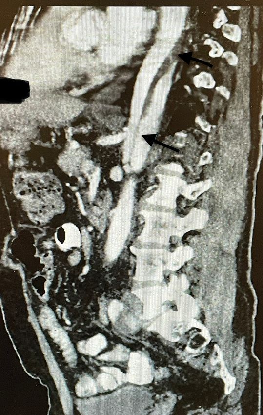
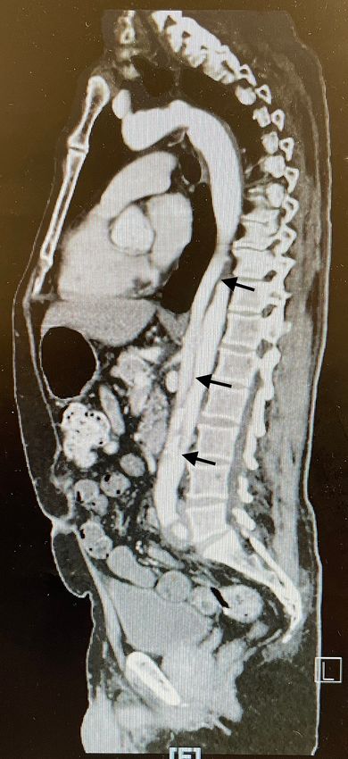
FIGURE 1: Focal arterial dissection along with thrombus extending
                                                   upward in the descending thoracic aorta approaching the distal arch as
                                                   seen on CT angiogram
                                                   CT: computed tomography

2022 Landa et al. Cureus 14(2): e22655. DOI 10.7759/cureus.22655                                                            2 of 8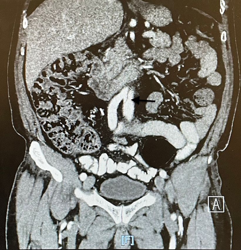
FIGURE 2: Aortic dissection as seen on CT angiogram
                                                   CT: computed tomography

2022 Landa et al. Cureus 14(2): e22655. DOI 10.7759/cureus.22655                                         3 of 8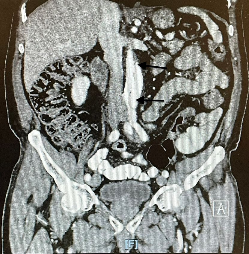
FIGURE 3: Extensive aortic dissection as seen on CT angiogram
                                                   CT: computed tomography

2022 Landa et al. Cureus 14(2): e22655. DOI 10.7759/cureus.22655                                                   4 of 8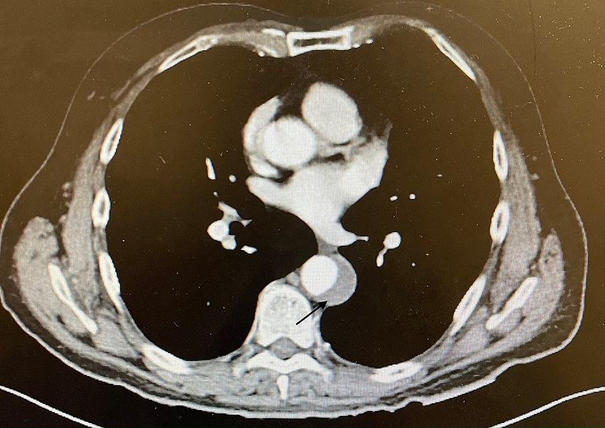
FIGURE 4: Extensive aortic dissection extending to the iliac arteries

2022 Landa et al. Cureus 14(2): e22655. DOI 10.7759/cureus.22655                                                           5 of 8
FIGURE 5: Extensive aortic dissection extending into the iliac arteries

                                                 The patient was asymptomatic without any evidence of malperfusion syndrome, but due to the extensive
                                                 nature of the dissection, he was sent to the emergency department for further evaluation by vascular
                                                 surgery. An echocardiogram was performed to evaluate the cardiac function, which revealed a normal
                                                 ejection fraction of 55-60% with no regional wall motion abnormalities. The decision was made by the
                                                 surgical team to perform a thoracic endovascular aortic repair (TEVAR) given how extensive the dissection
                                                 was. Stents were placed just distal to the left subclavian artery, proximal to the celiac artery. No
                                                 complications occurred during or after the procedure. He remained in the hospital for another two days.
                                                 After a successful procedure and recovery, the patient was discharged home on aspirin 81 mg and
                                                 atorvastatin 20 mg. The patient followed up at the clinic four weeks later and reported feeling good, and
                                                 denied any abdominal and/or back pain, nausea, or other symptoms.

                                                 Discussion
                                                 Aortic dissection is the most common and devastating manifestation of acute aortic syndrome. Its other
                                                 manifestations include penetrating aortic ulcer, intramural hematoma, and ruptured thoracic aortic
                                                 aneurysm [1].

                                                 Genetic or acquired diseases predispose individuals to aortic wall weakening, leading to acute aortic
                                                 syndromes [2]. The International Registry of Acute Aortic Dissection (IRAD) data review showed that aortic
                                                 dissection is more common in men and presents in them earlier than in women (60 vs. 67 years).
                                                 Hypertension, atherosclerosis, prior aortic surgeries, and connective tissue disorders are the most common
                                                 risk factors associated with aortic dissection, the latter being the genetic component, involving the younger
                                                 population [2-4]. Another review has shown that among people under the age of 40 years with aortic
                                                 dissection, only 40% had a history of hypertension, and 1% had a history of atherosclerosis [5].

                                                 The presentation of aortic dissection varies depending on the extent of involvement. Excruciating pain in
                                                 the chest, back, or abdomen is the typical presentation associated with aortic dissection. Chest pain is
                                                 common in ascending dissection (type A) while back and abdominal pain are more commonly associated
                                                 with descending aortic dissections (type B) [2,6-7]. Although uncommon, painless presentations of aortic

2022 Landa et al. Cureus 14(2): e22655. DOI 10.7759/cureus.22655                                                                                                 6 of 8
dissection have also been reported as in the case discussed above. Patients with painless presentation
usually have a history of diabetes, aortic aneurysm, or cardiac surgery [8]. Physical findings associated with
the dissection may include blood pressure discrepancies, pulse deficit, and heart murmurs. Focal
neurological deficits may also be appreciated with an extensive dissection propagating to the branch
arteries.
The diagnosis of acute aortic dissection requires a high index of clinical suspicion based on the presenting
symptoms and is an important one to make as a missed diagnosis could lead to a high mortality risk [9-11].
The clinical triad of sharp tearing pain, blood pressure discrepancy (>20-mmHg difference between the right
and left arms), and mediastinal widening on chest radiograph could characterize 96% of cases, but
cardiovascular imaging remains the ultimate method of diagnosis [12]. CT angiography, MR angiography,
and transesophageal echocardiography are the imaging modalities used to diagnose aortic dissection. One
may be preferred over the other depending on the availability and patient characteristics. Due to its
relatively wider availability, CT angiography is usually preferred as the initial modality of choice [13]. The
creation of a false lumen and intimal flap separating it from the true lumen suggests dissection of the aorta.
Transesophageal echocardiography is preferred for hemodynamically unstable patients since it has the
advantage of bedside availability and being a portable procedure [13]. Digital subtraction aortography is
usually reserved for cases where the suspicion of aortic dissection is very high and the noninvasive
modalities are inconclusive.
The management of type A aortic dissection involves emergent open-heart surgery. The timely transfer to
the operating room should not be interrupted by administering anti-impulse and antihypertensive therapy.
The surgical approach involves the excision of the intimal tear and graft replacement of the dissected aorta
with or without aortic valve repair. Although limited, endovascular stent grafting is an alternative to surgery
for ischemic complications. Treatment of type B aortic dissection depends on the severity of the disease [13-
14]. Uncomplicated type B dissection can be successfully managed with antihypertensive and anti-pulsatile
therapy. Endovascular or surgical intervention is usually reserved for complicated dissections that involve
the progression of the dissection leading to end-organ ischemia, hematoma formation, or frank rupture of
the aorta. These complications are associated with a higher mortality rate of 60% [14]. A similar yet different
case has been reported in the literature, and it involves an asymptomatic dissection that required immediate
action; the patient was a 39-year-old pregnant woman at 35 weeks' gestation who was found to have an
aortic dissection on echocardiogram. She had to deliver immediately; however, she refused aortic surgery
and never followed up after discharge [15]. Our case involved an incidental finding of an extensive aortic
dissection.
Conclusions
Although type B aortic dissections are not as severe or acute as the type A variant, they require immediate
attention as they can evolve into an emergency. Patients are often asymptomatic but can also present
acutely, and in such cases, the imaging modality of choice is a CT angiogram in order to visualize the culprit
lesion. In our case, the patient was supposed to undergo a CT without contrast for the evaluation of an
abdominal hernia but erroneously received one with contrast, which led to the discovery of an extensive
aortic dissection extending from the thoracic aorta to the iliac arteries. This report presents an extreme case
of an asymptomatic incidental finding of a type B aortic dissection where the patient required emergent
surgical repair.
Additional Information
Disclosures
Human subjects: Consent was obtained or waived by all participants in this study. Conflicts of interest: In
compliance with the ICMJE uniform disclosure form, all authors declare the following: Payment/services
info: All authors have declared that no financial support was received from any organization for the
submitted work. Financial relationships: All authors have declared that they have no financial
relationships at present or within the previous three years with any organizations that might have an
interest in the submitted work. Other relationships: All authors have declared that there are no other
relationships or activities that could appear to have influenced the submitted work.
References
1. Vilacosta I, San Román JA: Acute aortic syndrome. Heart. 2001, 85:365-8. 10.1136/heart.85.4.365
2. Larson EW, Edwards WD: Risk factors for aortic dissection: a necropsy study of 161 cases . Am J Cardiol.
1984, 53:849-55. 10.1016/0002-9149(84)90418-1
3. Hagan PG, Nienaber CA, Isselbacher EM, et al.: The International Registry of Acute Aortic Dissection
(IRAD): new insights into an old disease. JAMA. 2000, 283:897-903. 10.1001/jama.283.7.897
4. Spittell PC, Spittell JA Jr, Joyce JW, Tajik AJ, Edwards WD, Schaff HV, Stanson AW: Clinical features and
differential diagnosis of aortic dissection: experience with 236 cases (1980 through 1990). Mayo Clin Proc.
1993, 68:642-51. 10.1016/s0025-6196(12)60599-0
5. Januzzi JL, Isselbacher EM, Fattori R, et al.: Characterizing the young patient with aortic dissection: results
from the International Registry of Aortic Dissection (IRAD). J Am Coll Cardiol. 2004, 43:665-9.
10.1016/j.jacc.2003.08.054
2022 Landa et al. Cureus 14(2): e22655. DOI 10.7759/cureus.22655 7 of 86. Pape LA, Awais M, Woznicki EM, et al.: Presentation, diagnosis, and outcomes of acute aortic dissection: 17-
year trends from the International Registry of Acute Aortic Dissection. J Am Coll Cardiol. 2015, 66:350-8.
10.1016/j.jacc.2015.05.029
7. Evangelista A, Isselbacher EM, Bossone E, et al.: Insights from the International Registry of Acute Aortic
Dissection: a 20-year experience of collaborative clinical research. Circulation. 2018, 137:1846-60.
10.1161/CIRCULATIONAHA.117.031264
8. Park SW, Hutchison S, Mehta RH, et al.: Association of painless acute aortic dissection with increased
mortality. Mayo Clin Proc. 2004, 79:1252-7. 10.4065/79.10.1252
9. Huynh N, Thordsen S, Thomas T, et al.: Clinical and pathologic findings of aortic dissection at autopsy:
review of 336 cases over nearly 6 decades. Am Heart J. 2019, 209:108-15. 10.1016/j.ahj.2018.11.006
10. Hansen MS, Nogareda GJ, Hutchison SJ: Frequency of and inappropriate treatment of misdiagnosis of acute
aortic dissection. Am J Cardiol. 2007, 99:852-6. 10.1016/j.amjcard.2006.10.055
11. Chua M, Ibrahim I, Neo X, Sorokin V, Shen L, Ooi SB: Acute aortic dissection in the ED: risk factors and
predictors for missed diagnosis. Am J Emerg Med. 2012, 30:1622-6. 10.1016/j.ajem.2011.11.017
12. von Kodolitsch Y, Schwartz AG, Nienaber CA: Clinical prediction of acute aortic dissection . Arch Intern Med.
2000, 160:2977-82. 10.1001/archinte.160.19.2977
13. Kienzl D, Prosch H, Töpker M, Herold C: Imaging of non-cardiac, non-traumatic causes of acute chest pain .
Eur J Radiol. 2012, 81:3669-74. 10.1016/j.ejrad.2011.02.042
14. Nienaber CA, von Kodolitsch Y, Nicolas V, et al.: The diagnosis of thoracic aortic dissection by noninvasive
imaging procedures. N Engl J Med. 1993, 328:1-9. 10.1056/NEJM199301073280101
15. Abo-Salem E, López-Candales A: Diagnosis of asymptomatic aortic dissection during pregnancy using
contrast echocardiography. J Cardiol Cases. 2014, 9:200-2. 10.1016/j.jccase.2014.01.007
2022 Landa et al. Cureus 14(2): e22655. DOI 10.7759/cureus.22655 8 of 8You can also read