Impact of Two Neuronal Sigma-1 Receptor Modulators, PRE084 and DMT, on Neurogenesis and Neuroinflammation in an Aβ1-42-Injected, Wild-Type Mouse ...
←
→
Page content transcription
If your browser does not render page correctly, please read the page content below
International Journal of
               Molecular Sciences

Article
Impact of Two Neuronal Sigma-1 Receptor Modulators, PRE084
and DMT, on Neurogenesis and Neuroinflammation in an
Aβ1–42-Injected, Wild-Type Mouse Model of AD
Emőke Borbély † , Viktória Varga † , Titanilla Szögi                  , Ildikó Schuster, Zsolt Bozsó         , Botond Penke
and Lívia Fülöp *

                                           Department of Medical Chemistry, University of Szeged, Dóm Tér 8, H-6720 Szeged, Hungary;
                                           emokeborbely@gmail.com (E.B.); vargaviki666@gmail.com (V.V.); szogititi@gmail.com (T.S.);
                                           schuster.ildiko@med.u-szeged.hu (I.S.); bozso.zsolt@med.u-szeged.hu (Z.B.);
                                           penke.botond@med.u-szeged.hu (B.P.)
                                           * Correspondence: fulop.livia@med.u-szeged.hu; Tel.: +36-62-545-698
                                           † These authors contributed equally to this work.

                                           Abstract: Alzheimer’s disease (AD) is the most common form of dementia characterized by cognitive
                                           dysfunctions. Pharmacological interventions to slow the progression of AD are intensively studied. A
                                           potential direction targets neuronal sigma-1 receptors (S1Rs). S1R ligands are recognized as promising
                                           therapeutic agents that may alleviate symptom severity of AD, possibly via preventing amyloid-β-
                                           (Aβ-) induced neurotoxicity on the endoplasmic reticulum stress-associated pathways. Furthermore,
                                 S1Rs may also modulate adult neurogenesis, and the impairment of this process is reported to be

                                           associated with AD. We aimed to investigate the effects of two S1R agonists, dimethyltryptamine
Citation: Borbély, E.; Varga, V.; Szögi,
                                           (DMT) and PRE084, in an Aβ-induced in vivo mouse model characterizing neurogenic and anti-
T.; Schuster, I.; Bozsó, Z.; Penke, B.;
                                           neuroinflammatory symptoms of AD, and the modulatory effects of S1R agonists were analyzed by
Fülöp, L. Impact of Two Neuronal
                                           immunohistochemical methods and western blotting. DMT, binding moderately to S1R but with high
Sigma-1 Receptor Modulators,
                                           affinity to 5-HT receptors, negatively influenced neurogenesis, possibly as a result of activating both
PRE084 and DMT, on Neurogenesis
and Neuroinflammation in an
                                           receptors differently. In contrast, the highly selective S1R agonist PRE084 stimulated hippocampal
Aβ1–42 -Injected, Wild-Type Mouse          cell proliferation and differentiation. Regarding neuroinflammation, DMT and PRE084 significantly
Model of AD. Int. J. Mol. Sci. 2022, 23,   reduced Aβ1–42 -induced astrogliosis, but neither had remarkable effects on microglial activation. In
2514. https://doi.org/10.3390/             summary, the highly selective S1R agonist PRE084 may be a promising therapeutic agent for AD.
ijms23052514                               Further studies are required to clarify the multifaceted neurogenic and anti-neuroinflammatory roles
                                           of these agonists.
Academic Editors: Antonella
Scorziello and Babak Baban
                                           Keywords: Alzheimer’s disease; Aβ1–42 -induce mouse model; neurogenesis; neuroinflammation;
Received: 21 December 2021                 sigma-1 receptor; dimethyltryptamine; PRE084
Accepted: 23 February 2022
Published: 24 February 2022

Publisher’s Note: MDPI stays neutral
with regard to jurisdictional claims in    1. Introduction
published maps and institutional affil-         Alzheimer’s disease (AD) is the most common form of dementia, characterized by
iations.                                   progressive memory loss, impaired learning, and cognitive dysfunction. The main patho-
                                           logical hallmarks of AD are extracellular amyloid plaques and intracellular neurofibrillary
                                           tangles accumulated in the cerebral tissue [1], which first appear in the hippocampal and
                                           entorhinal regions of the brain, explaining the impairment of cognitive functions [2]. These
Copyright: © 2022 by the authors.
                                           changes are accompanied by the damage of synaptic connections, and neuronal death.
Licensee MDPI, Basel, Switzerland.
                                           The abnormal cleavage of amyloid precursor protein (APP) by β- and γ-secretases pre-
This article is an open access article
distributed under the terms and
                                           dominantly yields 40 to 43 amino acid long amyloid-β (Aβ) peptides, which aggregate,
conditions of the Creative Commons
                                           and manifest as cerebral deposits. Besides forming plaques, these oligomeric forms of
Attribution (CC BY) license (https://      Aβ are also thought to be neurotoxic [3–6]. These short oligomers might interfere with
creativecommons.org/licenses/by/           crucial intracellular mechanisms and signaling pathways. Thus, they may affect cell home-
4.0/).                                     ostasis, proliferation, differentiation, and survival [7–10]. Another significant symptom

Int. J. Mol. Sci. 2022, 23, 2514. https://doi.org/10.3390/ijms23052514                                         https://www.mdpi.com/journal/ijms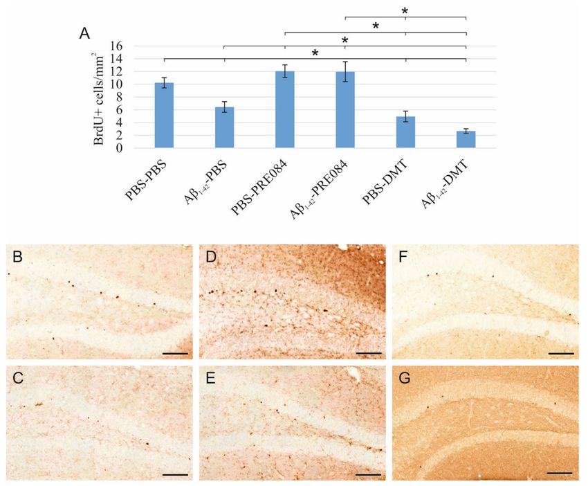
Int. J. Mol. Sci. 2022, 23, 2514                                                                                           2 of 20

                                   of AD is neuroinflammation, which involves various inflammatory components, such as
                                   immune cells, cytokines, and chemokines. Neuroinflammation might significantly alter
                                   neurogenesis, as well as enhancing Aβ production and plaque formation [11–13]. Cur-
                                   rently, there is no cure for AD, and its progression cannot be prevented; at present, only
                                   symptomatic treatments of mild to moderate efficiency are available. Therefore, effective
                                   disease-modifying therapeutics that may halt the progression of AD and contribute to
                                   the protection of neuronal integrity are eagerly awaited. A potentially new direction of
                                   the research aiming to find novel disease-modulating agents targets the sigma receptors
                                   (SRs). SRs have received considerable attention for their potential role in the prevention
                                   of Aβ-induced neurotoxicity, as well as in the regulation of the pathophysiology of AD.
                                   Furthermore, SRs may be essential for modulating neurogenesis in adulthood, and the
                                   stimulation of this process has been linked to AD. Thus, SR ligands are being recognized as
                                   promising therapeutic agents for treating or alleviating AD [6,14–16].
                                         Two subtypes of SRs are distinguished, sigma-1 receptor (S1R) and sigma-2 recep-
                                   tor [17–19]. S1R is broadly expressed in the central nervous system (CNS), especially in the
                                   dentate gyrus (DG) region of the hippocampus (HC), both in neurons and glial cells. S1Rs
                                   are mainly located in a specific part of the cell where the endoplasmic reticulum (ER) and
                                   the mitochondria establish a tight interplay; this area is called the mitochondria-associated
                                   ER membrane (MAM) [16,20–23]. S1R is known to influence neuronal survival, prolifera-
                                   tion, neurite growth, plasticity, as well as learning and memory functions [24–27]. It has
                                   been reported that the expression level of S1R decreases in patients with neurodegenerative
                                   diseases like AD [16,22,23,28–33].
                                         S1R binds a diverse set of molecules, for example, antipsychotics, antidepressants, and
                                   neurosteroids [34–37]. A non-specific endogenous ligand of S1R is N,N-dimethyltryptamine
                                   (DMT), a hallucinogenic agent assumed to be produced in small quantities and accumu-
                                   lated in the CNS [16,38–40]. Previous studies have shown that the administration of DMT
                                   modulates many ion channels [39], protects against hypoxia-induced damage [41], alle-
                                   viates neuroinflammation [42,43], increases the density of dendritic spines [44], as well
                                   as promotes neurogenesis and neuritogenesis [45–49]. However, DMT might also exert
                                   anxiogenic, neuro- and cytotoxic effects [47,50–52]. DMT is known to bind to several
                                   receptors with different affinities: 5-hydroxytryptamine (5-HT)1A-B, 5-HT1D , 5-HT2A-C ,
                                   5-HT5A , 5-HT6 , 5-HT7 receptors, S1R, SERT, dopamine (D)1-5 receptors, α1 AR, I1-3 , TAAR,
                                   NMDA [53–55]. Several adverse effects of DMT are primarily associated with the stimu-
                                   lation of 5-HT2A receptors [47,50,51,53,56], while its positive impacts are rather related to
                                   the activation of S1Rs [40–44,46,49,50,52,57]. Moreover, the inflammation regulatory and
                                   plasticity promoting activities of DMT are also considered to result from its binding to both
                                   the S1Rs and 5-HT receptors. Identifying the valid contributor molecules and signaling
                                   pathways behind this assumption requires more convincing evidence.
                                         Many exogenous ligands of S1R have been identified, including (+)-pentazocine,
                                   fluvoxamine, ANAVEX2-73, and 2-(4-morpholinethyl)-1-phenylcyclohexanecarboxylate
                                   (PRE084) [16,22,24,58]. The antidepressant and nootropic properties of PRE084 are also rec-
                                   ognized [59]. Based on our current knowledge, PRE084 may promote neuroprotection and
                                   neurite growth by stimulating the expression of different neurotrophic factors, as well as by
                                   activating signaling pathways involved in cell survival [60–65]. Previous studies suggest
                                   that this S1R-agonist might positively impact learning and memory, as demonstrated in
                                   animal models of neurodegenerative diseases or traumatic brain injuries [63,64,66]. It is also
                                   reported that after the administration of Aβ25–35 infusion into the right lateral ventricle of
                                   mice, PRE084 administration has moderated the adverse behavioral effects of Aβ25–35 [27]
                                   via reducing neurotoxicity-induced cell death [32,64]. Moreover, PRE084 may also promote
                                   neurogenesis [9] and cell survival by attenuating excitotoxicity and reducing microglial
                                   activity, as well as diminishing the expression of proinflammatory factors [67,68].
                                         As mentioned above, in addition to its ability to support cell survival under stress
                                   conditions, activated S1Rs may also stimulate the formation of new neurons, even in the
                                   adult brain. In adulthood, mammalian neurogenesis is derived from neuronal stem cells
Int. J. Mol. Sci. 2022, 23, 2514 3 of 20
(NSCs) located in the subgranular zone (SGZ) of the dentate gyrus (DG) in the hippocampus
(HC), as well as from NSCs in the subventricular region of the lateral ventricles [69,70].
After differentiation and migration, these newly formed neurons can integrate into local
neuronal circuits of the HC; thus, they might have a significant role in plasticity, cognitive
functions, learning, and memory processes [71]. An optimal microenvironment is essential
for the division, differentiation, migration, and maturation of NSCs. Physiologically, the
activity of adult hippocampal neurogenesis decreases with aging, leading to a usually
mild, age-associated cognitive decline. However, a growing body of evidence indicates
that the extent of adult neurogenesis is sharply diminished in the early stages of AD,
even before the appearance of senile plaques [72–78]. This finding raises the question of
whether impaired neurogenesis may initiate and/or contribute to more severe cognitive
deficits, thus mediating AD’s pathogenesis. Furthermore, these findings suggest that the
stimulation of neurogenesis might serve as a therapeutic target in AD, with a potential to
improve cognitive functions and promote neural adaptability, thereby it might prevent or
even treat AD.
In this study, two main objectives were addressed. First, to induce early acute AD-
like impairments in neurogenesis and generate neuroinflammation in adult wild-type
C57BL/6 mice by the intracerebroventricular (ICV) administration of Aβ1–42 oligomers.
In this experimental paradigm, we followed the administration protocol described by
Li et al., who examined the effects of Aβ25–35 on the same processes [9]. They reported
that Aβ25–35 stimulated the proliferation of neuronal progenitor cells, while enhancing the
death of newly formed neurons and impaired neurite growth. Secondly, we attempted to
restore the normal functioning of adult neurogenesis and reduce neuroinflammation by
activating S1Rs with two different ligands, PRE084 and DMT. The intraperitoneally-(IP-)-
injected compounds were tested in wild-type mice, either treated with Aβ1–42 -oligomers or
injected with vehicle (phosphate buffered saline (PBS)) as a control. Based on previously
published articles on the beneficial effects of these S1R modulators, we expected to detect
an obvious positive impact of the tested agents on the Aβ1–42 -induced impairments in
adult neurogenesis and neuroinflammation [41–43,49,52,57,60–64,79].
2. Results
2.1. Effects of PRE084 and DMT on Adult Neurogenesis in Aβ1–42 and Vehicle-Treated Mice
Aβ1–42 and DMT impair, while PRE084 promotes the survival of progenitor cells in DG.
Proliferating cells were labeled by three IP injections of 5-Bromo-20 -Deoxyuridine
(BrdU) with a 6 h interval, which was administered 24 h after the stereotaxic surgery.
BrdU is a synthetic thymidine analog, which incorporates into the DNA strand, and can
be detected by specific antibodies. We counted BrdU+ cells 14 days after the surgery.
According to our results, the quantity of BrdU+ stem cells in the SGZ of the DG significantly
differed among the six groups (ANOVA: p ≤ 0.0001). Aβ1–42 infusion significantly reduced
the number of progenitor cells compared to the respective control group (PBS-PBS vs.
Aβ1–42 -PBS p = 0.001). Interestingly, significantly more severe negative changes were
detected in animals treated with DMT. In those co-treated with both Aβ1–42 and DMT,
hardly any BrdU+ stem cells were detected in the SGZ (Aβ1–42 -DMT vs. PBS-PBS p ≤ 0.0001,
vs. Aβ1–42 -PBS p = 0.005, vs. Aβ1–42 -PRE084 p ≤ 0.0001; PBS-DMT vs. PBS-PBS p = 0.001,
vs. PBS-PRE084 p ≤ 0.0001). PRE084 treatment increased the amount of BrdU+ cells; the
difference between the Aβ1–42 -infused groups was significant (Aβ1–42 -PBS vs. Aβ1–42 -
PRE084 p ≤ 0.0001) (Figure 1).
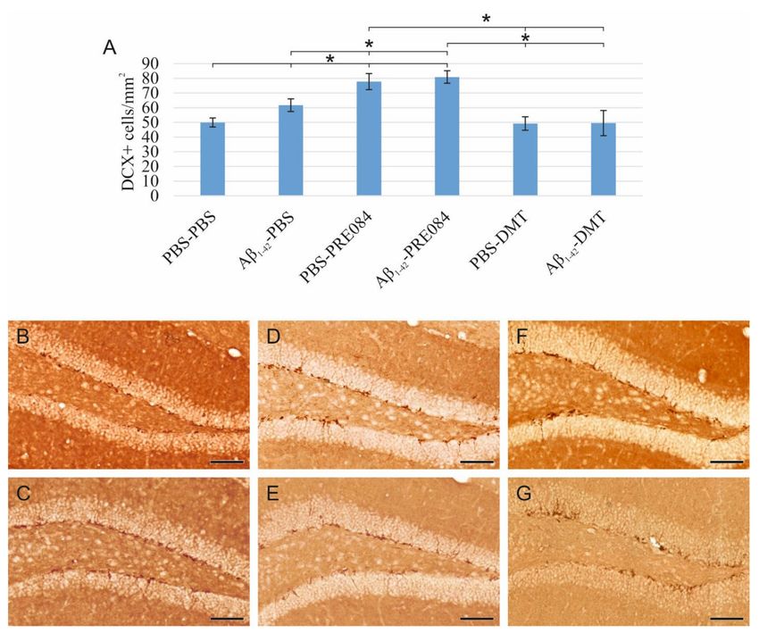
Aβ1–42 and PRE084 increase the number of premature cells, while DMT does not affect
their quantity.
To understand the effects of PRE084 and DMT on the maturation of granule cells, we
quantified immature neurons in the SGZ of DG. To label premature cells, we stained a
microtubule-associated protein called doublecortin (DCX), which is expressed specifically
in migrating neuronal precursors. The measured DCX densities were significantly differ-
ent among the six groups (ANOVA: p ≤ 0.0001). In those treated with Aβ1–42 -PBS andInt. J. Mol. Sci. 2022, 23, 2514 4 of 20
PBS-PRE084, the number of immature neurons was significantly higher compared to the
control group (PBS-PBS vs. Aβ1–42 -PBS p = 0.037, vs. PBS-PRE084 p ≤ 0.0001, vs. Aβ1–42 -
PRE084 p ≤ 0.0001). We also detected a significant difference between the Aβ1–42 -PBS and
Aβ1–42 -PRE084 mice groups (p = 0.007). DMT administration did not affect the number of
premature neurons compared to PBS-PBS mice (Figure 2).
Figure 1. (A) Results for 5-Bromo-20 -Deoxyuridine (BrdU) immunolabeling. We observed significant
differences in the quantity of stem cells between the six groups (ANOVA: p ≤ 0.0001). Significantly
fewer BrdU+ cells were detected in the Aβ1–42 -PBS, PBS-DMT, and in the Aβ1–42 -DMT treated
animals compared to the PBS-PBS group (PBS-PBS vs. Aβ1–42 -PBS p = 0.001, vs. PBS-DMT p = 0.001,
vs. Aβ1–42 -DMT p ≤ 0.0001). The difference between the Aβ1–42 -PBS and Aβ1–42 -DMT treatment
groups was also significant (p = 0.005). PRE084-treatment increased the number of stem cells detected
in the SGZ; this change was significant in the Aβ1–42 -administered group compared to its vehicle-
treated control (Aβ1–42 -PBS vs. Aβ1–42 -PRE084 p ≤ 0.0001). The differences between the following
groups in pairwise comparisons also reached significance: PBS-PRE084 vs. Aβ1–42 -PBS p ≤ 0.0001,
vs. PBS-DMT p ≤ 0.0001, vs. Aβ1–42 -DMT p ≤ 0.0001; Aβ1–42 -PRE084 vs. PBS-DMT p ≤ 0.0001, vs.
Aβ1–42 -DMT p ≤ 0.0001. (B–G) Representative images of BrdU staining: (B) PBS-PBS, (C) Aβ1–42 -PBS,
(D) PBS-PRE084, (E) Aβ1–42 -PRE084, (F) PBS-DMT, (G) Aβ1–42 -DMT. Scale bars represent 100 µm.
*: p ≤ 0.05.Int. J. Mol. Sci. 2022, 23, 2514 5 of 20
Figure 2. (A) Results for doublecortin (DCX) immunostaining. Detected DCX densities significantly
differed among the six groups (ANOVA: p ≤ 0.0001). Compared to the control (PBS-PBS) animals,
a significantly higher amount of DCX+ cells were detected in the Aβ1–42 -PBS, PBS-PRE084 and
Aβ1–42 -PRE084-treated groups (PBS-PBS vs. Aβ1–42 -PBS p = 0.037, vs. PBS-PRE084 p ≤ 0.0001, vs.
Aβ1–42 -PRE084 p ≤ 0.0001). Similarly, a significant difference was detected between the groups treated
with Aβ1–42 -PBS and Aβ1–42 -PRE084 (p = 0.007). DMT treatment did not alter the number of immature
neurons in the SGZ. Furthermore, significant differences were found when the groups were compared
to the PBS-PRE084-treated group: PBS-PRE084 vs. Aβ1–42 -PBS p = 0.023, vs. PBS-DMT p = 0.001, vs.
Aβ1–42 -DMT p = 0.001. Additional significant results were detected: Aβ1–42 -PRE084 vs. PBS-DMT
p ≤ 0.0001, vs. Aβ1–42 -DMT p ≤ 0.0001. (B–G) Representative images of DCX immunolabeling:
(B) PBS-PBS, (C) Aβ1–42 -PBS, (D) PBS-PRE084, (E) Aβ1–42 -PRE084, (F) PBS-DMT, (G) Aβ1–42 -DMT.
Scale bars represent 100 µm. *: p ≤ 0.05.
The density of mature granule cells is unaffected by Aβ1–42 or PRE084 administration,
while DMT induces a decrease in neuronal density.
To detect and evaluate mature granule cells in the HC, we performed neuronal nuclei
(NeuN) immunostaining (Figure 3). Again, significant differences were observed among
the groups (ANOVA: p = 0.001). In DMT-treated animals, significantly lower NeuN+ cell
densities were evident in the HC compared to the PBS-PBS and Aβ1–42 -PBS group (PBS-PBS
vs. PBS-DMT p = 0.001, vs. Aβ1–42 -DMT p = 0.022; Aβ1–42 -PBS vs. PBS-DMT p ≤ 0.0001, vs.
Aβ1–42 -DMT p = 0.003).Int. J. Mol. Sci. 2022, 23, 2514 6 of 20
Figure 3. (A) Results for neuronal nuclei (NeuN) immunostaining. Significant differences were
detected among the groups as follows (ANOVA: p = 0.001): in DMT-treated animals, significantly
lower NeuN densities were evident compared to the PBS-PBS and Aβ1–42 -PBS groups (PBS-DMT vs.
PBS-PBS p = 0.001, vs. Aβ1–42 -PBS p ≤ 0.0001; Aβ1–42 -DMT vs. PBS-PBS p = 0.022, vs. Aβ1–42 -PBS
p = 0.003). Furthermore, significant differences were found when the groups were compared to the
PBS-DMT-treated group: PBS-DMT vs. PBS-PRE084 p = 0.001, vs. Aβ1–42 -PRE084 p = 0.006; Aβ1–42 -
DMT vs. PBS-PRE084 p = 0.024. (B–G) Representative photomicrographs of NeuN immunolabeling:
(B) PBS-PBS, (C), Aβ1–42 -PBS (D) PBS-PRE084, (E) Aβ1–42 -PRE084, (F) PBS-DMT, (G) Aβ1–42 -DMT.
Scale bars represent 200 µm. *: p ≤ 0.05.
2.2. Effects of PRE084 and DMT on Neuroinflammation Induced by Aβ1–42
Aβ1–42 stimulates microglia activation, and neither PRE084, nor DMT alleviate this
effect, while DMT alone significantly decreases microglial density.
Neuroinflammation results from the activation of an immune response in the CNS,
mediated by microglia and astrocytes. This process is induced by infective agents, neu-
rodegenerative diseases, or injuries. To identify activated microglia in the HC, we stained
ionized calcium-binding adapter molecule 1 (Iba1), expressed explicitly by monocyte-
derived and resident macrophages, including microglia. Our results showed a significant
difference in the density of Iba1+ microglia among the groups (ANOVA: p = 0.002). Aβ1–42
administration significantly increased the density of activated microglia compared to the
vehicle-treated control groups (PBS-PBS vs. Aβ1–42 -PBS p = 0.015; PBS-PRE084 vs. Aβ1–42 -
PRE084 p = 0.035; PBS-DMT vs. Aβ1–42 -DMT p = 0.039). In the PBS-DMT group, the
density of Iba1+ microglia was significantly reduced compared to PBS-PBS-treated animals
(PBS-PBS vs. PBS-DMT p = 0.031). Still, none of the treatments were found to be able to
alleviate the proinflammatory effect of Aβ1–42 (Figure 4).Int. J. Mol. Sci. 2022, 23, 2514 7 of 20
Figure 4. (A) Results for ionized calcium-binding adapter molecule 1 (Iba1) immunolabeling. Sig-
nificant differences were observed among the groups (ANOVA: p = 0.002). Aβ1–42 increased the
density of Iba1+ microglia significantly compared to PBS-PBS, PBS-PRE084, and PBS-DMT treated
mice, respectively (PBS-PBS vs. Aβ1–42 -PBS p = 0.015; PBS-PRE084 vs. Aβ1–42 -PRE084 p = 0.035;
PBS-DMT vs. Aβ1–42 -DMT p = 0.039). The difference between the PBS-PBS and PBS-DMT groups was
also significant (PBS-PBS vs. PBS-DMT p = 0.031). Moreover, significant differences were detected
between the following groups: Aβ1–42 -PBS vs. PBS-PRE084 p = 0.005, vs. PBS-DMT p ≤ 0.0001;
Aβ1–42 -PRE084 vs. PBS-DMT p = 0.002. (B–G) Representative images of Iba1 immunostaining:
(B) PBS-PBS, (C) Aβ1–42 -PBS, (D) PBS-PRE084, (E) Aβ1–42 -PRE084, (F) PBS-DMT, (G) Aβ1–42 -DMT.
Scale bars represent 100 µm. *: p ≤ 0.05.
Aβ1–42 stimulates astrocyte reactivation, while the administration of DMT or PRE084
reduces this effect.
Reactive astrocytes were immunostained for glial fibrillary acidic protein (GFAP), an
intermediate filament protein expressed by different cell types, mainly reactive astrocytes, in
the CNS. Significantly different GFAP+ cell densities were detected in the HC of the different
groups (ANOVA: p = 0.002). A significant increase in the rate of reactivated astrocytes
was detected in the Aβ1–42 -PBS group compared to PBS-PBS-treated mice (p ≤ 0.0001).
Furthermore, GFAP+ cell densities were significantly lower in all other groups compared
to Aβ1–42 -PBS-treated mice (Aβ1–42 -PBS vs. PBS-PRE084 p = 0.013, vs. Aβ1–42 -PRE084
p = 0.013, vs. PBS-DMT p ≤ 0.0001, vs. Aβ1–42 -DMT, p = 0.001). The stimulatory effect
of Aβ1–42 on astrocyte reactivation was alleviated by PRE084 and DMT administration
(Figure 5).Int. J. Mol. Sci. 2022, 23, 2514 8 of 20
Figure 5. (A) Results of glial fibrillary acidic protein (GFAP) immunostaining. The densities of GFAP+
astrocytes differed among the groups (ANOVA: p ≤ 0.0001). A significantly higher GFAP+ density
was detected in the Aβ1–42 -PBS group compared to those treated with PBS-PBS (p ≤ 0.0001), PBS-
PRE084 (p = 0.013), Aβ1–42 -PRE084 (p = 0.013), PBS-DMT (p ≤ 0.0001), and Aβ1–42 -DMT (p = 0.001).
(B–G) Representative images of GFAP immunolabeling: (B) PBS-PBS, (C) Aβ1–42 -PBS, (D) PBS-
PRE084, (E) Aβ1–42 -PRE084, (F) PBS-DMT, (G) Aβ1–42 -DMT. Scale bars represent 100 µm. *: p ≤ 0.05.
The activation of inflammatory processes was assessed by the determination of certain
proinflammatory cytokines (IL1β and TNFα). The levels of both pro- IL1β and soluble
IL1β, as well as membrane-bound TNFα and soluble TNFα, were determined by western
blot analyses (see Supplement Figure S1). These results corroborate our findings regarding
the activation of the glial immunodefense system in response to the Aβ1–42 stimulus. The
production of the active cytokine forms could be modulated by DMT-treatment; however,
only the change in TNFα-level was significant.
2.3. S1R Protein Level Is Elevated by Aβ1–42 Treatment, as Well as by the Co-Administration of
Aβ1–42 and PRE084 or DMT
To determine the effects of Aβ1–42 and PRE084 or DMT on the expression of S1R,
a western blot (WB) analysis using GAPDH loading control was performed on HC and
cerebral cortex samples of three animals per group. Our findings revealed a significant
difference in the S1R levels among the groups (ANOVA: p ≤ 0.0001). S1R protein levels were
significantly elevated in all groups, except in PBS-DMT-treated animals, as compared to
control subjects (PBS-PBS vs. Aβ1–42 -PBS p ≤ 0.0001, vs. PBS-PRE084 p = 0.018, vs. Aβ1–42 -Int. J. Mol. Sci. 2022, 23, 2514 9 of 20
PRE084 p ≤ 0.0001, vs. PBS-DMT p = 0.540; vs. Aβ1–42 -DMT p ≤ 0.0001, respectively).
In comparison with Aβ1–42 -PBS-treated mice, the Aβ1–42 -PRE084 (p = 0.004) and Aβ1–42 -
DMT (p = 0.673) groups showed higher protein levels, while significantly lower levels of
S1R were detected in PBS-PRE084 (p = 0.032) and PBS-DMT (p = 0.001) treated mice. As
expected, the co-administration of Aβ1–42 and either of the S1R agonists increased the S1R
protein level compared to the respective control group (Aβ1–42 -PRE084 vs. PBS-PRE084
p ≤ 0.0001; Aβ1–42 -DMT vs. PBS-DMT p = 0.015). Notably, the expression of S1R was
significantly increased in Aβ1–42 -PRE084-treated animals compared to the Aβ1–42 -DMT
group (p ≤ 0.0001). (Figure 6).
Figure 6. (A) Results for the western blot (WB) analysis. Significant differences were observed in the
S1R levels among the groups (ANOVA: p ≤ 0.0001). Compared to PBS-PBS-treated mice, the S1R
protein levels were significantly elevated in the Aβ1–42 -PBS (p ≤ 0.0001), PBS-PRE084 (p = 0.018),
Aβ1–42 -PRE084 (p ≤ 0.0001), and Aβ1–42 -DMT (p ≤ 0.0001) groups. In PBS-DMT-treated mice, the S1R
protein expression remained close to the control level (p = 0.540), while S1R levels were somewhat
higher in the PRE084-treated groups (PBS-PBS vs. PBS-PRE084 p = 0.018; Aβ1–42 -PBS vs. Aβ1–42 -
PRE084 p = 0.004; PBS-PRE084 vs. Aβ1–42 -PRE084 p ≤ 0.0001). In contrast, the co-administration of
Aβ1–42 and DMT induced a significant increase in the quantity of S1R (PBS-DMT vs. Aβ1–42 -DMT
p ≤ 0.0001). Furthermore, significant differences were detected in the S1R expression upon the
pairwise comparisons of the following groups: Aβ1–42 -PBS vs. PBS-PRE084 p = 0.032, vs. PBS-DMT
p = 0.001; Aβ1–42 -PRE084 vs. PBS-DMT p ≤ 0.0001, vs. Aβ1–42 -DMT p ≤ 0.0001, respectively. (B) WB
gel electrophoresis images of S1R and GAPDH lines of the experimental groups. *: p ≤ 0.05.
3. Discussion
During neurogenesis in adulthood, new neurons continuously develop and differentiate
from hippocampal stem cells, and are integrated into existing neuronal networks to maintain
plasticity of the CNS, and thereby preserve learning and memory functions. It has been
recognized that the formation of new neurons reduces with age, manifesting in impaired
cognitive functions [80]. In certain neurodegenerative diseases this cluster of mental symptoms
is much more pronounced due to a decreased rate of neurogenesis, increased destruction of
mature neurons, and enhanced neuroinflammatory responses. The most prevalent disease of
this kind is AD, characterized by progressive dementia. Early alternations in adult neurogenesisInt. J. Mol. Sci. 2022, 23, 2514 10 of 20
and neuroinflammation may appear several years or even a decade before the diagnosis of AD,
and probably contributes to the onset of neurological symptoms. It is hypothesized that an
intensive stimulation of hippocampal neurogenesis and the reduction in neuroinflammation
in adulthood could slow down the rate of decline of cognitive skills. Moreover, the uniquely
structured S1R protein, functioning as a ligand-operated chaperone, is known to play a major
role in both neurogenesis and neuroinflammation. Thus, it is assumed that the activation of
S1Rs may be a promising therapeutic strategy to stimulate adult neurogenesis and alleviate
neuroinflammatory processes.
The first objective of our study was to model these early alternations appearing in AD.
Our experimental paradigm was based on the work of Li et al., in a modified way: instead
of Aβ25–35 , we injected Aβ1–42 ICV to induce early AD-like changes [9]. The reason for this
modification is that Aβ25–35 is a non-natural, truncated sequence, and although it is prone to
aggregation, its kinetics for aggregation differ from that of the native Aβ1–42 peptide. There-
fore, using this latter peptide should yield biologically more relevant findings [81]. In the
work of Li et al., neurogenesis was assessed 14 and 28 days after the peptide injections, and
significant differences were detected on day 28 in neurogenic markers compared to baseline
(reduced proliferation and neurite growth, increased death of newly formed cells) [9]. In our
experimental model, AD-like cerebral neurogenic and neuroinflammatory changes could
be detected as early as two weeks after the administration of Aβ1–42 . We demonstrated that
a single administration of Aβ1–42 , directly into the lateral ventricles, significantly impaired
the proliferation and increased the number of immature cells in mice. The effects of Aβ on
neurogenesis are highly controversial in the literature. Numerous reports indicate that Aβ
significantly decreases the formation of new neurons, possibly by impairing their ability to
divide, as well as by diminishing the survival of neuronal stem cells in DG [7–9,75–77,82].
However, some research groups have published that Aβ can induce the initial proliferation
step of neuron formation in different transgenic mouse strains [9,78,83–85] or in cellular
models of AD [86–91]. In our experiments, an increase in the number of differentiating
immature neurons was observed in Aβ1–42 -treated animals, which may be explained by a
compensatory cerebral mechanism [77,92]. Specifically, this enhancement of neuronal cell
differentiation may be a response to the disturbed homeostasis resulting from the decrease
in the stem cell population, aiming to restore the balance within the CNS. As we expected,
in our experimental model, no significant reduction was detected in the density of mature,
functional neurons in HC two weeks after the administration of Aβ1–42 , indicating that
the existing neuronal system may remain unaffected. Regarding neuroinflammation, we
found that a single administration of Aβ1–42 stimulated neuroinflammatory processes,
causing a significant increase in the densities of activated microglia and hyperreactive
astrocytes. In line with our observations, several in vivo experiments have demonstrated
the neuroinflammation-inducing effects of Aβ fibrils and oligomers injected into the brain
tissue in different experimental models [93–95]. This neuroinflammatory environment may
affect adult neurogenesis either positively or negatively [11,12,96–101]. It is known that cy-
tokines and chemokines produced by activated microglia and astrocytes play an important
role in neuroinflammatory processes. Certain anti-(IL-4, IL-10) and proinflammatory (IL-6,
TNF-α) factors substantially influence neurogenesis, e.g., they can diminish proliferation
and cell survival, while they may also stimulate cell differentiation [13]. Thus, beyond its
direct effects on immature neurons, Aβ1–42 may also affect neurogenesis by generating a
relatively mild, but chronic neuroinflammatory environment. Further research is needed
to clarify the relative contribution of these two processes (direct and indirect) to the final
decline of adult neurogenesis in AD.
Since the S1R protein plays a major role in neurogenesis and neuroinflammation, and
changes in S1R expression levels have not been studied in exogenous Aβ-induced AD
models, we examined the expression levels of this protein. In our case, the expression of
S1R increased after a single administration of Aβ1–42 . This finding may contradict some
literature data, which report on the down-regulation of S1R in the early stage of human
AD [24]. In the reported cases, both the amount and the binding potential of S1R were foundInt. J. Mol. Sci. 2022, 23, 2514 11 of 20
to be decreased, presumably as a consequence of hippocampal neuronal death [24,102–105].
In contrast, other studies indicate that AD-related ER-stress can lead to an up-regulation
of S1R [16,29,106,107], which, serving as a chaperon, modulates the canonical unfolded
protein response (UPR) pathways (PERK, IRE1a, ATF6) [16,108]. In our study, the observed
elevation of the level of S1R may be a consequence of the cytotoxic effect of Aβ1–42 , which
induces ER stress, and thus activates the UPR pathways and upregulates S1R expression.
To date, the biological effects of DMT and PRE084 have not been studied in an Aβ-
induced model of early AD with demonstrated changes in neurogenesis and S1R expression
levels, as well as neuroinflammation. Therefore, we aimed to assess whether the modulation
of S1Rs with selected ligands can restore Aβ1–42 -induced alternations in adult neurogenesis
and reduce neuroinflammation.
In our study, DMT significantly reduced the number of neuronal stem cells and densi-
ties of neurons. Similar to this finding, another tryptamine, psilocybin (4-phosphoryloxy-N,
N-dimethyltryptamine) with a chemical structure close to that of DMT and a high binding
affinity to 5-HT2A receptors (Kd = 6 nM), was also found to impair synaptic growth and
neurogenesis (proliferation and neuronal survival) [109]. However, the neuroprotective and
neurogenesis stimulating effects of DMT and its analog, 5-methoxy-DMT, exerted via S1Rs,
were also described in in vitro cell cultures and in a wild-type rodent model [44,46,49,54].
In our study, DMT was administered at a concentration of 1 mg kg–1 , thus it is supposed to
have occupied both receptor types, so their mixed effects could have been observed. Com-
parison of the Kd values (DMT-S1R Kd = 14.75 µM, DMT-5-HT2A receptor Kd = 130 nM)
indicates that DMT binds to the 5-HT2A receptor with higher affinity than to S1R; thus, it is
more likely to act on the 5-HT2A receptors than on S1R [39,53]. Therefore, we suppose that
DMT exerted its negative effect on neurogenesis via the 5-HT2A receptors. The results of
our WB analysis support this hypothesis, since the expression of the S1R protein was only
slightly elevated after DMT treatment.
Regarding the relation of DMT and neuroinflammation, conflicting findings are pub-
lished in the literature. Some of them support the theory that DMT can alleviate neuroin-
flammatory processes, thus it may reduce the density of reactive astrocytes [41–43,52,57].
This effect may be related to the ability of DMT to bind to S1R [41–43,52], but the serotoner-
gic receptors may also have roles in this process [110]. Morales-Garcia et al. reported that
DMT induces a significant increase in the density of GFAP+ astrocytes via the activation
of S1Rs, but these researchers conclude that this elevated GFAP level promotes neuroge-
nesis [49]. In our experiments, DMT treatment was found to exert a positive effect on
activated microglia and hyperreactive astrocytes against the Aβ1–42 -induced neurotoxicity,
but it was not detected to promote neurogenesis.
These contradictory results may be explained by the application of different protocols
(injection and doses of BrdU and DMT, different survival times). It is also known that
although DMT can penetrate the blood-brain barrier, upon exogenous administration its
concentration in the CNS is elevated for a relatively short time only (elimination half-life
~15 min [44]). Therefore, it is also possible that in our model, the concentration of DMT in
the CNS after IP administration was not sufficient to exert its effects on S1R as Morales-
Garcia reported [49]. Further experiments are required to elucidate the exact mode of action
of DMT regarding neurogenesis and neuroinflammation.
To study the effect of an exogenous S1R agonist on neurogenesis and neuroinflam-
mation, we applied PRE084 (Kd = 2.2 nM, [111]). Similarly, as Li et al. reported in an
Aβ25–35 -induced mouse model of AD, we have demonstrated that PRE084 promotes neu-
rogenesis upon treatment with Aβ1–42 , as it is indicated by the quantitative increase in
stem cells and immature neurons after PRE084 administration. Furthermore, PRE084 per
se activates cell proliferation, possibly by stimulating S1R.
Regarding neuroinflammation, the density of hyperreactive astrocytes and the degree
of Aβ1–42 -induced astrogliosis were reduced by the administration of PRE084. However,
the substance neither per se, nor in combination with Aβ1–42 could impair microglial
activation. It is known that in case of CNS tissue damage, activated microglia may behaveInt. J. Mol. Sci. 2022, 23, 2514 12 of 20
either neurotoxic or neuroprotective, depending on their morphological and functional
states. According to the literature, PRE084 can stimulate the proliferation of the anti-
inflammatory type of microglia (M2), while it suppresses pro-inflammatory M1 microglia,
thus it maintains the delicate balance between functional restorative and inflammatory
glial phenotypes [62,112]. As we did not analyze the distribution and morphology of the
microglia, we assume that the apparent ineffectiveness of PRE084 treatment on microglial
activation may result from the above mentioned two mutual processes.
PRE084 binds to S1R with high affinity, either alone (compared to PBS and DMT
controls) and when co-administered with Aβ1–42 (compared to Aβ1–42 -PBS or Aβ1–42 -DMT
animals), and significantly induces the expression of this receptor protein. These results
may confirm that PRE084 activates the S1R receptors effectively, so its neurogenic impact is
more pronounced than that of DMT.
4. Materials and Methods
4.1. Animals
Male C57BL/6 wild-type mice (n = 80) from in-house breeding, weighing 23–28 g
and aged 12 weeks at the beginning of the study, were used for the experiments. All
animals, divided into groups, were kept under constant circumstances, including constant
temperature (23 ± 0.5 ◦ C), lighting (12:12 h light/dark cycle, lights on at 7 a.m.), and
humidity (~50%). Standard mouse chow and tap water were supplied ad libitum. All
behavioral experiments were performed in the light period. Handling was executed daily,
at the same time, started one week before the experiments. All efforts were made to
minimize the number of animals used, and their suffering throughout the experiments.
All experiments were performed in accordance with the European Communities
Council Directive of 22 September 2010 (2010/63/EU on protecting animals used for
scientific purposes). The experimental protocols were approved by the National Food
Chain Safety and Animal Health Directorate of Csongrad County, Hungary (project li-
cense: XXVI./3644/2017). Formal approval to conduct the experiments was obtained from
the Animal Welfare Committee of the University of Szeged (project No. I-74-16/2017,
04.07.2017).
4.2. Preparation and Structure Analysis of Aβ1–42 Peptide Oligomers
The iso-Aβ1–42 peptide was synthesized in the solid phase using tert-butyloxycarbonyl
(Boc)-chemistry in-house, as reported earlier [113]. A stock solution of this peptide was
prepared using distilled water, to yield a concentration of 1 mg/mL (200 µM, pH = 7), and
it was sonicated for 3 min. The solution was incubated for 10 min at room temperature
(RT), then the pH level was adjusted (pH = 11), and it was further incubated for 2 h.
After a 3-min-long sonication process, the Aβ1–42 solution was diluted in phosphate buffer
(PBS, 20 mM) to a final peptide concentration of 50 µM (26.67 mM phosphate, 1.2% NaCl,
pH = 7.4). The solution was stored at 4 ◦ C until further use on the same day.
The oligomeric state of the Aβ peptide was verified by a transmission electron micro-
scope (JEM-1400, JEOL USA Inc., Peabody, MA, USA) operating at 120 kV. Images were
taken by an EM-15300SXV system, routinely at a magnification of 25,000 and 50,000, and
were processed by the SightX Viewer Software (EM-15300SXV Image Edit Software, JEOL
Ltd., Tokyo, Japan).
4.3. Surgery, Solutions, and Drug Administration
Mice were anesthetized by an IP injection of a mixture of ketamine (10.0 mg/0.1 kg)
and xylazine (0.8 mg/0.1 kg). The animals were then placed into a stereotaxic apparatus
(David Kopf Instruments, Tujunga, CA, USA; Stoelting Co., Wood Dale, IL, USA), a midline
incision of the scalp was made, the skin and muscles were carefully retracted to expose
the skull, and a hole was drilled above the target area. A single intracerebroventricular
injection of either Aβ1–42 (50 µM) or PBS (20 mM) was administered at the right side using
a Hamilton syringe (32 G), injected at a rate of 0.5 µL/min. The following coordinates wereInt. J. Mol. Sci. 2022, 23, 2514 13 of 20
used (from Bregma point): AP: −0.3; ML: −1.0; DV: −2.5. All animals were treated with
antibiotics and analgesics after the surgery.
To detect stem cells, the animals were injected IP with BrdU (50 mg kg–1 ; Sigma-
Aldrich, Saint Louis, MO, USA) dissolved in physiological saline, 3 times, 24 h after the
surgery as described previously by Li et al. [9].
PRE084 (1 mg kg–1 , Sigma-Aldrich, Saint Louis, MO, USA) and DMT (1 mg kg–1 ,
Lipomed AG, Arlesheim, Switzerland) were also administered IP on a daily basis between
postsurgery days 7–12. Both substances were dissolved in PBS (sterile-filtered, 20 mM)
complemented with 1% dimethyl sulfoxide (DMSO, Sigma-Aldrich, Saint Louis, MO, USA).
Six groups of animals (with 18 mice in the control group, whereas 11 mice per group
in the other groups) were developed to represent a control for each of the Aβ1–42 -treated
groups (i.e., those PBS-treated after the development of AD-like symptoms of impaired
neurogenesis and neuroinflammation and those treated with DMT or PRE084 after the
induction of neurogenic and neuroinflammatory changes). In the nomenclature of the
groups, the first term refers the ICV administered solution (PBS or Aβ1–42 ), while the
second one indicates the IP injected agent with potential disease-modifying activity (PBS
again as a control, or PRE084 or DMT). Based on this nomenclature, the six groups were
the following:
ICV: PBS-IP: PBS (PBS-PBS, i.e., PBS-treated, non-diseased control; n = 18),
ICV: Aβ1–42 -IP: PBS (Aβ1–42 -PBS, i.e., Aβ1–42 -treated, PBS-treated control; n = 18),
ICV: PBS-IP: PRE084 (PBS-PRE084, i.e., PRE084-treated, non-diseased control; n = 11),
ICV: Aβ1–42 -IP: PRE084 (Aβ1–42 -PRE084, i.e., Aβ1–42 -treated, PRE084-treated group; n = 11),
ICV: PBS-IP: DMT (PBS-DMT, i.e., DMT-treated, non-diseased control; n = 11),
ICV: Aβ1–42 -IP: DMT (Aβ1–42 -DMT, i.e., Aβ1–42 -treated, DMT-treated group; n = 11).
4.4. Immunohistochemistry
Two weeks after the surgery, mice (n = 8-8 from the PRE084- and DMT-treated, and
n = 15-15 from the control groups) were anesthetized with chloral hydrate (1 mg kg–1 ) and
were perfused transcardially with PBS, followed by 4% paraformaldehyde (PFA, Sigma-
Aldrich, St. Louis, MO, USA). All procedures after perfusion, including the post-fixation
and the preparation of the slides, were executed the same way as described previously [114].
Immunohistochemical analysis was carried out on 20 µM formalin fixed cryosections.
All immunohistochemical procedures were performed according to Szogi et al. [114]. All
chemicals used in the immunohistochemical procedures, except the antibodies (Ab), were
purchased from Sigma-Aldrich (St. Louis, MO, USA). Briefly, for BrdU staining, the sections
were incubated in 2 M HCl for 2 h at RT to denature DNA. For the evaluation of BrdU-
stained and NeuN-positive cells, the sections were blocked in a mixture of 8% normal
goat serum, 0.3% bovine serum albumin (BSA), and 0.3% Triton X-100 in PBS for 1 h at
RT. For DCX, Iba1 and GFAP labeling, the sections were blocked in a mixture of 0.1% BSA
and 0.3% Triton X-100 in PBS for 1 h at RT. After this step, the slices were incubated at
4 ◦ C overnight with primary antibodies added to the samples in the following dilutions:
mouse anti-BrdU Ab (1:800; Santa Cruz Biotechnology, Dallas, TX, USA), goat anti-DCX Ab
(1:4000; Santa Cruz Biotechnology, Dallas, TX, USA), mouse anti-NeuN Ab (1:500; Merck
Millipore, Darmstadt, Germany), rabbit anti-Iba1 Ab (1:3600; Wako Chemicals GmbH,
Neuss, Germany), and mouse anti-GFAP Ab (1:1500; Santa Cruz Biotechnology, Dallas, TX,
USA). For BrdU, DCX, and NeuN stainings, the sections were treated with a polymer-based
HRP-amplifying system (Super SensitiveTM One-Step Polymer-HRP Detection System,
BioGenex, Fremont, CA, USA), according to the manufacturer’s instructions. For Iba1 and
GFAP labeling, the slices were incubated with the corresponding secondary antibodies:
biotinylated goat anti-rabbit Ab (1:400; Jackson ImmunoResearch, West Grove, PA, USA),
and biotinylated goat anti-mouse Ab (1:400; ThermoFisher Scientific, Waltham, MA, USA)
for 60 min. Next, the sections were rinsed 3 times in PBS, and were incubated with avidin-
biotin-complex (ABC Elite Kit; Vector Laboratories, Burlingame, CA, USA) for Iba1 in
1:1000 and for GFAP stainings in 1:1500, for 60 min at RT. The peroxidase immunolabelingInt. J. Mol. Sci. 2022, 23, 2514 14 of 20
was developed in 0.5 M Tris-HCl buffer (pH 7.7) with 3,30 -diaminobenzidine (10 mM) at RT
in 30 min. The sections were mounted with dibutyl phthalate xylene onto the slides and
were coverslipped.
4.5. Quantification of the Immunohistochemical Data
Slides were scanned by a digital slide scanner (Mirax Midi, 3DHistech Ltd., Budapest,
Hungary), equipped with a Panoramic Viewer 1.15.4, a CaseViewer 2.1 program and a
QuantCenter, HistoQuant module (3DHistech Ltd., Budapest, Hungary). For quantifica-
tions, all sections derived from each animal were analyzed. In DG and HC, the regions
of interest (ROI) were manually outlined. Antibody-positive cell types were counted and
quantified from ROIs. The number of stem cells (BrdU+) and neuroblasts (DCX+) were
assessed by the observers. The densities (%) of neurons (NeuN+), microglia (Iba1+), and
astrocytes (GFAP+) were calculated by the quantification software. To assess cell densi-
ties, we divided the total number of counted cells per animal with the DG/HC area, and
represented them as cells/mm2 (BrdU+, DCX+) or % (NeuN+, Iba+, GFAP+).
4.6. Western Blot Analysis
To determine the effects of Aβ1–42 and PRE084 or DMT on the expression of S1R,
the receptor protein samples of 3 animals per group (n = 18) were identically prepared,
separated, and transferred to nitrocellulose membranes. The membranes were washed
and treated as described by Szogi et al. [54]. The levels of S1R (mouse S1R antibody,
Santa Cruz, Dallas, TX, USA, 1:1000) were analyzed in each group. For the analysis, we
used glyceraldehyde 3-phosphate dehydrogenase (GAPDH, rabbit GAPDH antibody, Cell
Signaling, Danvers, MA, USA, 1:200,000) as the loading control.
4.7. Statistical Analysis
The data obtained from the immunohistochemistry analyses were evaluated with a
one-way ANOVA, followed by Fisher’s LSD post hoc tests. The WB data did not follow
normal distribution; thus, they were analyzed with Kruskal-Wallis nonparametric tests,
followed by Mann–Whitney U tests for multiple comparisons. Data were analyzed with the
SPSS software (IBM SPSS Statistics 24), and the results were expressed as mean ± (SEM).
Statistical significance was set at p ≤ 0.05.
5. Conclusions
Adult neurogenesis is essential for CNS plasticity. In early AD, neurogenic impairment
can be observed, accompanied by hyperreactive astrogliosis. During the treatment of
AD, neurogenesis should be promoted, while neuroinflammation should be suppressed.
S1R plays a role in both processes. In our experiments, we established a model of early
AD induced by Aβ1–42 , in which acute neuroinflammation, impaired neurogenesis and
elevated S1R levels were detected. In this model, two S1R agonists were tested. DMT,
binding moderately to S1R but with a high affinity to 5-HT receptors, negatively influenced
neurogenesis in the Aβ1–42 -induced rodent model, probably explained by its acting on the
latter receptor class. In contrast, the highly selective S1R agonist, PRE084 improved the
proliferation and differentiation of hippocampal stem cells, manifesting in a quantitative
increase in progenitor cells and immature neurons. Further experiments are required
to investigate the main molecular pathways targeted by DMT, through which it affects
neurogenesis and the survival of mature neurons. Moreover, DMT and PRE084 were found
to significantly reduce Aβ1–42 -induced hyperreactive astrogliosis. However, none of these
ligands had a remarkable effect on microglial activation. Therefore, further studies are
needed to clarify the role of DMT and PRE084 in neuroinflammatory processes induced by
Aβ1–42 , resembling the changes characteristic of AD.
Supplementary Materials: The following supporting information can be downloaded at: https:
//www.mdpi.com/article/10.3390/ijms23052514/s1.Int. J. Mol. Sci. 2022, 23, 2514 15 of 20
Author Contributions: Conceptualization: L.F. and B.P.; methodology: V.V., E.B., T.S. and I.S.;
statistical analysis: E.B.; investigation: V.V., E.B. and T.S.; preparation and structure analysis of Aβ1–42
peptide oligomers: Z.B. and T.S.; resources: L.F. and B.P.; data curation: T.S. and E.B.; writing/original
draft preparation: V.V. and E.B.; writing/review/editing: L.F.; visualization: E.B.; supervision: L.F.;
funding acquisition: L.F. and B.P. All authors have read and agreed to the published version of
the manuscript.
Funding: This project was supported by the National Research, Development, and Innovation Office
(GINOP-2.3.2-15-2016-00060) and by the Hungarian Brain Research Program I and II—Grant No.
KTIA_13_NAP-A-III/7, and 2017-1.2.1-NKP-2017-00002. Support by the Ministry of Innovation and
Technology of Hungary from the National Research, Development and Innovation Fund (TKP2021-
EGA-32) is acknowledged.
Institutional Review Board Statement: The study was conducted according to the guidelines of the
Declaration of Helsinki, and approved by the Institutional Review Board (or Ethics Committee) of
University of Szeged (project license: XXVI./3644/2017).
Informed Consent Statement: Not applicable.
Data Availability Statement: Not applicable.
Acknowledgments: The technical assistance of Szilvia Dénes and Péter Sütő is greatly acknowledged.
The authors thank Dora Bokor, for proofreading the manuscript.
Conflicts of Interest: The authors declare no conflict of interest. The funders had no role in the
design of the study, nor in the collection, analyses, or interpretation of data, nor in the writing of the
manuscript, nor in the decision to publish the results.
Abbreviations
5-HT 5-hydroxytryptamine
AD Alzheimer’s disease
APP amyloid precursor protein
Aβ amyloid-β
BrdU 5-Bromo-20 -Deoxyuridine
BSA bovine serum albumin
CNS central nervous system
DCX doublecortin
DG dentate gyrus
DMT N,N-dimethyltryptamine
ER endoplasmic reticulum
GAPDH glyceraldehyde 3-phosphate dehydrogenase
GFAP glial fibrillary acidic protein
HC hippocampus
Iba1 ionized calcium-binding adapter molecule 1
ICV intracerebroventricular
IP intraperitoneal injection
MAM mitochondria-associated ER membrane
NeuN neuronal nuclei
NMDA N-methyl-d-aspartate receptor
NSC neuronal stem cell
PFA paraformaldehyde
PRE084 2-(4-morpholinethyl)-1-phenylcyclohexanecarboxylate
SR sigma receptor
S1R sigma-1 receptor
SGZ subgranular zone
UPR unfolded protein response
WB western blot analysisInt. J. Mol. Sci. 2022, 23, 2514 16 of 20
References
1. Alzheimer, A.; Stelzmann, R.A.; Schnitzlein, H.N.; Murtagh, F.R. An English translation of Alzheimer’s 1907 paper, “Uber eine
eigenartige Erkankung der Hirnrinde”. Clin. Anat. 1995, 8, 429–431. [PubMed]
2. Karlawish, J. 2017 Alzheimer’s Disease Facts and Figures; Alzheimer’s Association: Chicago, IL, USA, 2017; p. 88.
3. Mu, Y.; Gage, F.H. Adult hippocampal neurogenesis and its role in Alzheimer’s disease. Mol. Neurodegener. 2011, 6, 85. [CrossRef]
[PubMed]
4. Winner, B.; Winkler, J. Adult neurogenesis in neurodegenerative diseases. Cold Spring Harb. Perspect. Biol. 2015, 7, a021287.
[CrossRef] [PubMed]
5. Weintraub, S.; Wicklund, A.H.; Salmon, D.P. The neuropsychological profile of Alzheimer disease. Cold Spring Harb. Perspect.
Med. 2012, 2, a006171. [CrossRef]
6. Penke, B.; Bogár, F.; Fülöp, L. β-Amyloid and the Pathomechanisms of Alzheimer’s Disease: A Comprehensive View. Molecules
2017, 22, 1692. [CrossRef]
7. Ramírez, E.; Mendieta, L.; Flores, G.; Limón, I.D. Neurogenesis and morphological-neural alterations closely related to amyloid
β-peptide (25-35)-induced memory impairment in male rats. Neuropeptides 2018, 67, 9–19. [CrossRef]
8. Zheng, M.; Liu, J.; Ruan, Z.; Tian, S.; Ma, Y.; Zhu, J.; Li, G. Intrahippocampal injection of Aβ1-42 inhibits neurogenesis and
down-regulates IFN-γ and NF-κB expression in hippocampus of adult mouse brain. Amyloid 2013, 20, 13–20. [CrossRef]
9. Li, L.; Xu, B.; Zhu, Y.; Chen, L.; Sokabe, M. DHEA prevents Aβ25-35-impaired survival of newborn neurons in the dentate gyrus
through a modulation of PI3K-Akt-mTOR signaling. Neuropharmacology 2010, 59, 323–333. [CrossRef]
10. Russo, I.; Caracciolo, L.; Tweedie, D.; Choi, S.H.; Greig, N.H.; Barlati, S.; Bosetti, F. 3,6’-Dithiothalidomide, a new TNF-α synthesis
inhibitor, attenuates the effect of Aβ1-42 intracerebroventricular injection on hippocampal neurogenesis and memory deficit. J.
Neurochem. 2012, 122, 1181–1192. [CrossRef]
11. Minter, M.R.; Taylor, J.M.; Crack, P.J. The contribution of neuroinflammation to amyloid toxicity in Alzheimer’s disease. J.
Neurochem. 2016, 136, 457–474. [CrossRef]
12. Sung, P.S.; Lin, P.Y.; Liu, C.H.; Su, H.C.; Tsai, K.J. Neuroinflammation and Neurogenesis in Alzheimer’s Disease and Potential
Therapeutic Approaches. Int. J. Mol. Sci. 2020, 21, 701. [CrossRef] [PubMed]
13. Fuster-Matanzo, A.; Llorens-Martín, M.; Hernández, F.; Avila, J. Role of neuroinflammation in adult neurogenesis and Alzheimer
disease: Therapeutic approaches. Mediat. Inflamm. 2013, 2013, 260925. [CrossRef] [PubMed]
14. Fish, P.V.; Steadman, D.; Bayle, E.D.; Whiting, P. New approaches for the treatment of Alzheimer’s disease. Bioorg. Med. Chem.
Lett. 2019, 29, 125–133. [CrossRef]
15. Ma, W.H.; Chen, A.F.; Xie, X.Y.; Huang, Y.S. Sigma ligands as potent inhibitors of Aβ and AβOs in neurons and promising
therapeutic agents of Alzheimer’s disease. Neuropharmacology 2020, 190, 108342. [CrossRef] [PubMed]
16. Penke, B.; Fulop, L.; Szucs, M.; Frecska, E. The Role of Sigma-1 Receptor, an Intracellular Chaperone in Neurodegenerative
Diseases. Curr. Neuropharmacol. 2018, 16, 97–116. [CrossRef]
17. Bowen, W.D.; Hellewell, S.B.; McGarry, K.A. Evidence for a multi-site model of the rat brain sigma receptor. Eur. J. Pharm. 1989,
163, 309–318. [CrossRef]
18. Hellewell, S.B.; Bowen, W.D. A sigma-like binding site in rat pheochromocytoma (PC12) cells: Decreased affinity for (+)-
benzomorphans and lower molecular weight suggest a different sigma receptor form from that of guinea pig brain. Brain Res.
1990, 527, 244–253. [CrossRef]
19. Hellewell, S.B.; Bruce, A.; Feinstein, G.; Orringer, J.; Williams, W.; Bowen, W.D. Rat liver and kidney contain high densities of
sigma 1 and sigma 2 receptors: Characterization by ligand binding and photoaffinity labeling. Eur. J. Pharm. 1994, 268, 9–18.
[CrossRef]
20. Schmidt, H.R.; Zheng, S.; Gurpinar, E.; Koehl, A.; Manglik, A.; Kruse, A.C. Crystal structure of the human σ1 receptor. Nature
2016, 532, 527–530. [CrossRef]
21. Yang, K.; Wang, C.; Sun, T. The Roles of Intracellular Chaperone Proteins, Sigma Receptors, in Parkinson’s Disease (PD) and
Major Depressive Disorder (MDD). Front. Pharm. 2019, 10, 528. [CrossRef]
22. Hayashi, T.; Su, T.P. An update on the development of drugs for neuropsychiatric disorders: Focusing on the sigma 1 receptor
ligand. Expert Opin. Ther. Targets 2008, 12, 45–58. [CrossRef] [PubMed]
23. Tesei, A.; Cortesi, M.; Zamagni, A.; Arienti, C.; Pignatta, S.; Zanoni, M.; Paolillo, M.; Curti, D.; Rui, M.; Rossi, D.; et al. Sigma
Receptors as Endoplasmic Reticulum Stress “Gatekeepers” and their Modulators as Emerging New Weapons in the Fight Against
Cancer. Front. Pharm. 2018, 9, 711. [CrossRef] [PubMed]
24. Jin, J.L.; Fang, M.; Zhao, Y.X.; Liu, X.Y. Roles of sigma-1 receptors in Alzheimer’s disease. Int. J. Clin. Exp. Med. 2015, 8, 4808–4820.
[PubMed]
25. Maurice, T.; Meunier, J.; Feng, B.; Ieni, J.; Monaghan, D.T. Interaction with sigma(1) protein, but not N-methyl-D-aspartate
receptor, is involved in the pharmacological activity of donepezil. J. Pharm. Exp. Ther. 2006, 317, 606–614. [CrossRef] [PubMed]
26. Maurice, T.; Privat, A. SA4503, a novel cognitive enhancer with sigma1 receptor agonist properties, facilitates NMDA receptor-
dependent learning in mice. Eur. J. Pharm. 1997, 328, 9–18. [CrossRef]
27. Meunier, J.; Ieni, J.; Maurice, T. The anti-amnesic and neuroprotective effects of donepezil against amyloid beta25-35 peptide-
induced toxicity in mice involve an interaction with the sigma1 receptor. Br. J. Pharm. 2006, 149, 998–1012. [CrossRef]You can also read


